. 2010 Sep 28;49(38):8240-50.
doi: 10.1021/bi101052d.

Oncogenic CARD11 mutations induce hyperactive signaling by disrupting autoinhibition by the PKC-responsive inhibitory domain

Affiliations

Oncogenic CARD11 mutations induce hyperactive signaling by disrupting autoinhibition by the PKC-responsive inhibitory domain

Rebecca L Lamason et al. Biochemistry. .

Abstract

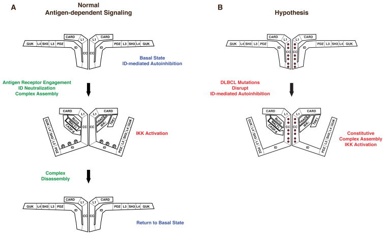
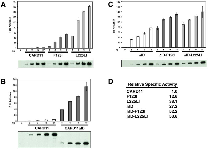
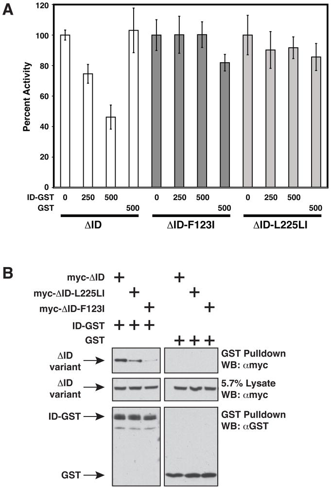
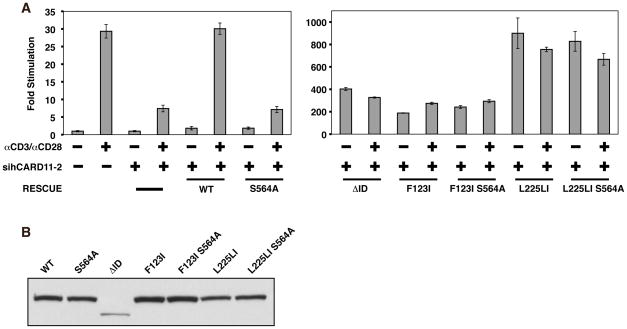
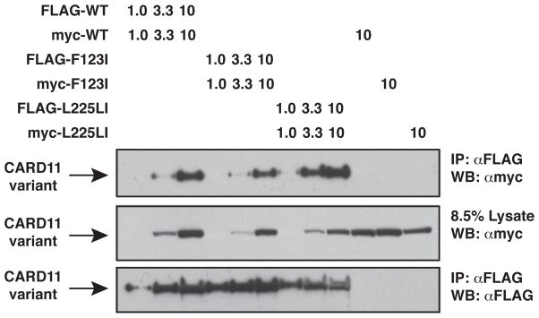
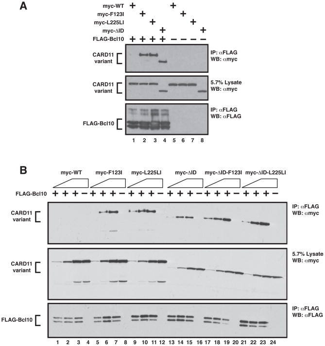
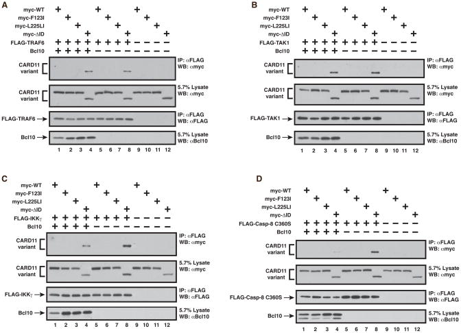
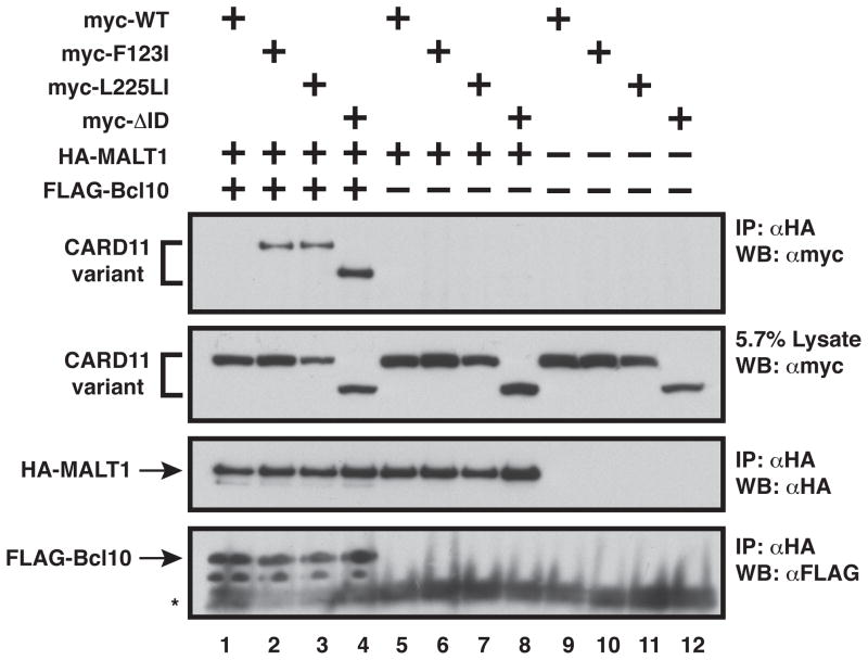
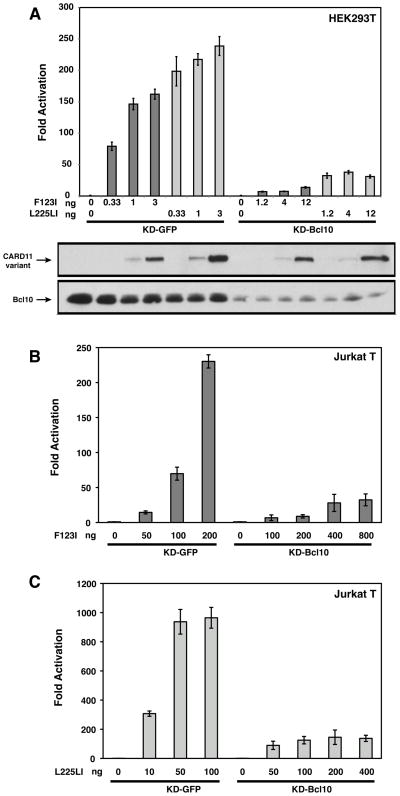
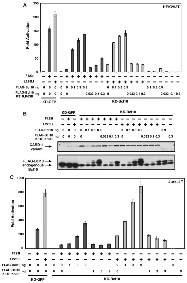
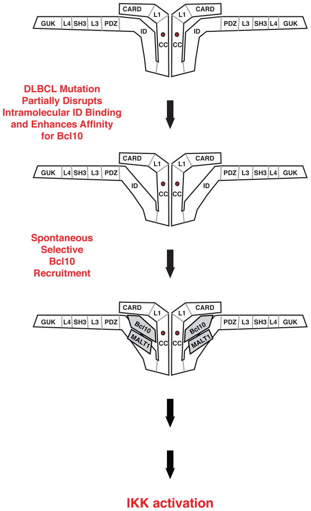
The regulated activation of NF-κB by antigen receptor signaling is required for normal B and T lymphocyte activation during the adaptive immune response. Dysregulated NF-κB activation is associated with several types of lymphoma, including diffuse large B cell lymphoma (DLBCL). During normal antigen receptor signaling, the multidomain scaffold protein CARD11 undergoes a transition from a closed, inactive state to an open, active conformation that recruits several signaling proteins into a complex, leading to IKK kinase activation. This transition is regulated by the CARD11 inhibitory domain (ID), which participates in intramolecular interactions that prevent cofactor binding to CARD11 prior to signaling, but which is neutralized after receptor engagement by phosphorylation. Several oncogenic CARD11 mutations have been identified in DLBCL that enhance activity and that are mostly found in the coiled-coil domain. However, the mechanisms by which these mutations cause CARD11 hyperactivity and spontaneous NF-κB activation are poorly understood. In this report, we provide several lines of evidence that oncogenic mutations F123I and L225LI induce CARD11 hyperactivity by disrupting autoinhibition by the CARD11 ID. These mutations disrupt ID-mediated intramolecular interactions and ID-dependent inhibition and bypass the requirement for ID phosphorylation during T cell receptor signaling. Intriguingly, these mutations selectively enhance the apparent affinity of CARD11 for Bcl10, but not for other signaling proteins that are recruited to CARD11 in an ID-dependent manner during normal antigen receptor signaling. Our results establish a mechanism that explains how DLBCL-associated mutations in CARD11 can initiate spontaneous, receptor-independent activation of NF-κB.


Figures

Figure 1
Figure 1
Conformational changes in CARD11 during signaling. (A) Prior to TCR or BCR engagement, CARD11 rests in an inactive, “closed” conformation promoted by the interaction of the ID with the CARD and Coiled-coil domains. Receptor engagement results in ID phosphorylation, which causes the ID to disengage from the CARD and Coiled-coil domains, converting CARD11 to an active, ”open” conformation, and allowing the depicted signaling proteins to associate with CARD11, leading to IKK activation. The signaling complex is rapidly disassembled to return CARD11 to the basal state. (B) Hypothesized action of oncogenic CARD11 mutations to spontaneously convert CARD11 into the “open” active conformation. The mutations are predicted to disrupt ID-mediated autoinhibition, thus allowing CARD11 to associate with signaling cofactors independently of receptor engagement and ID phosphorylation.
Figure 2
Figure 2
Effect of F123I and L225LI oncogenic mutations on CARD11 specific signaling activity. (A–C) HEK293T cells were transfected with 6 ng of pCSK-LacZ and 20 ng of Igκ2-IFN-LUC in the presence of the indicated amounts (in ng) of expression vectors for the indicated myc-tagged CARD11 variants. The panel below each titration displays western blots of corresponding lysates probed with anti-myc primary antibody to indicate the relative expression level of each variant. β-galactosidase activity, driven by pCSK-LacZ, was used to normalize luciferase activity and to calculate equivalent amounts of transfected cell-lysate for western analysis. (D) The relative specific activity of each variant is shown, normalized to that of wild-type CARD11, determined as described in Experimental Procedures.
Figure 3
Figure 3
F123I and L225LI oncogenic mutations confer resistance to ID inhibition and impair ID binding in trans. (A) Jurkat T cells were transfected with 200ng pCSK-LacZ and 1800ng Igκ2-IFN-LUC in the presence of expression vectors for ΔID (100 ng), ΔID-F123I (200 ng), or ΔID-L225LI (150 ng) and the indicated amounts (in ng) of expression vectors for GST (pEBG) or ID-GST (pEBB-HA-ID-GST). Fold reporter activation was normalized on a percent basis to that observed with each CARD11 variant in the absence of ID-GST or GST. (B) HEK293T cells were transfected with the following amounts in ng of expression vectors for ΔID (165–660 ng), ΔID-F123I (235–940 ng), ΔID-L225LI (125–500 ng), ID-GST(380–420 ng), or GST(200 ng), and glutathione-sepharose pulldowns were performed as described in Experimental Procedures and analyzed by western analysis with the indicated primary antibodies. WB, western blot; α, anti.
Figure 4
Figure 4
F123I and L225LI oncogenic mutations bypass the need for ID neutralization in T cells. (A) Wild-type Jurkat T cells, or Jurkat T cells stably expressing the sihCARD11-2 hairpin that targets human CARD11, were transfected with 200ng pCSK-LacZ, 1800 ng Igκ2-IFN-LUC, and expression vectors for wild-type murine CARD11 (150 ng), S564A (150 ng), ΔID (150 ng), F123I (150ng), F123I S564A (150 ng), L225LI (75 ng), or L225LI S564A (75 ng). Cells were stimulated with anti-CD3/anti-CD28 crosslinking for 4.5 hours as indicated. (B) HEK293T cells were transfected with 200ng pCSK-LacZ, 1800 ng Igκ2-IFN-LUC, and expression vectors for wild-type murine CARD11 (150 ng), S564A (150 ng), ΔID (150 ng), F123I (150ng), F123I S564A (150 ng), L225LI (75 ng), or L225LI S564A (75 ng). Lysates were probed by western blot using anti-myc primary to indicate the relative expression level of each variant. β-galactosidase activity, driven by pCSK-LacZ, was used to calculate equivalent amounts of transfected cell-lysate for western analysis. α, anti
Figure 5
Figure 5
Effect of F123I and L225LI oncogenic mutations on CARD11:CARD11 association. HEK293T cells were transfected with the indicated amounts (in ng) of the indicated FLAG- and myc-tagged CARD11 variants and anti-FLAG immunoprecipitations were performed and analyzed by western blot using the indicated primary antibodies. IP, Immunoprecipitate; WB, western blot; α, anti
Figure 6
Figure 6
F123I and L225LI mutations enhance the apparent affinity of CARD11 for Bcl10. (A) HEK293T cells were transfected with expression vectors for wild-type CARD11 (120 ng), F123I (120 ng), L225LI (120 ng), ΔID (200 ng), and FLAG-Bcl10 (100 ng), as indicated and anti-FLAG immunoprecipitations were performed as described in Experimental Procedures and analyzed by western analysis with the indicated primary antibodies. (B) Anti-FLAG immunoprecipitations were performed as in (A) using expression vectors for wild-type CARD11 (10–100 ng), F123I (15–150 ng), L225LI (10–100 ng), ΔID (10–100 ng), ΔID-F123I (14–140 ng), ΔID-L225LI (7–70 ng), and FLAG-Bcl10 (40–150 ng), as indicated. IP, Immunoprecipitate; WB, western blot; α, anti
Figure 7
Figure 7
The ID-mediated prevention of binding of TRAF6, TAK1, IKKγ, and Caspase-8 is intact in the F123I and L225LI oncogenic mutants. Anti-FLAG immunoprecipitations were performed as described in Experimental Procedures using the following expression vectors for each panel, as indicated: (A) wild-type CARD11 (200–400 ng), F123I (300–400 ng), L225LI (400 ng), ΔID (400 ng), FLAG-TRAF6 (350–450 ng), and Bcl10 (150–600 ng); (B) wild-type CARD11 (50–600 ng), F123I (280–600 ng), L225LI (300–600 ng), ΔID (500–850 ng), FLAG-TAK1 (100–200 ng), and Bcl10 (150–800 ng); (C) wild-type CARD11 (200–800 ng), F123I (350–800 ng), L225LI (400–800 ng), ΔID (400–800 ng), FLAG-IKKγ (1 ug), and Bcl10 (120–800 ng); (D) wild-type CARD11 (200–400 ng), F123I (350–400 ng), L225LI (400 ng), ΔID (400 ng), FLAG-Caspase-8 C360S (1 ug), and Bcl10 (150–600 ng). IP, Immunoprecipitate; WB, western blot; α, anti.
Figure 8
Figure 8
Bcl10 bound to F123I and L225LI mutants is competent to recruit MALT1 to CARD11. Anti-HA immunoprecipitations were performed as described in Experimental Procedures and analyzed by western blot with the indicated primary antibodies using the following expression vectors as indicated: wild-type CARD11 (400–600 ng), F123I (600 ng), L225LI (600 ng), ΔID (900 ng), HA-MALT1 (500–800 ng), and FLAG-Bcl10 (300–500 ng). The asterisk indicates Protein G. IP, Immunoprecipitate; WB, western blot; α, anti,
Figure 9
Figure 9
F123I and L225LI oncogenic mutants require Bcl10 for signaling to NF-κB. (A) HEK293T cells stably expressing either a hairpin that targets human Bcl10 (KD-Bcl10), or a control hairpin that targets GFP (KD-GFP), were transfected with 6 ng of pCSK-LacZ and 20 ng of Igκ2-IFN-LUC in the presence of the indicated amounts (in ng) of expression vectors for the indicated myc-tagged CARD11 variants. The lower panels display western blots of corresponding lysates probed with anti-myc and anti-Bcl10 primary antibodies to indicate relative expression levels. β-galactosidase activity, driven by pCSK-LacZ, was used to normalize luciferase activity and to calculate equivalent amounts of transfected cell-lysate for western analysis. (B, C) Jurkat T cells stably expressing either a hairpin that targets human Bcl10 (KD-Bcl10), or a control hairpin that targets GFP (KD-GFP), were transfected with 200 ng of pCSK-LacZ and 1500 ng of Igκ2-IFN-LUC in the presence of the indicated amounts (in ng) of expression vectors for myc-tagged F123I (B) or L225LI (C) CARD11 variants.
Figure 10
Figure 10
Bcl10 residues K31 and K63 are required for maximal signaling by F123I and L225LI oncogenic mutants. (A) KD-GFP or KD-Bcl10 HEK293T cell lines were transfected as in Figure 9A with 2–8 ng of myc-F123I or 1–8 ng of myc-L225LI, and the indicated amounts of expression vectors for either FLAG-Bcl10 or the FLAG-Bcl10 K31R, K63R mutant. (B) The corresponding lysates from (A) were analyzed by western blot and probed with anti-myc or anti-Bcl10 primary antibodies to indicate relative expression level. β-galactosidase activity, driven by pCSK-LacZ, was used to normalize luciferase activity and to calculate equivalent amounts of transfected cell-lysate for western analysis. (C) KD-GFP or KD-Bcl10 Jurkat T cell lines were transfected as in Figure 9B with 200 ng (for KD-GFP) or 800 ng (for KD-Bcl10) of myc-F123I or 50 ng (for KD-GFP) or 200 ng (for KD-Bcl10) of myc-L225LI, and the indicated amount (in ng) of either FLAG-Bcl10 or FLAG-Bcl10 K31R, K63R.
Figure 11
Figure 11
Model of the mechanism by which oncogenic mutations initiate dysregulated signaling to NF-κB in Diffuse Large B Cell Lymphoma.

Similar articles

Cited by

References

    1. Schulze-Luehrmann J, Ghosh S. Antigen-receptor signaling to nuclear factor kappa B. Immunity. 2006;25:701–715. - PubMed
    1. Gerondakis S, Siebenlist U. Roles of the NF-kappaB pathway in lymphocyte development and function. Cold Spring Harb Perspect Biol. 2:a000182. - PMC - PubMed
    1. Blonska M, Lin X. CARMA1-mediated NF-kappaB and JNK activation in lymphocytes. Immunol Rev. 2009;228:199–211. - PMC - PubMed
    1. Thome M, Weil R. Post-translational modifications regulate distinct functions of CARMA1 and BCL10. Trends Immunol. 2007;28:281–288. - PubMed
    1. Rawlings DJ, Sommer K, Moreno-Garcia ME. The CARMA1 signalosome links the signalling machinery of adaptive and innate immunity in lymphocytes. Nat Rev Immunol. 2006;6:799–812. - PubMed

Publication types

MeSH terms