

Quantitative fluorescence tomography using a trimodality system: in vivo validation
- PMID: 20799770
- PMCID: PMC2937044
- DOI: 10.1117/1.3467495
Quantitative fluorescence tomography using a trimodality system: in vivo validation
Abstract
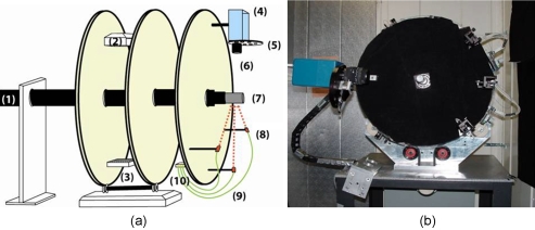
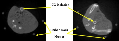
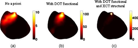
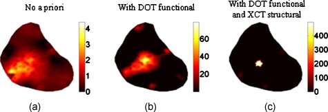
A fully integrated trimodality fluorescence, diffuse optical, and x-ray computed tomography (FT/DOT/XCT) system for small animal imaging is reported in this work. The main purpose of this system is to obtain quantitatively accurate fluorescence concentration images using a multimodality approach. XCT offers anatomical information, while DOT provides the necessary background optical property map to improve FT image accuracy. The quantitative accuracy of this trimodality system is demonstrated in vivo. In particular, we show that a 2-mm-diam fluorescence inclusion located 8 mm deep in a nude mouse can only be localized when functional a priori information from DOT is available. However, the error in the recovered fluorophore concentration is nearly 87%. On the other hand, the fluorophore concentration can be accurately recovered within 2% error when both DOT functional and XCT structural a priori information are utilized together to guide and constrain the FT reconstruction algorithm.
Figures
Similar articles
-
In vivo validation of quantitative frequency domain fluorescence tomography.J Biomed Opt. 2012 Dec;17(12):126021. doi: 10.1117/1.jbo.17.12.126021. J Biomed Opt. 2012. PMID: 23323291 Free PMC article.
-
Quantitative fluorescence tomography using a combined tri-modality FT/DOT/XCT system.Opt Express. 2010 Apr 12;18(8):7835-50. doi: 10.1364/OE.18.007835. Opt Express. 2010. PMID: 20588625 Free PMC article.
-
Hybrid small animal imaging system combining magnetic resonance imaging with fluorescence tomography using single photon avalanche diode detectors.IEEE Trans Med Imaging. 2011 Jun;30(6):1265-73. doi: 10.1109/TMI.2011.2112669. Epub 2011 Feb 10. IEEE Trans Med Imaging. 2011. PMID: 21317083
-
Introduction to clinical and laboratory (small-animal) image registration and fusion.Conf Proc IEEE Eng Med Biol Soc. 2006;2006:1580-3. doi: 10.1109/IEMBS.2006.259649. Conf Proc IEEE Eng Med Biol Soc. 2006. PMID: 17946907 Review.
-
Radiologic and near-infrared/optical spectroscopic imaging: where is the synergy?AJR Am J Roentgenol. 2010 Aug;195(2):321-32. doi: 10.2214/AJR.10.5002. AJR Am J Roentgenol. 2010. PMID: 20651186 Free PMC article. Review.
Cited by
-
Fast ICCD-based temperature modulated fluorescence tomography.Appl Opt. 2023 Oct 1;62(28):7420-7430. doi: 10.1364/AO.499281. Appl Opt. 2023. PMID: 37855510 Free PMC article.
-
Simulation-based evaluation of the resolution and quantitative accuracy of temperature-modulated fluorescence tomography.Appl Opt. 2015 Sep 1;54(25):7612-21. doi: 10.1364/AO.54.007612. Appl Opt. 2015. PMID: 26368884 Free PMC article.
-
A compact frequency-domain photon migration system for integration into commercial hybrid small animal imaging scanners for fluorescence tomography.Phys Med Biol. 2012 Dec 21;57(24):8135-52. doi: 10.1088/0031-9155/57/24/8135. Epub 2012 Nov 22. Phys Med Biol. 2012. PMID: 23171509 Free PMC article.
-
Parallel multigrid solver of radiative transfer equation for photon transport via graphics processing unit.J Biomed Opt. 2012 Sep;17(9):96004-1. doi: 10.1117/1.JBO.17.9.096004. J Biomed Opt. 2012. PMID: 23085905 Free PMC article.
-
Hybrid FMT-MRI applied to in vivo atherosclerosis imaging.Biomed Opt Express. 2014 Apr 28;5(5):1664-76. doi: 10.1364/BOE.5.001664. eCollection 2014 May 1. Biomed Opt Express. 2014. PMID: 24877023 Free PMC article.
References
-
- Bremer C., Ntziachristos V., and Weissleder R., “Optical-based molecular imaging: contrast agents and potential medical applications,” Eur. Radiol. ZZZZZZ 13(2), 231–243 (2003). - PubMed
Publication types
MeSH terms
Grants and funding
LinkOut - more resources
Full Text Sources
Other Literature Sources
