. 2010;12(4):R165.
doi: 10.1186/ar3124. Epub 2010 Aug 27.

High mobility group box 1 potentiates the pro-inflammatory effects of interleukin-1β in osteoarthritic synoviocytes

Affiliations

High mobility group box 1 potentiates the pro-inflammatory effects of interleukin-1β in osteoarthritic synoviocytes

Isabel García-Arnandis et al. Arthritis Res Ther. 2010.

Abstract

Introduction: High mobility group box 1 (HMGB1) is released by necrotic cells or secreted in response to inflammatory stimuli. Extracellular HMGB1 may act as a pro-inflammatory cytokine in rheumatoid arthritis. We have recently reported that HMGB1 is released by osteoarthritic synoviocytes after activation with interleukin-1beta (IL-1β) The present study investigated the role of HMGB1 in synovial inflammation in osteoarthritis (OA).

Methods: HMGB1 was determined in human synovium using immunohistochemistry, comparing normal to OA. OA synoviocytes were incubated with HMGB1 at 15 or 25 ng/ml in the absence or presence of IL-1β (10 ng/ml). Gene expression was analyzed by quantitative PCR and protein expression by Western Blot and ELISA. Matrix metalloproteinase (MMP) activity was studied by fluorometric procedures and nuclear factor (NF)-κB activation by transient transfection with a NF-κB-luciferase plasmid.

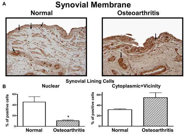
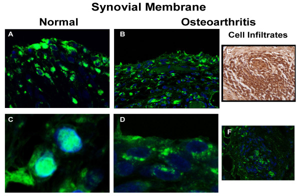
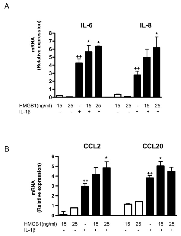
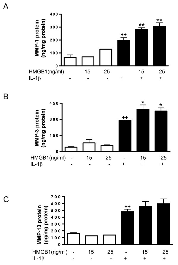
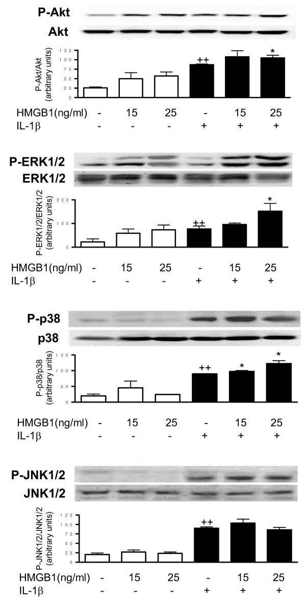
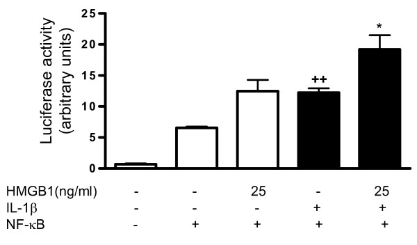
Results: In the normal synovium, HMGB1 was found in the synovial lining cells, sublining cells, and in the vascular wall cells. The distribution of HMGB1 in OA synovium was similar but the number of HMGB1 positive cells was higher and HMGB1 was also present in infiltrated cells. In normal synovial membrane cells, HMGB1 was found mostly in the nuclei, whereas in OA, HMGB1 was generally found mostly in the cytoplasm. In OA synoviocytes, HMGB1 alone at concentrations of 15 or 25 ng/ml did not affect the production of IL-6, IL-8, CCL2, CCL20, MMP-1 or MMP-3, but in the presence of IL-1β, a significant potentiation of protein and mRNA expression, as well as MMP activity was observed. HMGB1 also enhanced the phosphorylated ERK1/2 and p38 levels, with a lower effect on phosphorylated Akt. In contrast, JNK1/2 phosphorylation was not affected. In addition, HMGB1 at 25 ng/ml significantly potentiated NF-κB activation in the presence of IL-1β.

Conclusions: Our results indicate that HMGB1 is overexpressed in OA synovium and mostly present in extracellular form. In OA synoviocytes, HMGB1 cooperates with IL-1β to amplify the inflammatory response leading to the production of a number of cytokines, chemokines and MMPs. Our data support a pro-inflammatory role for this protein contributing to synovitis and articular destruction in OA.


Figures

Figure 1
Figure 1
Expression of HMGB1 protein in human synovium. (a) Representative immunohistochemical sections of normal (n = 3) and osteoarthritic (n = 3) human synovium with high mobility group box 1 (HMGB1) revealed in 3,3'-diaminobenzidine (DAB). Arrows indicate HMGB1 positive cells. Original magnification × 100. (b) Histograms of normal and osteoarthritic positive cells (%) in the synovial lining cell nucleus and cytoplasm and vicinity. Data are expressed as mean ± standard error of the mean (n = 3). *P < 0.05 with respect to normal.
Figure 2
Figure 2
Confocal analysis of HMGB1 expression in human synovium. Representative immunohistochemical sections of human (a, c) normal (n = 3) and (b, d) osteoarthritic (n = 3) synovium with high mobility group box 1 (HMGB1) revealed in fluorescence and read with confocal microscopy. (e) represents immunohistochemistry revealed in 3,3'-diaminobenzidine (DAB) of human osteoarthritic synovium infiltrates, and (f) is as (e) but revealed with the fluorescent antibody. The colour green represents HMGB1 and blue the nucleus. Original magnification × 100. (c) and (d) were amplified by the image processor software.
Figure 3
Figure 3
Effect of HMGB1 and IL-1β on the levels of cytokine and chemokine released into the medium by osteoarthritic synoviocytes. (a) IL-6, (b) IL-8, (c) CCL2 and (d) CCL20 protein levels. Cells were stimulated with IL-1b (10 ng/ml) for 24 hours in the presence or absence of high mobility group box 1 (HMGB1) at 15 and 25 ng/ml. Protein levels were determined in supernatants by ELISA. Data are expressed as mean ± standard error of the mean. Duplicate samples from six patients were used. ++P < 0.01 with respect to nonstimulated cells. *P < 0.05, **P < 0.01 with respect to IL-1β.
Figure 4
Figure 4
Effect of HMGB1 and IL-1β on cytokine and chemokine mRNA levels in OA synoviocytes. (a) IL-6, IL-8, and (b) CCL2 and CCL20 mRNA relative expression. Cells were stimulated with IL-1b (10 ng/ml) for 24 hours in the presence or absence of high mobility group box 1 (HMGB1) at 15 and 25 ng/ml. mRNA expression was determined by real-time PCR. Data are expressed as mean ± standard error of the mean. Duplicate samples from four patients were used. ++P < 0.01 with respect to nonstimulated cells. *P < 0.05 with respect to IL-1β.
Figure 5
Figure 5
Effect of HMGB1 and IL-1β on the levels of MMP released into the medium by osteoarthritic synoviocytes. (a) Matrix metalloproteinase (MMP)-1, (b) MMP-3 and (c) MMP-13 protein levels in the medium. Cells were stimulated with IL-1b (10 ng/ml) for 24 hours in the presence or absence of high mobility group box 1 (HMGB1) at 15 and 25 ng/ml. MMP protein was measured by ELISA in supernatants. Data are expressed as mean ± standard error of the mean. Duplicate samples from six to eight patients were used. ++P < 0.01 with respect to nonstimulated cells. *P < 0.05, **P < 0.01 with respect to IL-1β.
Figure 6
Figure 6
Effect of HMGB1 and IL-1β on MMP mRNA levels in osteoarthritic synoviocytes and MMP activity released into the medium. (a) Matrix metalloproteinase (MMP)-1, MMP-3 and MMP-13 mRNA levels and (b) MMP activity. Cells were stimulated with IL-1b (10 ng/ml) for 24 hours in the presence or absence of high mobility group box 1 (HMGB1) at 15 and 25 ng/m. mRNA was determined by real-time PCR and MMP activity was measured in supernatants by a fluorometric procedure, as indicated in Materials and Methods. Data are expressed as mean ± standard error of the mean. Duplicate samples from four patients were used. ++P < 0.01 with respect to nonstimulated cells. *P < 0.05, **P < 0.01 with respect to IL-1β.
Figure 7
Figure 7
Effect of HMGB1 and IL-1β on Akt and MAPK phosphorylation. Cells were stimulated with IL-1b (10 ng/ml) for five minutes in the presence or absence of high mobility group box 1 (HMGB1) at 15 and 25 ng/ml. Protein level was determined in cell lysates by western blotting by using specific antibodies against phosphorylated or total proteins. Relative expression of phosphorylated and total protein bands was calculated after densitometric analysis. Data are expressed as mean ± standard error of the mean (samples from three patients). ++P < 0.01 with respect to nonstimulated cells. *P < 0.05, with respect to IL-1β. ERK, extracellular signal-regulated kinase; JNK, c-Jun N-terminal kinase; MAPK, mitogen-activated protein kinase.
Figure 8
Figure 8
Effect of HMGB1 and IL-1β on NF-κB activation. Transient transfection was performed with the reporter construct nuclear factor (NF)-κB-luc and the internal control pRL-TK, as indicated in Materials and Methods. Cells were treated for 24 hours with high mobility group box 1 (HMGB1) at 25 ng/ml in the absence or presence of IL-1β (10 ng/ml). Firefly luciferase activity was normalized to Renilla luciferase activity. Data are expressed as mean ± standard error of the mean. Duplicate samples from six patients were used. ++P < 0.01 with respect to nonstimulated cells; *P < 0.05 with respect to IL-1β.

References

    1. Pisetsky DS, Erlandsson-Harris H, Andersson U. High-mobility group box protein 1 (HMGB1): an alarmin mediating the pathogenesis of rheumatic disease. Arthritis Res Ther. 2008;10:209. doi: 10.1186/ar2440. - DOI - PMC - PubMed
    1. Sundberg E, Grundtman C, af Klint E, Lindberg J, Ernestam S, Ulfgren AK, Harris HE, Andersson U. Systemic TNF blockade does not modulate synovial expression of the pro-inflammatory mediator HMGB1 in rheumatoid arthritis patients - a prospective clinical study. Arthritis Res Ther. 2008;10:R33. doi: 10.1186/ar2387. - DOI - PMC - PubMed
    1. Tang D, Shi Y, Kang R, Li T, Xiao W, Wang H, Xiao X. Hydrogen peroxide stimulates macrophages and monocytes to actively release HMGB1. J Leukoc Biol. 2007;81:741–747. doi: 10.1189/jlb.0806540. - DOI - PMC - PubMed
    1. Andersson U, Wang H, Palmblad K, Aveberger AC, Bloom O, Erlandsson-Harris H, Janson A, Kokkola R, Zhang M, Yang H, Tracey KJ. High mobility group 1 protein (HMG-1) stimulates proinflammatory cytokine synthesis in human monocytes. J Exp Med. 2000;192:565–570. doi: 10.1084/jem.192.4.565. - DOI - PMC - PubMed
    1. Dumitriu IE, Bianchi ME, Bacci M, Manfredi AA, Rovere-Querini P. The secretion of HMGB1 is required for the migration of maturing dendritic cells. J Leukoc Biol. 2007;81:84–91. doi: 10.1189/jlb.0306171. - DOI - PubMed

Publication types

MeSH terms