Review
. 2011 Feb;1809(2):67-79.
doi: 10.1016/j.bbagrm.2010.08.008. Epub 2010 Aug 26.

Structure characterization of the 26S proteasome

Affiliations
Review

Structure characterization of the 26S proteasome

Ho Min Kim et al. Biochim Biophys Acta. 2011 Feb.

Abstract

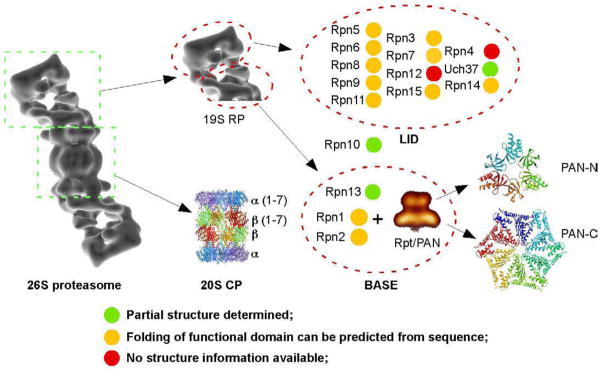
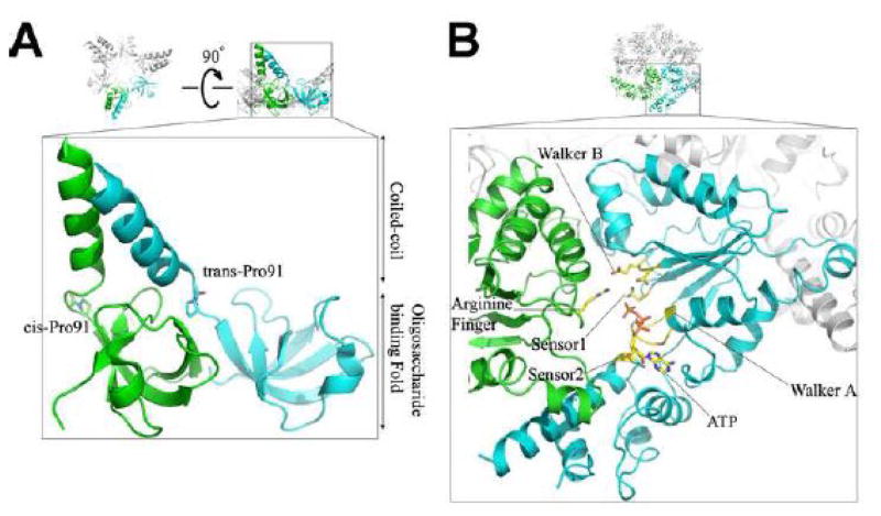
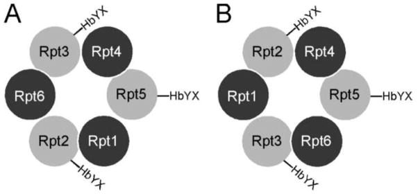
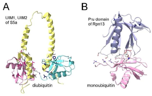
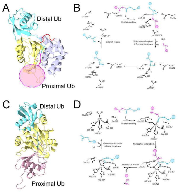
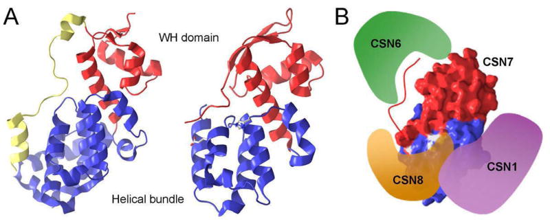
In all eukaryotic cells, 26S proteasome plays an essential role in the process of ATP-dependent protein degradation. In this review, we focus on structure characterization of the 26S proteasome. Although the progress towards a high-resolution structure of the 26S proteasome has been slow, the recently solved structures of various proteasomal subcomplexes have greatly enhanced our understanding of this large machinery. In addition to having an ATP-dependent proteolytic function, the 26S proteasome is also involved in many non-proteolytic cellular activities, which are often mediated by subunits in its 19S regulatory complex. Thus, we include a detailed discussion of the structures of 19S subunits, including proteasomal ATPases, ubiquitin receptors, deubiquitinating enzymes and subunits that contain PCI domain. This article is part of a Special Issue entitled The 26S Proteasome: When degradation is just not enough!


Figures

Figure 1
Figure 1
Schematic representation of the 26S proteasome. The components of 26S proteasome are divided into functional groups and subunits. The availability of structure information at different levels is also shown in different colors.
Figure 2
Figure 2
Atomic structures of domains from archaeal PAN ATPase. (A) The CC-OB domain of the PAN assembles into a hexameric ring that has a 3-fold symmetry. The cis- and trans-proline residues in the neighboring CC-OB domain are shown in the enlarged view that shows how the coiled-coil is formed. (B) A model of a hexameric ring formed by PAN’s ATPase domain. The ATP binding site of this model is shown in the enlarged view.
Figure 3
Figure 3
Two possible arrangements of Rpt-subunits in the base subcomplex. They are viewed from the top of the hexameric ring of ATPases sits on top of the 20S CP. Rpt2, Rpt3 and Rpt5 have the conserved C-terminal HbYX motif that is required to induce gate opening in the 20S CP. They also have the cis-proline that is required to form the coiled-coil interaction with its adjacent subunit on the right side.
Figure 4
Figure 4
Structures of ubiquitin binding domains. (A) The NMR structure of S5a UIM domain and diubiquitin (pdb code: 2KDE). Pink is the proximal ubiquitin interacting with the UIM2, and cyan is the distal ubiquitin that interacts with the UIM1. Magenta, blue and gray side chains on the interaction surface belong to proximal ubiquitin, distal ubiquitin and UIM, respectively. (B) Complex structure of Rpn13 with monoubiquitin (pdb code: 2Z59). Red side chain is the ubiquitin K48 residue. Blue side chains on the opposite side of ubiquitin represent the residues interacting with the Rpn2.
Figure 5
Figure 5
The catalytic mechanisms of deubiquitinating enzymes. (A) A structure model of UCH37 in complex with a diubiquitin. Two lobes of the UCH37 (pdb code: 3IHR) are colored in yellow and light blue, side chains of the catalytic residues are shown. The loop that links the two lobes is disordered, and its speculated structure is drawn in red. The distal ubiquitin is shown in cyan, which is derived by superimposing UCH37 with the complex structure of UCH-L3 and UbVME (pdb code: 1XD3). The expected position of proximal ubiquitin is drawn as a pink disk. (B) The catalytic mechanism of UCH37 predicted from similar cysteine protease: Three residues Cys88, His164 and Asp179 form a catalytic triad in which the His164 activates the thiol group of Cys88 to a nucleophile. While the main chain amide of Cys88 and Gln82 form an oxyanion hole, the nucleophilic attack to the carbonyl carbon of the ubiquitin cleaves off the proximal ubiquitin. (C) Crystal structure of the AMSH-LP and K64-ubiquitin complex (pdb code: 2ZNV). In this atomic structure Glu292 was mutated to alanine and the zinc-binding site was not occupied. The side chain of the conserved catalytic residue and zinc molecule are taken from another crystal structure of apo AMSH-LP (pdb code: 2ZNR). (D) The catalytic mechanism of AMSH-LP: A water molecule is polarized by Glu292 and Zn2+ in the catalytic site. It acts as a nucleophile to attack the carbonyl carbon of Gly76 in the distal ubiquitin. The proton from the 292Glu is transferred to the nitrogen of K49 ε-amine. The tetrahedral intermediate is then generated, where zinc is penta-coordinated, while Ser357 involves the stabilization of this intermediate by the hydrogen bond with carbonyl oxygen. Finally, the isopeptide bond is cleaved and the proximal ubiquitin is released from the distal ubiquitin [130].
Figure 6
Figure 6
Crystal structures of the PCI domains. (A) The eIF3 (left, pdb code: 1RZ4) and CSN7 (right, pdb code: 3CHM) have a similar architecture. Blue and red ribbons represent N-terminal helix bundle and C-terminal WH domain. Glu44 and His71, shown as stick in the N-terminal helical bundle of CNS7, are critical for the binding of CSN8. (B) Partial interactions between CSN subunits.

References

    1. Ciechanover A, Schwartz AL. The ubiquitin-proteasome pathway: the complexity and myriad functions of proteins death. Proc Natl Acad Sci U S A. 1998;95:2727–2730. - PMC - PubMed
    1. Hershko A, Ciechanover A. The ubiquitin system. Annu Rev Biochem. 1998;67:425–479. - PubMed
    1. Glickman MH, Ciechanover A. The ubiquitin-proteasome proteolytic pathway: destruction for the sake of construction. Physiol Rev. 2002;82:373–428. - PubMed
    1. Glickman MH, Adir N. The proteasome and the delicate balance between destruction and rescue. PLoS Biol. 2004;2:E13. - PMC - PubMed
    1. Coux O, Tanaka K, Goldberg AL. Structure and functions of the 20S and 26S proteasomes. Annu Rev Biochem. 1996;65:801–847. - PubMed

Publication types

Substances

LinkOut - more resources