Review
. 2010 Dec;35(12):684-90.
doi: 10.1016/j.tibs.2010.07.012.

Emerging complex pathways of the actomyosin powerstroke

Affiliations
Review

Emerging complex pathways of the actomyosin powerstroke

András Málnási-Csizmadia et al. Trends Biochem Sci. 2010 Dec.

Abstract

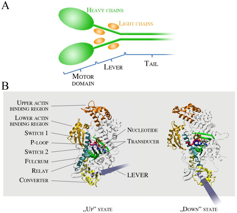
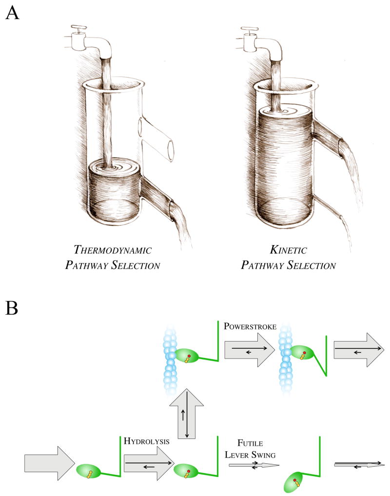
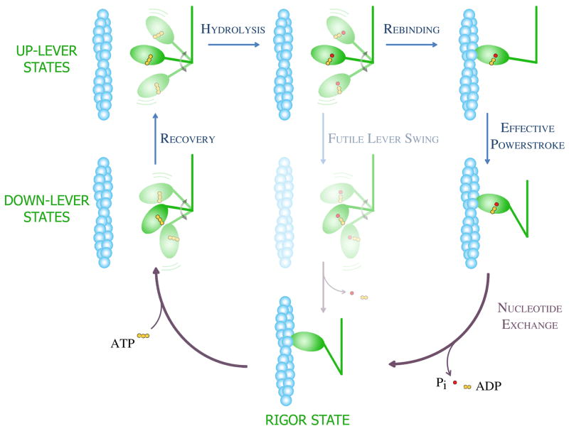
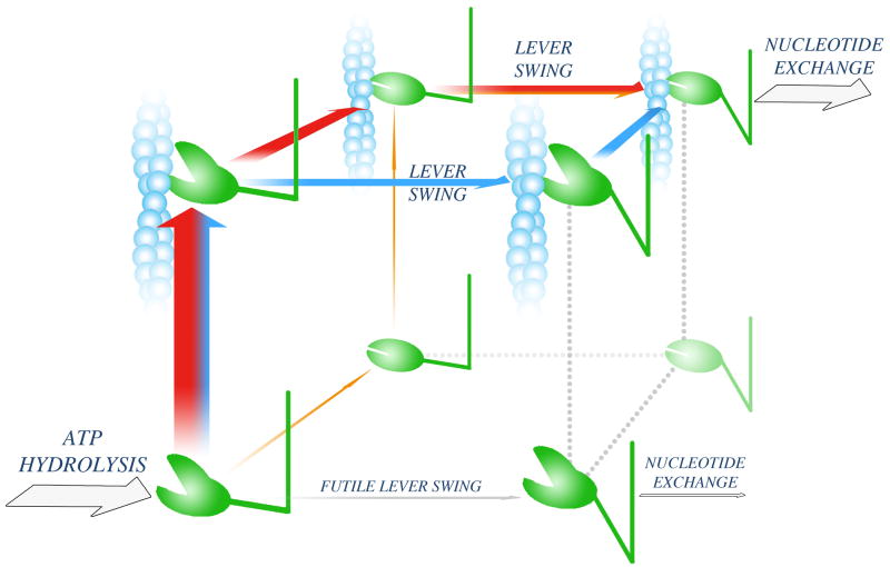
Actomyosin powers muscle contraction and various cellular activities, including cell division, differentiation, intracellular transport and sensory functions. Despite their crucial roles, key aspects of force generation have remained elusive. To perform efficient force generation, the powerstroke must occur while myosin is bound to actin. Paradoxically, this process must be initiated when myosin is in a very low actin-affinity state. Recent results shed light on a kinetic pathway selection mechanism whereby the actin-induced activation of the swing of myosin's lever enables efficient mechanical functioning. Structural elements and biochemical principles involved in this mechanism are conserved among various NTPase-effector (e.g. kinesin-microtubule, G protein exchange factor and kinase-scaffold protein) systems that perform chemomechanical or signal transduction.


Figures

Box 1, Figure I
Box 1, Figure I. Structure of the myosin holoenzyme and the motor domain
Box 3, Figure I
Box 3, Figure I. Pathway selection mechanisms
Figure 1
Figure 1. Mechanochemical cycle of the actomyosin motor
The mechanism shown incorporates the Lymn-Taylor model in conjunction with major conceptual advances gained from recent structural and kinetic investigations. The myosin head and the actin filament are shown in green and blue, respectively. During the working cycle, ATP binding to the myosin head dissociates the strongly-bound actomyosin “rigor” complex (lowest panel). During the recovery step (left two panels), which occurs in ATP-bound myosin, the myosin lever moves from a “down” to an “up” position. Note the change in lever orientation relative to the motor domain (see also Box 1, Fig. IB). In the actin-detached states (upper left four panels), the myosin head (motor domain plus lever) is shown in several different orientations to indicate its free rotation about a flexible joint that connects it to the distal part of the molecule (and the thick filament in muscle). The hydrolysis of ATP to ADP and Pi (inorganic phosphate) occurs only in the up-lever state (upper left panel). The post-hydrolytic up-lever complex (upper middle panel) can continue the cycle in two pathways. If the lever swings back to a down position when the head is detached from actin (futile lever swing, middle panels), the ATP hydrolysis cycle is completed without work production. In order to undergo an effective powerstroke leading to force generation, the head must rebind to actin (upper right panel) before the lever swing (right two panels). When the lever swing is completed, the hydrolysis products are exchanged to a new ATP molecule. Note that this scheme does not indicate changes in actin affinity and motor domain structure.
Figure 2
Figure 2. Powerstroke pathways
Following ATP hydrolysis, the myosin head adopts a weak actin-binding (open-cleft), up-lever state (lower left corner). Three possible pathways leading to an effective powerstroke are shown as orange, red and blue arrows. On the orange pathway, cleft closure is followed by association to actin and subsequent lever swing. The other two pathways (red and blue) start with actin attachment. On the red pathway, cleft closure precedes lever swing. On the blue pathway, lever swing occurs while the cleft is still open, and the pathway is completed with cleft closure. Experimental data indicate that the flux through the orange pathway is limited, whereas the red and blue pathways might both convey significant fluxes. A futile lever swing, which would lead to an ATP-wasting cycle (grey arrow), is kinetically blocked (Box 3).

References

    1. Pollard TD. Reflections on a quarter century of research on contractile systems. Trends Biochem Sci. 2000;25:607–611. - PubMed
    1. Geeves MA, Holmes KC. The molecular mechanism of muscle contraction. Adv Protein Chem. 2005;71:161–193. - PubMed
    1. Smith DA, et al. Towards a unified theory of muscle contraction. I: foundations. Ann Biomed Eng. 2008;36:1624–1640. - PubMed
    1. Sweeney HL, Houdusse A. Structural and Functional Insights into the Myosin Motor Mechanism. Annu Rev Biophys. 2010;39:539–557. - PubMed
    1. Odronitz F, Kollmar M. Drawing the tree of eukaryotic life based on the analysis of 2,269 manually annotated myosins from 328 species. Genome Biol. 2007;8:R196. - PMC - PubMed

Publication types

LinkOut - more resources