. 2010 Dec;101(4):324-31.
doi: 10.1016/j.ymgme.2010.08.001. Epub 2010 Aug 7.

Differences in the predominance of lysosomal and autophagic pathologies between infants and adults with Pompe disease: implications for therapy

Affiliations

Differences in the predominance of lysosomal and autophagic pathologies between infants and adults with Pompe disease: implications for therapy

Nina Raben et al. Mol Genet Metab. 2010 Dec.

Abstract

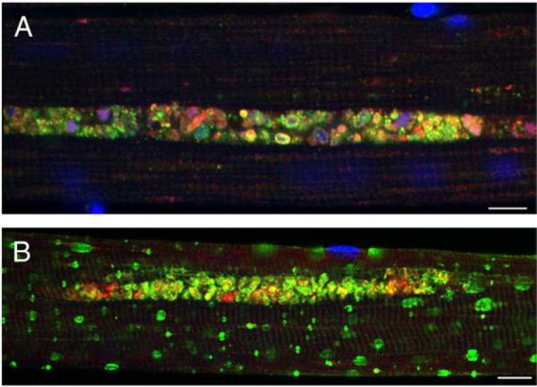
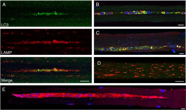
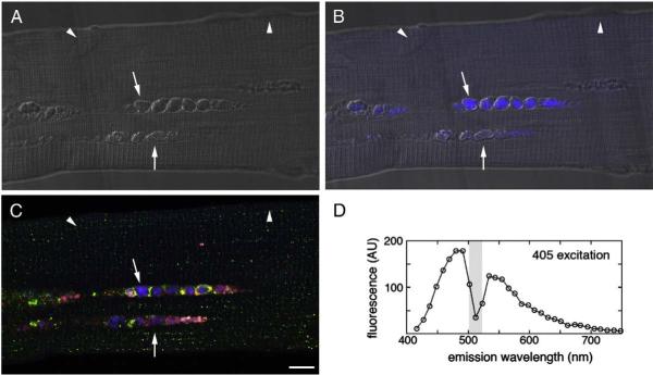
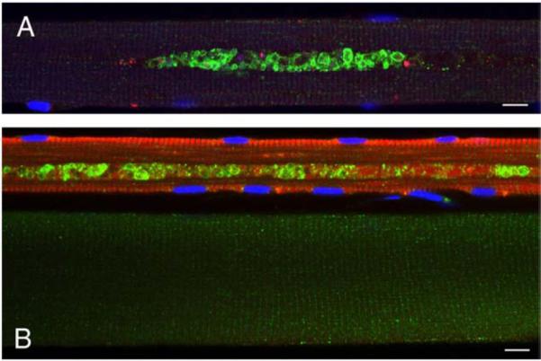
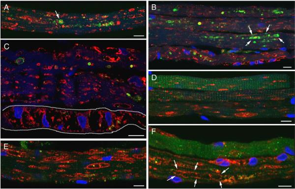
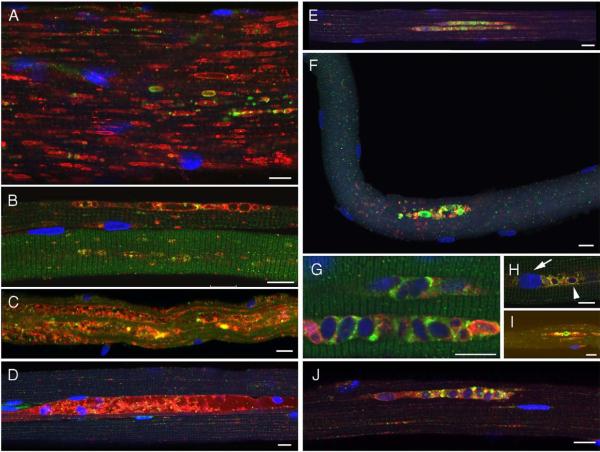
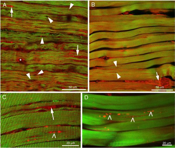
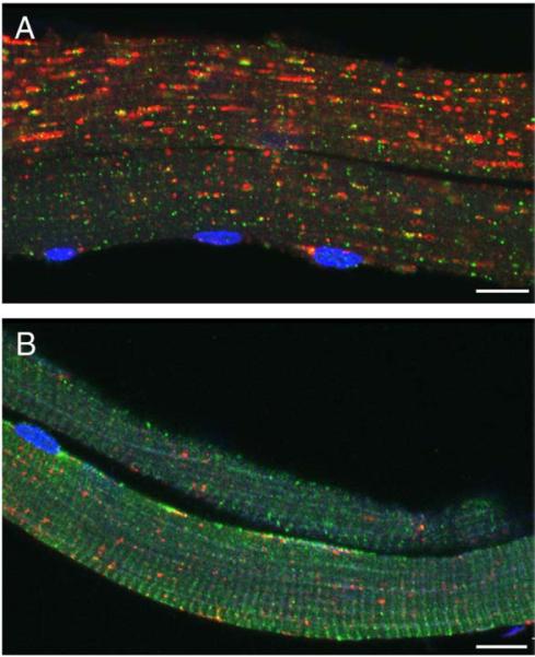
Pompe disease is a lysosomal storage disorder caused by the deficiency of acid alpha-glucosidase, the enzyme that degrades glycogen in the lysosomes. The disease manifests as a fatal cardiomyopathy and skeletal muscle myopathy in infants; in milder late-onset forms skeletal muscle is the major tissue affected. We have previously demonstrated that autophagic inclusions in muscle are prominent in adult patients and the mouse model. In this study we have evaluated the contribution of the autophagic pathology in infants before and 6 months after enzyme replacement therapy. Single muscle fibers, isolated from muscle biopsies, were stained for autophagosomal and lysosomal markers and analyzed by confocal microscopy. In addition, unstained bundles of fixed muscles were analyzed by second harmonic imaging. Unexpectedly, the autophagic component which is so prominent in juvenile and adult patients was negligible in infants; instead, the overwhelming characteristic was the presence of hugely expanded lysosomes. After 6 months on therapy, however, the autophagic buildup becomes visible as if unmasked by the clearance of glycogen. In most fibers, the two pathologies did not seem to coexist. These data point to the possibility of differences in the pathogenesis of Pompe disease in infants and adults.


Figures

Fig. 1
Fig. 1
Single fibers from an adult Italian patient (A) and a Pompe mouse (B) were stained for LAMP-2 (green) and LC3 (red). Nuclei were stained with DAPI. Autophagic accumulation appears to be the only pathology in human fibers. Bar: 10 microns. (For interpretation of the references to colour in this figure legend, the reader is referred to the web version of this article.)
Fig. 2
Fig. 2
Single fibers from Taiwanese juvenile patients (before ERT) were stained with LAMP-2 (red) and LC3 (green). Autophagic accumulation is a predominant pathology in juvenile patients (shown for NBSL9a in A, B, C, E); some fibers exhibit only lysosomal pathology (shown for NBSL2 in D). Bar: 10 microns. (For interpretation of the references to colour in this figure legend, the reader is referred to the web version of this article.)
Fig. 3
Fig. 3
Fiber from juvenile patient NBSL9a was stained for LAMP-2 (red)/LC3 (green), but not with DAPI. Autophagic areas contain autofluorescent structures (blue; arrows); arrowheads—autofluorescence-negative nuclei. (A) DIC (Differential Interference Contrast), (B) DIC and autofluorescence, (C) LAMP-2/LC3 staining and autofluorescence, (D) Characterization of autofluorescence signal in autophagic area. Bar: 10 microns. (For interpretation of the references to colour in this figure legend, the reader is referred to the web version of this article.)
Fig. 4
Fig. 4
Autophagic accumulation in slow fibers; fibers from an adult Italian patient were stained for LC3 (green) and fast myosin (red) in A or slow myosin in B. Bar: 10 microns. (For interpretation of the references to colour in this figure legend, the reader is referred to the web version of this article.)
Fig. 5
Fig. 5
Lysosomal pathology is predominant in infants. Fibers were stained for LAMP-2 (red)/LC3 (green). Clusters of autophagosomes are seen in some fibers [A (projection) and B]. Fibers from CLIN4 are shown in A, B, C, and F; fibers from NBS6 are shown in D and E. The dashed line marks a fiber that appears to be devoid of contractile elements. Bar: 10 microns. (For interpretation of the references to colour in this figure legend, the reader is referred to the web version of this article.)
Fig. 6
Fig. 6
Autophagic accumulation is noticeable on ERT. Fibers from infants were stained for LAMP-2 (red)/LC3 (green). Left: a variable response to ERT in NBS6 (A), CLIN3 (B, D), and CLIN5 (C). Right: autophagic buildup emerges upon ERT [CLIN3 (E, G, H, J) and NBS4 (F, I)]. Bar: 10 microns. (For interpretation of the references to colour in this figure legend, the reader is referred to the web version of this article.)
Fig. 7
Fig. 7
Combination of SHG (green) and 2PEF (red) microscopies highlights lysosomal holes (arrowheads), autophagic accumulations (arrows, except in B, damaged fiber), nucleus (asterisk) and autofluorescent particles (tick marks) in 3 mo-old pre-ERT infant (A; CLIN4), post-ERT infants (B; CLIN3, C; NBS3) and juvenile (D; NBLS2). (For interpretation of the references to colour in this figure legend, the reader is referred to the web version of this article.)
Fig. 8
Fig. 8
Effect of ERT in a patient (NBSL9) who started therapy at 1 month of age although his genetic defect would predict a late-onset form of the disease. Fibers were stained with LAMP-2 (red) and LC3 (green). (A) Before ERT. (B) 6m after ERT. Bar: 10 microns. (For interpretation of the references to colour in this figure legend, the reader is referred to the web version of this article.)

References

    1. Klionsky DJ. Autophagy revisited: a conversation with Christian de Duve. Autophagy. 2008;4:740–743. - PubMed
    1. Hirschhorn R, Reuser AJ. Glycogen storage disease type II: acid alpha-glucosidase (acid maltase) deficiency. In: Scriver C, Beaudet A, Sly W, Valle D, editors. The Metabolic and Molecular Basis of Inherited Disease. McGraw-Hill; New York, NY: 2001. pp. 3389–3420.
    1. Engel AG, Hirschhorn R, Huie ML. Acid Maltase Deficiency. In: Engel AG, Franzini-Armstrong C, editors. Myology. McGraw-Hill; New York, NY: 2003. pp. 1559–1586.
    1. Kishnani PS, Hwu WL, Mandel H, Nicolino M, Yong F, Corzo D. A retrospective, multinational, multicenter study on the natural history of infantile-onset Pompe disease. J. Pediatr. 2006;148:671–676. - PubMed
    1. Winkel LP, Hagemans ML, Van Doorn PA, Loonen MC, Hop WJ, Reuser AJ, Van der Ploeg AT. The natural course of non-classic Pompe's disease; a review of 225 published cases. J. Neurol. 2005;252:875–884. - PubMed

Publication types