

Projections from the hypothalamic paraventricular nucleus and the nucleus of the solitary tract to prechoroidal neurons in the superior salivatory nucleus: Pathways controlling rodent choroidal blood flow
- PMID: 20801105
- PMCID: PMC2949519
- DOI: 10.1016/j.brainres.2010.08.065
Projections from the hypothalamic paraventricular nucleus and the nucleus of the solitary tract to prechoroidal neurons in the superior salivatory nucleus: Pathways controlling rodent choroidal blood flow
Abstract
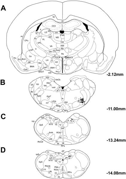
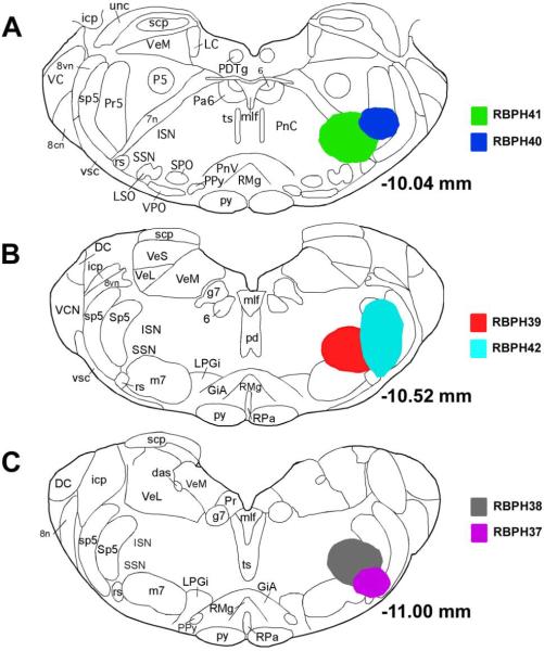
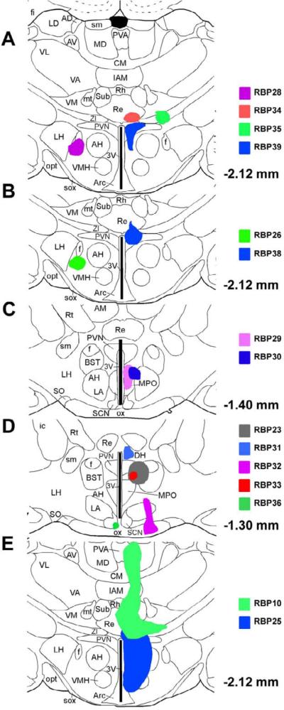
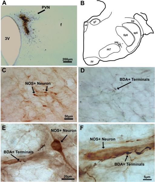
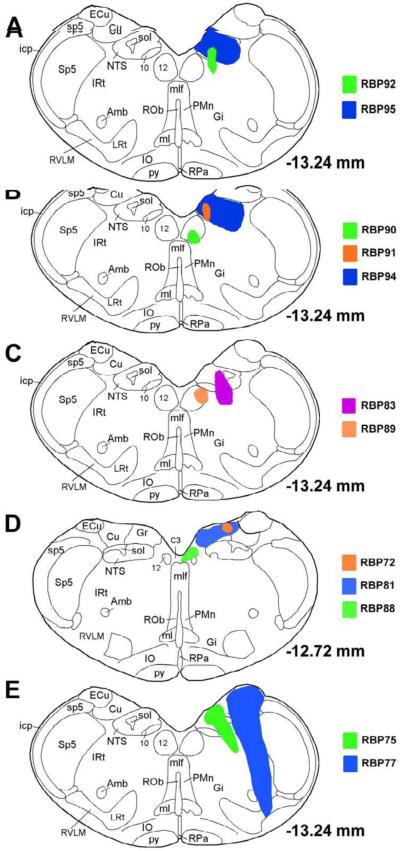
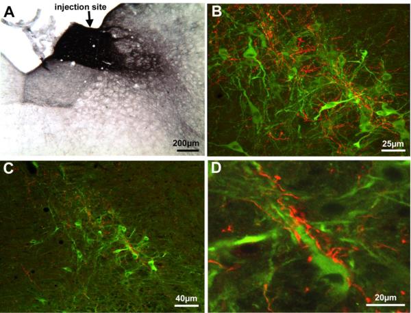
Using intrachoroidal injection of the transneuronal retrograde tracer pseudorabies virus (PRV) in rats, we previously localized preganglionic neurons in the superior salivatory nucleus (SSN) that regulate choroidal blood flow (ChBF) via projections to the pterygopalatine ganglion (PPG). In the present study, we used higher-order transneuronal retrograde labeling following intrachoroidal PRV injection to identify central neuronal cell groups involved in parasympathetic regulation of ChBF via input to the SSN. These prominently included the hypothalamic paraventricular nucleus (PVN) and the nucleus of the solitary tract (NTS), both of which are responsive to systemic BP and are involved in systemic sympathetic vasoconstriction. Conventional pathway tracing methods were then used to determine if the PVN and/or NTS project directly to the choroidal subdivision of the SSN. Following retrograde tracer injection into SSN (biotinylated dextran amine 3K or Fluorogold), labeled perikarya were found in PVN and NTS. Injection of the anterograde tracer, biotinylated dextran amine 10K (BDA10K), into PVN or NTS resulted in densely packed BDA10K+terminals in prechoroidal SSN (as defined by its enrichment in nitric oxide synthase-containing perikarya). Double-label studies showed these inputs ended directly on prechoroidal nitric oxide synthase-containing neurons of SSN. Our study thus establishes that PVN and NTS project directly to the part of SSN involved in parasympathetic vasodilatory control of the choroid via the PPG. These results suggest that control of ChBF may be linked to systemic blood pressure and central control of the systemic vasculature.
Published by Elsevier B.V.
Figures

Similar articles
-
The identification and neurochemical characterization of central neurons that target parasympathetic preganglionic neurons involved in the regulation of choroidal blood flow in the rat eye using pseudorabies virus, immunolabeling and conventional pathway tracing methods.Front Neuroanat. 2015 Jun 2;9:65. doi: 10.3389/fnana.2015.00065. eCollection 2015. Front Neuroanat. 2015. PMID: 26082687 Free PMC article.
-
Disinhibition of neurons of the nucleus of solitary tract that project to the superior salivatory nucleus causes choroidal vasodilation: Implications for mechanisms underlying choroidal baroregulation.Neurosci Lett. 2016 Oct 28;633:106-111. doi: 10.1016/j.neulet.2016.09.029. Epub 2016 Sep 20. Neurosci Lett. 2016. PMID: 27663135 Free PMC article.
-
Localization of preganglionic neurons that innervate choroidal neurons of pterygopalatine ganglion.Invest Ophthalmol Vis Sci. 2003 Sep;44(9):3713-24. doi: 10.1167/iovs.02-1207. Invest Ophthalmol Vis Sci. 2003. PMID: 12939284
-
Role of the superior salivatory nucleus in parasympathetic control of choroidal blood flow and in maintenance of retinal health.Exp Eye Res. 2021 May;206:108541. doi: 10.1016/j.exer.2021.108541. Epub 2021 Mar 16. Exp Eye Res. 2021. PMID: 33736985 Free PMC article.
-
Neural control of choroidal blood flow.Prog Retin Eye Res. 2018 May;64:96-130. doi: 10.1016/j.preteyeres.2017.12.001. Epub 2017 Dec 8. Prog Retin Eye Res. 2018. PMID: 29229444 Free PMC article. Review.
Cited by
-
Cluster headache pathophysiology - insights from current and emerging treatments.Nat Rev Neurol. 2021 May;17(5):308-324. doi: 10.1038/s41582-021-00477-w. Epub 2021 Mar 29. Nat Rev Neurol. 2021. PMID: 33782592 Review.
-
Ablation of the sphenopalatine ganglion does not attenuate the infarct reducing effect of vagus nerve stimulation.Auton Neurosci. 2013 Mar;174(1-2):31-5. doi: 10.1016/j.autneu.2012.12.001. Epub 2012 Dec 27. Auton Neurosci. 2013. PMID: 23273773 Free PMC article.
-
Optic nerve head and retinal blood flow regulation during isometric exercise as assessed with laser speckle flowgraphy.PLoS One. 2017 Sep 12;12(9):e0184772. doi: 10.1371/journal.pone.0184772. eCollection 2017. PLoS One. 2017. PMID: 28898284 Free PMC article. Clinical Trial.
-
Vagal Nerve Stimulation Attenuates Ischemia-Reperfusion Induced Retina Dysfunction in Acute Ocular Hypertension.Front Neurosci. 2019 Feb 11;13:87. doi: 10.3389/fnins.2019.00087. eCollection 2019. Front Neurosci. 2019. PMID: 30804746 Free PMC article.
-
Cardiovascular and autonomic reactivity to psychological stress: Neurophysiological substrates and links to cardiovascular disease.Auton Neurosci. 2017 Nov;207:2-9. doi: 10.1016/j.autneu.2017.03.003. Epub 2017 Mar 16. Auton Neurosci. 2017. PMID: 28391987 Free PMC article. Review.
References
-
- Agassandian K, Fazan VP, Adanina V, Talman WT. Direct projections from the cardiovascular nucleus tractus solitarii to pontine preganglionic parasympathetic neurons: a link to cerebrovascular regulation. J Comp Neurol. 2002;452:242–254. - PubMed
-
- Alm P, Uvelius B, Ekstrom J, Holmqvist B, Larsson B, Andersson KE. Nitric oxide synthase-containing neurons in rat parasympathetic, sympathetic and sensory ganglia: a comparative study. Histochem J. 1995;27:819–831. - PubMed
-
- Badoer E, Merolli J. Neurons in the hypothalamic paraventricular nucleus that project to the rostral ventrolateral medulla are activated by haemorrhage. Brain Res. 1998;791:317–320. - PubMed
-
- Bill A. The circulation in the eye. In: Renkin EM, Michel CC, editors. The microcirculation. Handbook of Physiology, section 2, Vol. American Physiological Society; Bethesda, MD, USA: 1984. pp. 1001–1035.
MeSH terms
Substances
Grants and funding
LinkOut - more resources
Full Text Sources
