

Plasticity of TRPM1 expression and localization in the wild type and degenerating mouse retina
- PMID: 20801142
- PMCID: PMC2975815
- DOI: 10.1016/j.visres.2010.08.034
Plasticity of TRPM1 expression and localization in the wild type and degenerating mouse retina
Abstract
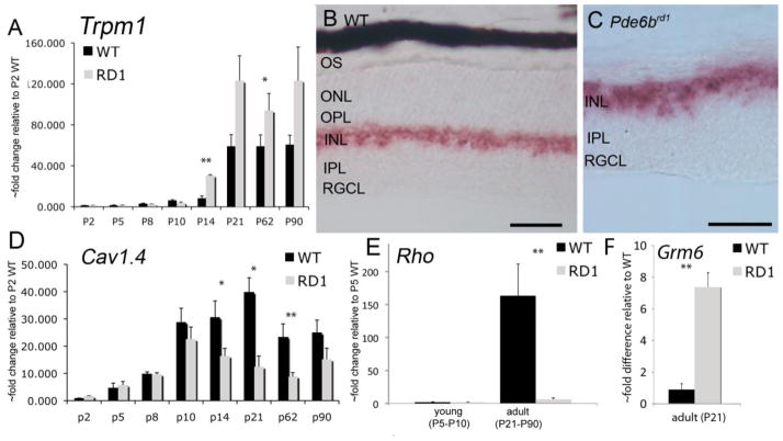
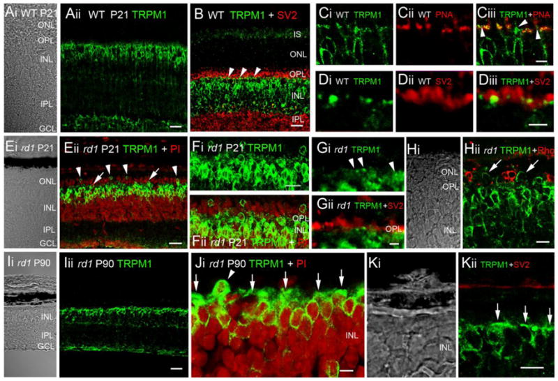
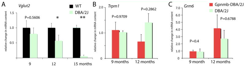
The light response in retinal ON bipolar cells is associated with disinhibition of current flow through cation channels recently identified as type 1 members of the melastatin transient receptor potential (TRPM) family. We determined the developmental expression of Trpm1 in the wild type C57BL/6, DBA/2J, DBA2J-Gpnmb mouse retinas and in Pde6brd1 retinas characterized by degeneration of rod photoreceptors. Trpm1 mRNA in wild type retinas was low at birth but exhibited progressive increases in abundance up to early adulthood at postnatal day 21 (P21). Retinal Trpm1 mRNA content did not decrease following loss of photoreceptors. At P21, TRPM1-immunopositive perikarya migrated into the outer nuclear layer. The TRPM1 protein was trafficked to discrete postsynaptic puncta in wild type retinas whereas in adult Pde6brd1 mouse retinas, TRPM1 translocated to bipolar perikarya and bar-like structures in the distal inner nuclear layer. These findings show that expression and localization of the TRPM1 in the mouse retina is plastic, modulated by use-dependence and availability of sustained excitatory input.
Copyright © 2010 Elsevier Ltd. All rights reserved.
Figures
References
-
- Armata IA, Giompres P, Smith A, Stasi K, Kouvelas ED, Mitsacos A. Genetically induced retinal degeneration leads to changes in metabotropic glutamate receptor expression. Neurosci Lett. 2006;393:12–17. - PubMed
-
- Berntson A, Taylor WR, Morgans CW. Molecular identity, synaptic localization, and physiology of calcium channels in retinal bipolar cells. J Neurosci Res. 2003;71:146–151. - PubMed
Publication types
MeSH terms
Substances
Grants and funding
LinkOut - more resources
Full Text Sources
Other Literature Sources
Miscellaneous
