

Association of visual function and ganglion cell layer thickness in patients with diabetes mellitus type 1 and no or minimal diabetic retinopathy
- PMID: 20801146
- PMCID: PMC3017636
- DOI: 10.1016/j.visres.2010.08.024
Association of visual function and ganglion cell layer thickness in patients with diabetes mellitus type 1 and no or minimal diabetic retinopathy
Abstract
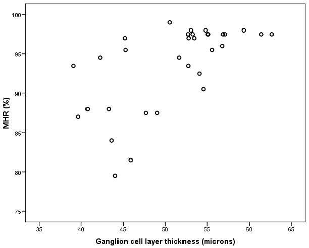
Diabetic retinopathy (DR) classically presents with micro-aneurysms, small haemorrhages and/or lipoprotein exudates. Several studies have indicated that neural loss occurs in DR even before vascular damage can be observed. This study evaluated the possible relationship between structure (spectral domain-optical coherence tomography) and function (Rarebit visual field test) in patients with type 1 diabetes mellitus and no or minimal diabetic retinopathy (DR). Results demonstrated loss of macular visual function and corresponding thinning of the ganglion cell layer (GCL) in the pericentral area of the macula of diabetic patients (Rs = 0.65, p < 0.001). In multivariable logistic regression analysis, GCL thickness remained an independent predictor of decreased visual function (OR 1.5, 95% CI 1.1-2.1). Early DR seems to include a neurodegenerative component.
Copyright © 2010 Elsevier Ltd. All rights reserved.
Figures
References
-
- Early Treatment Diabetic Retinopathy Study Research Group. Grading diabetic retinopathy from stereoscopic color fundus photographs--an extension of the modified Airlie House classification. ETDRS report number 10. Ophthalmology. 1991;98:786–806. - PubMed
-
- Abràmoff M, Magalhâes P, Ram S. Image Processing with ImageJ. Biophotonics. 2004;11:36–42.
-
- Abu-El-Asrar AM, Dralands L, Missotten L, Al-Jadaan IA, Geboes K. Expression of apoptosis markers in the retinas of human subjects with diabetes. Invest Ophthalmol Vis Sci. 2004;45:2760–2766. - PubMed
-
- Antonetti DA, Barber AJ, Bronson SK, Freeman WM, Gardner TW, Jefferson LS, Kester M, Kimball SR, Krady JK, LaNoue KF, Norbury CC, Quinn PG, Sandirasegarane L, Simpson IA. Diabetic retinopathy: seeing beyond glucose-induced microvascular disease. Diabetes. 2006;55:2401–2411. - PubMed
-
- Asefzadeh B, Fisch BM, Parenteau CE, Cavallerano AA. Macular thickness and systemic markers for diabetes in individuals with no or mild diabetic retinopathy. Clin Experiment Ophthalmol. 2008;36:455–463. - PubMed
Publication types
MeSH terms
Grants and funding
LinkOut - more resources
Full Text Sources
Medical
