Review
. 2010:283:93-128.
doi: 10.1016/S1937-6448(10)83003-5.

Axonal ensheathment and intercellular barrier formation in Drosophila

Affiliations
Review

Axonal ensheathment and intercellular barrier formation in Drosophila

Kevin Blauth et al. Int Rev Cell Mol Biol. 2010.

Abstract

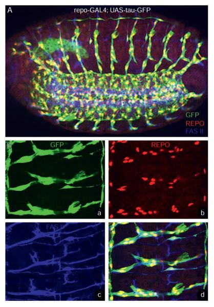
Glial cells are critical players in every major aspect of nervous system development, function, and disease. Other than their traditional supportive role, glial cells perform a variety of important functions such as myelination, synapse formation and plasticity, and establishment of blood-brain and blood-nerve barriers in the nervous system. Recent studies highlight the striking functional similarities between Drosophila and vertebrate glia. In both systems, glial cells play an essential role in neural ensheathment thereby isolating the nervous system and help to create a local ionic microenvironment for conduction of nerve impulses. Here, we review the anatomical aspects and the molecular players that underlie ensheathment during different stages of nervous system development in Drosophila and how these processes lead to the organization of neuroglial junctions. We also discuss some key aspects of the invertebrate axonal ensheathment and junctional organization with that of vertebrate myelination and axon-glial interactions. Finally, we highlight the importance of intercellular junctions in barrier formation in various cellular contexts in Drosophila. We speculate that unraveling the genetic and molecular mechanisms of ensheathment across species might provide key insights into human myelin-related disorders and help in designing therapeutic interventions.


Figures

Figure 3.1
Figure 3.1
Axonal ensheathment in the Drosophila embryonic PNS. (A, B) Whole mount stage 16 repo-Gal4; UAS-tauGFP (A) and higher magnification (Ba–d) of a portion of the embryo shown in (A) is stained with anti-GFP (green; A, Ba, Bd), anti-Repo (red; A, Bb, Bd), and anti-Fas II (blue; A, Bc, Bd). The GFP staining reveals the glial processes that surround the Fas II labeled motor axons. The glial nuclei expressing Repo show the arrangement of glial cells along the length of the axon trajectories.
Figure 3.2
Figure 3.2
Septate junctions and axonal ensheathment in the larval peripheral nerve fibers. (A–C) A portion of the nrx IV::GFP third instar larval ventral nerve cord (VNC) with peripheral nerves stained with anti-GFP (green) and anti-Repo (red). nrx IV::GFP expresses GFP in endogenous Nrx IV pattern. The peripheral nerves (A, B) reveal glial membrane expression and SJ localization of Nrx IV (arrowheads, A, B) along the length of the axon, while VNC shows localization of Nrx IV in surface glia (arrows, A, C), which are known to have SJs. Under the surface glial layer, there are Repo-positive glial cells (C, red). A wild-type third instar larval peripheral nerve (D) in cross section shows the presence of SJs (arrowheads) between outer and inner glial membranes. A large number of axons (a) are tightly fasciculated and ensheathed by glial processes (m).
Figure 3.3
Figure 3.3
Ensheathment of commissural axons in the Drosophila embryonic CNS. (A, B) sim-Gal4, UAS-tau-GFP embryo at a lower (Aa–d) and higher (Ba–d) magnifications show staining with anti-GFP (Aa, Ba, green), anti-Wrapper (Ab, Bb, red), and BP102 (Ac, Bc. blue). The GFP staining highlights the Sim-positive midline glia and neurons (Aa, Ba) while Wrapper expression is in the midline glia (Ab, Bb) and BP102 (Ac, Bc) labels the anterior commissure(AC) and posterior (PC) commissure. Note the midline glial processes (arrow, Aa, Ba) that ensheath the AC and PC (see merged panels, Ad, Bd). The midline glia express Wrapper (arrowheads, Bb).
Figure 3.4
Figure 3.4
Photoreceptor ensheathment and septate junctions in adult Drosophila eye. (A, B) A light microscopy image (A) and ultrastructural view in longitudinal section (B) of a single Drosophila ommatidium of the adult compound eye. Accessory cells (A), namely the cone cells (CC) and pigment cells (PC) express Nrx IV (green) while photoreceptors (PR) express the apical protein Crumbs (Crb). The ultrastructure at a lower magnification (B) reveals the anatomy of the ommatidium. On top of the pseudocone (PSC) is the lens (L) and at the bottom are the CC, PC, and PR. A higher magnification (C) reveals presence of extensive SJs (arrows) basal to the adherens junctions (arrowhead) that are formed between CC and PC. These SJs serve as protective barriers and seals the PR for proper phototransduction.

Similar articles

Cited by

References

    1. Abbott NJ, Bundgaard M, Cserr HF. Tightness of the blood–brain barrier and evidence for brain interstitial fluid flow in the cuttlefish, Sepia officinalis. J. Physiol. 1985;368:213–226. - PMC - PubMed
    1. Abbott NJ, Lane NJ, Bundgaard M. The blood–brain interface in invertebrates. Ann. N. Y. Acad. Sci. 1986;481:20–42. - PubMed
    1. Akiyama-Oda Y, Hosoya T, Hotta Y. Alteration of cell fate by ectopic expression of Drosophila glial cells missing in non-neural cells. Dev. Genes Evol. 1998;208:578–585. - PubMed
    1. Allen NJ, Barres BA. Neuroscience: glia—more than just brain glue. Nature. 2009;457:675–677. - PubMed
    1. Auld VJ, Fetter RD, Broadie K, Goodman CS. Gliotactin, a novel transmembrane protein on peripheral glia, is required to form the blood–nerve barrier in Drosophila. Cell. 1995;81:757–767. - PubMed

Publication types

LinkOut - more resources