. 2010 Dec;31(34):8953-63.
doi: 10.1016/j.biomaterials.2010.08.017.

Helicoidal multi-lamellar features of RGD-functionalized silk biomaterials for corneal tissue engineering

Affiliations

Helicoidal multi-lamellar features of RGD-functionalized silk biomaterials for corneal tissue engineering

Eun Seok Gil et al. Biomaterials. 2010 Dec.

Abstract

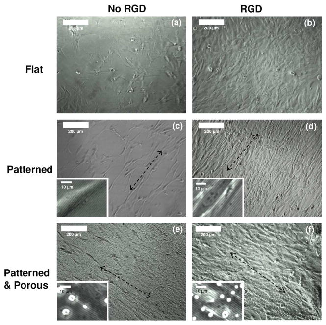
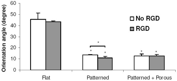
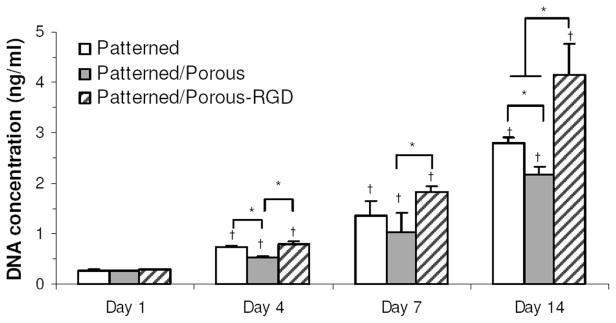
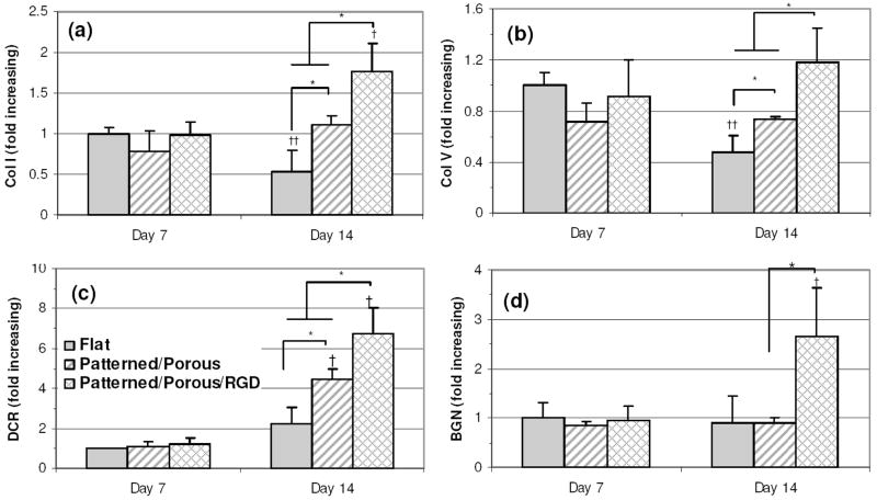
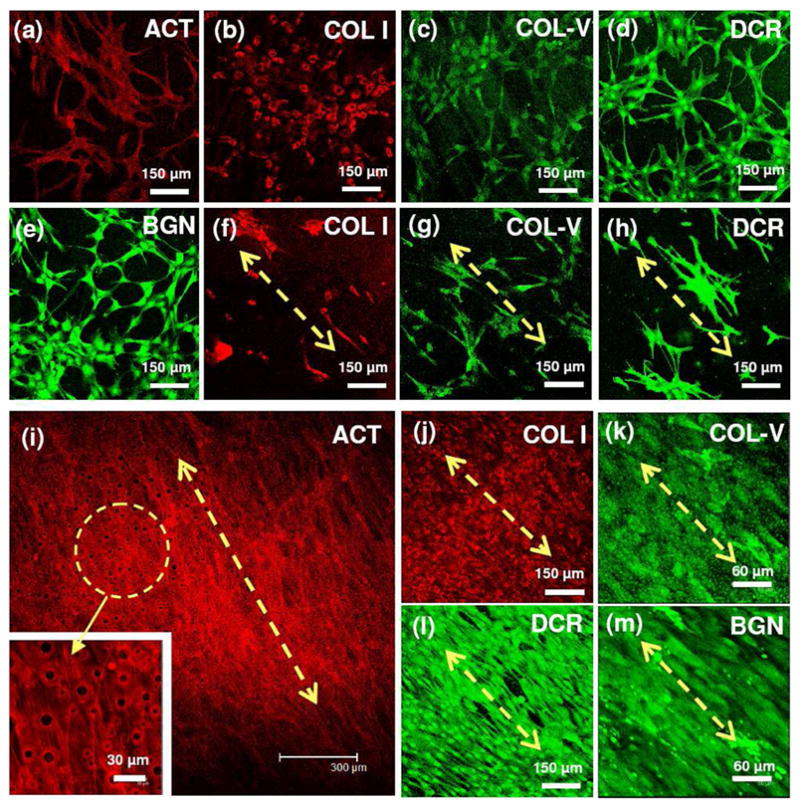
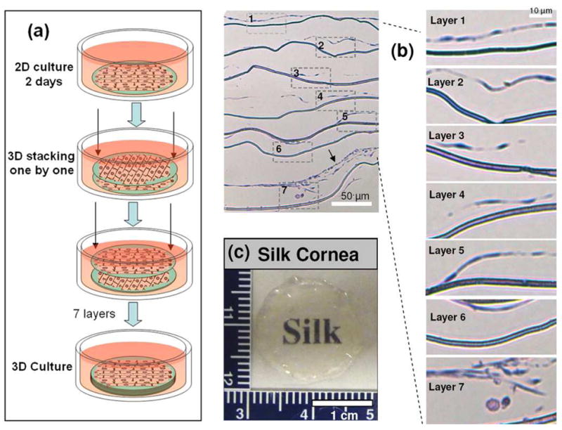
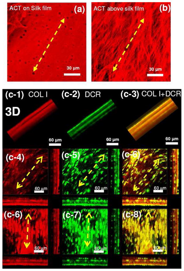
RGD-coupled silk protein-biomaterial lamellar systems were prepared and studied with human cornea fibroblasts (hCFs) to match functional requirements. A strategy for corneal tissue engineering was pursued to replicate the structural hierarchy of human corneal stroma within thin stacks of lamellae-like tissues, in this case constructed from scaffolds constructed with RGD-coupled, patterned, porous, mechanically robust and transparent silk films. The influence of RGD-coupling on the orientation, proliferation, ECM organization, and gene expression of hCFs was assessed. RGD surface modification enhanced cell attachment, proliferation, alignment and expression of both collagens (type I and V) and proteoglycans (decorin and biglycan). Confocal and histological images of the lamellar systems revealed that the bio-functionalized silk human cornea 3D constructs exhibited integrated corneal stroma tissue with helicoidal multi-lamellar alignment of collagen-rich and proteoglycan-rich extracellular matrix, with transparency of the construct. This biomimetic approach to replicate corneal stromal tissue structural hierarchy and architecture demonstrates a useful strategy for engineering human cornea. Further, this approach can be exploited for other tissue systems due to the pervasive nature of such helicoids in most human tissues.


Figures

Figure 1
Figure 1
Phase contrast optical micrographs of hCFs grown on silk films without (a, c, e) and with (b, d, f) RGD surface modification over 5 days in culture: flat (a, b), patterned (c, d) and patterned/porous (e, f). The magnified images reveal that hCFs are aligned on the patterned silk surface along with the groove axis and that hCFs are grown over pore structures (seen as white holes) within the silk film structure (2 ~ 10 μm in dia.).
Figure 2
Figure 2
Mean orientation angles of hCFs grown on the flat and patterned silk films. The orientation angle was determined by calculating the angle difference between the longest direction within the cell borders and the grooves. The mean orientation angle was defined by the mean±SD of three orientation angles averaged from three individual 10X images. (N=3, Cell population in each image varied from 25 to 58. Bars represent SD)
Figure 3
Figure 3
DNA content for hCFs grown on patterned, patterned/porous, and RGD coupled patterned/porous silk film substrates after 1~7 days in culture (p<0.05, N=4. Bars represent SD).
Figure 4
Figure 4
Expression of transcripts related to corneal stroma differentiation. Transcript expression of corneal stroma differentiation markers quantified by real-time RT-PCR; (a) Collagen Type I (Col I), (b) Collagen Type V (Col V), (c) Decorin (DCR), (d) Biglycan (BGN). Transcript levels were normalized to GAPDH within the linear range of amplification. *represents statistical significance between means of different substrate groups at similar time points; and †† indicate statistical significance (increase and decrease, respectively) between means of similar substrate groups at different time points (p<0.05, N=3. Bars represent SD)
Figure 5
Figure 5
Confocal images of hCFs after 6 days in culture on flat (a–e), patterned/porous (f–h), and RGD-coupled patterned/porous silk films (i–m); actin filaments (ACT) (a, i) and immunostained collagen type I (Col I) (a, f, j), immunostained collagen type V (Col V) (c, g, k), immunostained decorin (DCR) (d, h, l), immunostained biglycan (BGN) (e, m).
Figure 6
Figure 6
(a) Schematic of the assembly process for three-dimensional (3D) silk film corneal constructs. PVC rings (12 mm diameter) were placed on each silk film to hold the films before cell seeding at 100,000 cells/cm2 seeding density. After 2 days, hCFs layers with silk films were stacked one by one with applied pressure at each single film stacking step by using a 12 mm biopsy bunch and then the stacked constructs were cultured for 1 wk. Patterned and porous silk films with RGD surface modification were utilized for 3D corneal constructs. (b) Hematoxylin and eosin staining of cells on silk constructs after cultured for 1 wk. Arrow indicates three dimensional growth of corneal stroma tissue between the film layers. (c) Photo of stacked constructs after 1 wk. The constructs were visually clear enough to see the letters below.
Figure 7
Figure 7
Confocal images of hCFs in stacked 3D constructs after 7 d. Confocal images of stained actin filament near the silk film (a) and over the silk film (b) (both were obtained from same X-Y position) in the 3D constructs. 3D images (vertically projected images (c-1,2,3) and z-sectional images of two middle layers with cross section (c-4~8)). Red = immunostained collagen I (c-1,4,6), Green = immunostained decorin (c-2,5,7), and overlay images of decorin and collagen I (c-3,6,8).

References

    1. Morishige N, Petroll WM, Nishida T, Kenney MC, Jester JV. Noninvasive corneal stromal collagen imaging using two-photon-generated second-harmonic signals. J Cataract Refract Surg. 2006;32(11):1784–1791. - PMC - PubMed
    1. Torbet J, Malbouyres M, Builles N, Justin V, Roulet M, Damour O, et al. Orthogonal scaffold of magnetically aligned collagen lamellae for corneal stroma reconstruction. Biomaterials. 2007;28(29):4268–4276. - PubMed
    1. Griffith M, Osborne R, Munger R, Xiong XJ, Doillon CJ, Laycock NLC, et al. Functional human corneal equivalents constructed from cell lines. Science. 1999;286(5447):2169–2172. - PubMed
    1. Eye Bank Association of America. Annual report. 2005.
    1. Mannis MJ, McDonough G, Howard K, Morales R, Sugar J. Screening donor corneas that have undergone PRK. Cornea. 1997;16(6):683–685. - PubMed

Publication types

MeSH terms