. 2011 Jan;32(1):192-8.
doi: 10.3174/ajnr.A2216. Epub 2010 Aug 26.

Posterior circulation and high prevalence of ischemic stroke among young pediatric patients with Moyamoya disease: evidence of angiography-based differences by age at diagnosis

Affiliations

Posterior circulation and high prevalence of ischemic stroke among young pediatric patients with Moyamoya disease: evidence of angiography-based differences by age at diagnosis

S Mugikura et al. AJNR Am J Neuroradiol. 2011 Jan.

Abstract

Background and purpose: At diagnosis, the primary clinical manifestations of pediatric Moyamoya disease are TIA or CSs. CSs are reported to be more prevalent in younger than in older children. We sought to determine whether age-related differences in clinical manifestations are associated with age-related angiographic differences.

Materials and methods: We divided 78 patients diagnosed with Moyamoya disease before 16 years of age into four 4-year age groups and examined the relationships between age at diagnosis and clinical manifestations and angiographic and MR imaging findings.

Results: Among the 4 diagnostic age groups, in those younger than 4 years of age, the prevalence of CSs and of infarctions on MR images was highest, and along with severity of steno-occlusive lesions of the PCA, the prevalence was significantly higher than that in the next diagnostic age group (4-7 years), though the severity of steno-occlusive lesions in the ICA and the degree of transdural collaterals did not differ significantly. The prevalence of CSs and infarctions did not differ significantly in the 3 oldest diagnostic age groups, whereas ICA and PCA lesions and transdural collaterals correlated positively with diagnostic age.

Conclusions: The high prevalence of CSs and infarctions in patients diagnosed before 4 years of age is associated with advanced steno-occlusive lesions of the PCA. In patients 4 years of age and older at diagnosis, transdural collaterals develop in parallel with advancement of ICA and PCA lesions, which may contribute to the nearly constant prevalence of CSs.


Figures
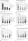
Fig 1.
Fig 1.
The relationship between the 4 diagnostic age groups and clinical manifestations (A), number of infarcted zones (B), distribution of corticosubcortical infarcted zones (C), ICA staging (D), PCA staging (E), and number of transdural collaterals (F). Asterisks indicate significant differences. Double asterisks indicate a significant difference and a positive relationship to diagnostic age among the 3 oldest diagnostic age groups.
Fig 2.
Fig 2.
A 15-month-old girl (diagnostic age group younger than 4 years of age) initially presented with CS with right upper motor paresis a month before and subsequently presented with contralateral hemiparesis CS. Moyamoya disease was diagnosed. Her clinical manifestation was CS. A and B, Coronal and axial time-of-flight MR angiograms show steno-occlusive bilateral lesions in the terminal part of the ICA and in the proximal parts of the ACA and MCA cerebral arteries (ICA stage II, bilaterally). The right PCA is almost completely occluded (short arrow), with well-developed dilated perforators around it (stage III); the proximal part of the left PCA is stenotic (long arrow, PCA stage II). Note that in this patient, the bilateral steno-occlusive changes involved the PCAs even in the hemispheres with less advanced ICA lesions (ICA stage II, bilaterally). C, Diffusion-weighted MR image shows hyperintense regions of recent infarction in the anterior and posterior watersheds and the boundary zone between the regions of the ACA and PCA in the right. Five infarcted zones are seen on the right. D, T2-weighted MR image additionally shows an old cortical infarction in the posterior MCA territory (arrow), seen on the left.
Fig 3.
Fig 3.
A 7-year-old girl (diagnostic age group, 4–7 years) who had recurrent transient left hemiparesis for 5 years was diagnosed with Moyamoya disease, which manifested clinically as TIA. A and B, Coronal and axial time-of-flight MR angiograms show advanced steno-occlusive changes at or around the terminal part of the right ICA, with poorly visualized ACA and MCA branches (ICA stage III, right). Moderate steno-occlusive changes at or around the terminal part of the left ICA with relatively good visualization of the ACA (small arrows) and MCA (large arrows) cortical branches (ICA stage II) are seen on the left. No steno-occlusive lesions are seen bilaterally in the posterior cerebral artery (PCA stage 1, bilaterally). C, Anteroposterior view of the vertebral angiogram shows no steno-occlusive lesions bilaterally in the PCA and well-developed leptomeningial collateral circulation to the anterior circulation. D, Left lateral external carotid angiogram shows dilated anterior branches of the middle meningeal artery providing transdural collaterals to the contralateral frontal region (large arrows), with the medial branches of the maxillary artery providing transdural collaterals to the right anterior basal region (small arrow). Two transdural collaterals can be seen on the right, and none are seen on the left (anteroposterior view of the left external carotid angiogram, not shown). E, Axial T2-weighted MR image shows no infarction bilaterally (ie, the number of infarcted regions is zero on either side). Note that in this patient, the steno-occlusive changes do not involve the PCA, even in the right hemisphere, where ICA lesions are advanced (stage III).
Fig 4.
Fig 4.
A 12-year-old boy (diagnostic age group, 12–15 years) with right and left transient motor paresis. His clinical manifestation was TIA. A, Axial time-of-flight MR angiogram shows advanced steno-occlusive changes bilaterally at or around the terminal parts of the ICAs, with no apparent ACA and MCA branches (ICA stage IV, bilaterally). Advanced steno-occlusive lesions bilaterally in the PCAs with well-developed Moyamoya vessels from the PCA are seen (PCA stage 3, bilaterally). B, Anteroposterior view of the vertebral angiogram shows advanced steno-occlusive lesions bilaterally in the PCAs (PCA stage III, bilaterally). C and D, Lateral view of the arterial (C) and capillary (D) phases of the right external carotid angiograms shows that the dilated posterior branch of the middle meningeal artery (large arrows) and the meningeal branch of the occipital artery (arrowhead) provide transdural collaterals mainly to the right posterior part of the cerebral hemispheres. The frontal branch of the superficial temporal artery (small arrow) and medial branches of the internal maxillary artery provide transdural collaterals to the frontobasal region. Three transdural collaterals are seen on the right. The right cerebral hemisphere is largely supplied through the transdural collaterals (D). Three transdural collaterals were seen on the left (left external carotid angiogram, not shown). E, Axial T2-weighted MR image shows an old small infarction in the left anterior MCA territory. No infarcted regions are seen on the right, but 1 is seen on the left. Note that this patient has severe steno-occlusive PCA changes that parallel the advanced ICA lesions. This progressive state should have provoked the development of transdural collaterals, which might have prevented a large infarction and CS.
Fig 5.
Fig 5.
The relationship between the 4 diagnostic age groups and PCA stage in the 117 hemispheres with less advanced steno-occlusive ICA lesions (ICA stage I or II). The asterisk indicates a significant difference between diagnostic age groups younger than 4 years and 4–7 years of age.

References

    1. Suzuki J, Takaku A. Cerebrovascular “Moyamoya” disease: disease showing abnormal net-like vessels in base of brain. Arch Neurol 1969;20:288–99 - PubMed
    1. Miyamoto S, Kikuchi H, Karasawa J, et al. . Study of the posterior circulation in Moyamoya disease: clinical and neuroradiological evaluation. J Neurosurg 1984;61:1032–37 - PubMed
    1. Yamada I, Himeno Y, Suzuki S, et al. . Posterior circulation in Moyamoya disease: angiographic study. Radiology 1995;197:239–46 - PubMed
    1. Mugikura S, Takahashi S, Higano S, et al. . The relationship between cerebral infarction and angiographic characteristics in childhood Moyamoya disease. AJNR Am J Neuroradiol 1999;20:336–43 - PMC - PubMed
    1. Mugikura S, Takahashi S, Higano S, et al. . Predominant involvement of ipsilateral anterior and posterior circulations in Moyamoya disease. Stroke 2002;33:1497–500 - PubMed

MeSH terms

LinkOut - more resources