. 2010 Nov 5;285(45):34729-40.
doi: 10.1074/jbc.M110.116756. Epub 2010 Aug 27.

Aberrant overexpression of the Rgl2 Ral small GTPase-specific guanine nucleotide exchange factor promotes pancreatic cancer growth through Ral-dependent and Ral-independent mechanisms

Affiliations

Aberrant overexpression of the Rgl2 Ral small GTPase-specific guanine nucleotide exchange factor promotes pancreatic cancer growth through Ral-dependent and Ral-independent mechanisms

Dominico Vigil et al. J Biol Chem. .

Abstract

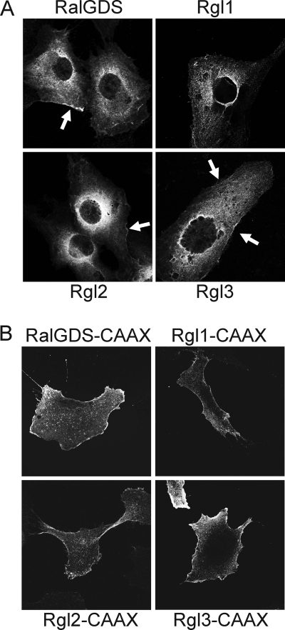
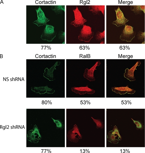
Our recent studies established essential and distinct roles for RalA and RalB small GTPase activation in K-Ras mutant pancreatic ductal adenocarcinoma (PDAC) cell line tumorigencity, invasion, and metastasis. However, the mechanism of Ral GTPase activation in PDAC has not been determined. There are four highly related mammalian RalGEFs (RalGDS, Rgl1, Rgl2, and Rgl3) that can serve as Ras effectors. Whether or not they share distinct or overlapping functions in K-Ras-mediated growth transformation has not been explored. We found that plasma membrane targeting to mimic persistent Ras activation enhanced the growth-transforming activities of RalGEFs. Unexpectedly, transforming activity did not correlate directly with total cell steady-state levels of Ral activation. Next, we observed elevated Rgl2 expression in PDAC tumor tissue and cell lines. Expression of dominant negative Ral, which blocks RalGEF function, as well as interfering RNA suppression of Rgl2, reduced PDAC cell line steady-state Ral activity, growth in soft agar, and Matrigel invasion. Surprisingly, the effect of Rgl2 on anchorage-independent growth could not be rescued by constitutively activated RalA, suggesting a novel Ral-independent function for Rgl2 in transformation. Finally, we determined that Rgl2 and RalB both localized to the leading edge, and this localization of RalB was dependent on endogenous Rgl2 expression. In summary, our observations support nonredundant roles for RalGEFs in Ras-mediated oncogenesis and a key role for Rgl2 in Ral activation and Ral-independent PDAC growth.


Figures

FIGURE 1.
FIGURE 1.
Domain structure and sequence identity of RA domain-containing RalGEFs. The four RalGEFs that can serve as effectors of Ras share the same domain topology, with an N-terminal Ras exchange motif (REM), followed by the CDC25 homology RalGEF catalytic domain and a C-terminal RA domain. The sequence identities to RalGDS are indicated below each domain. The precise function of the Ras exchange motif domain is not known, but deletion analysis of RalGDS indicates that it is dispensable for RalGEF catalytic activity in vivo. Unlike other CDC25 homology domain-containing GEFs, the RalGEFs are selective for the two Ral isoforms. The RA domain binds to GTP-bound Ral. Expression vectors encoding full-length mouse RalGDS (NM_009058), Rgl1 (NM_016846), Rgl2 (NM_009059), and Rgl3 (NM_023622) were generated that included either N-terminal HA epitope or GFP sequences. To generate plasma membrane-targeted versions that mimic constitutive association with activated K-Ras, the C-terminal 20 residues of human K-Ras4B were added to the C terminus of each RalGEF; this sequence includes the CAAX prenylation signal sequence and the polybasic second signal required for plasma membrane association. Rlf-CAAX contains an N-terminal HA epitope tag (MAYPYDVPDYASTD) followed by residues 2–532 of mouse RGL2 and then 12 vector-encoded residues (HAPGRHGRGIVN) and then terminating with 20 residues that include the K-Ras4B sequence (KMSKDGKKKKKKSKTKCVIM). The C-terminal 246 residues of Rlf, including the RA domain, are deleted.
FIGURE 2.
FIGURE 2.
RalGEFs exhibit similar cytosolic subcellular localization and plasma membrane association is enhanced by the addition of the K-Ras4B plasma membrane targeting sequence. A, wild type RalGEFs exhibit a cytosolic subcellular localization. Mass populations of HEK-HT cells were stably infected with pBabe-puro expression vectors expressing the indicated HA-tagged RalGEFs and were grown on glass coverslips. After labeling with anti-HA antibody followed by Alexa Fluor 488-conjugated secondary antibody, the cells were visualized by confocal microscopy. Arrows, plasma distinct plasma membrane localization. B, the addition of Ras membrane targeting sequence enhances RalGEF plasma membrane association. Analyses were done as described in A with chimeric RalGEFs terminating with the K-Ras4B plasma membrane targeting sequence. Data shown are representative of at least two independent experiments.
FIGURE 3.
FIGURE 3.
Membrane association promotes RalGEF transformation of HEK-HT cells. HEK-HT cells were suspended in soft agar as described (57), and the number of proliferating viable colonies of >30 cells were quantitated after 4 weeks. Data shown are the average ± S.D. (error bars) of triplicate plates and are representative of three independent experiments. *, p < 0.005 versus vector control.
FIGURE 4.
FIGURE 4.
Pancreatic carcinoma cells express multiple RalGEFs. A, RT-PCR detection of RalGEF mRNA colorectal and pancreatic ductal adenocarcinoma cell lines, with β-actin as a control. Total cellular RNA was isolated from the indicated cell lines. After reverse transcription, PCR was performed with RalGEF-specific and β-actin-specific primers. B, Rgl2 protein expression in PDAC cell lines. Lysates from the cell lines were separated by SDS-PAGE and transferred to a PVDF membrane and blotted for Rgl2 protein expression. A parallel blot for β-actin was done to ensure equivalent loading of total cellular protein. C, overexpression of Rgl2 protein in tumor (T) versus normal (N) matched PDAC patient samples, as analyzed by Western blot analysis, with γ-tubulin monoclonal antibody (Sigma) as a control for equivalent total protein.
FIGURE 5.
FIGURE 5.
Dominant negative Ral mutant inhibitors of RalGEFs impair PDAC soft agar growth and invasion through Matrigel. A, effect on the RalGEFs in RalA- and RalB-GTP levels in pancreatic ductal adenocarcinoma cell lines. The indicated cell lines were stably infected with retroviral pBabe empty vector or Ral dominant negatives (RalA(31N) and RalB(28N)) and selected with puromycin. RalA- and RalB-GTP were detected as previously described (29) by pull-down from the indicated PDAC lines, followed by immunoblot analysis with the RalA or RalB antibody. Total RalA and RalB were derived from immunoblotting total lysate and show stable expression of RalA(31N) or RalB(28N), respectively. Blot analysis with anti-β-actin antibody was done to verify equivalent total protein. B, role of the RalGEFs in PDAC anchorage-independent growth. The number of proliferating viable colonies of >30 cells were quantitated after 2 weeks. Data shown are the average ± S.D. (error bars) of triplicate plates and are representative of at least two independent experiments. C, role of the RalGEFs in PDAC invasion. The indicated cells were dissociated and resuspended in serum-free growth medium containing 1% BSA and incubated at 37 °C in the upper chamber of a Matrigel invasion chamber. Data shown are the percentage of invaded cells relative to vehicle and are the average ± S.D. of triplicate chambers and are representative of at least two independent experiments. *, p < 0.05 versus pBabe vector control. **, p < 0.005 versus pBabe vector control.
FIGURE 6.
FIGURE 6.
The RalGEF Rgl2 plays a crucial role in PDAC. The indicated cell lines were stably infected with a nonspecific shRNA vector (NS), which does target any human genes, or two independent shRNA vectors targeting Rgl2. A, Rgl2 is responsible for RalA and RalB activation in PDAC lines. RalA- and RalB-GTP were detected as described previously (29) by pull-down from the indicated PDAC lines, followed by immunoblot analysis with the RalA or RalB antibody. Total RalA and RalB were derived from immunoblotting total lysate. Blot analysis with anti-β-actin antibody was done to verify equivalent total protein. Data shown are representative of two independent experiments. B, role of the Rgl2 in PDAC anchorage-independent growth. The number of proliferating viable colonies of >30 cells was quantitated after 2 weeks. Data shown are the average ± S.D. (error bars) of triplicate plates and are representative of at least two independent experiments. C, role of Rgl2 in PDAC invasion. The indicated cells were dissociated and resuspended in serum-free growth medium in the upper chamber of a Matrigel invasion chamber. After 22 h, the non-invaded cells were removed, and the chambers were fixed, stained, and counted under a microscope. Data shown are the percentage of invaded cells relative to vehicle and are the average ± S.D. of triplicate chambers and are representative of at least two independent experiments. *, p < 0.05 versus nonspecific shRNA control.
FIGURE 7.
FIGURE 7.
Activated RalA cannot rescue the anchorage-independent growth defect of Rgl2 knockdown in MIA PaCa-2 cells. The indicated cell lines were stably infected with a puromycin-resistant nonspecific shRNA vector (NS), which does target any human genes, or puromycin-resistant Rgl2 shRNA 1 vector targeting Rgl2, without and with ectopic expression of constitutively activated RalA(Q75L). A, blot analysis showing expression levels of endogenous wild type Rgl2 and ectopic RalA(Q75L) in the indicated PDAC lines as well as anti-tubulin antibody to verify equivalent total protein. Data shown are representative of two independent experiments. B, the number of proliferating viable colonies of >30 cells was quantitated after 2 weeks. Data shown are the average ± S.D. (error bars) of triplicate plates and are representative of at least two independent experiments. C, blot analysis demonstrating phosphorylated and total AKT and ERK levels upon Rgl2 knockdown in MIA PaCa-2 cells. Blot data for Rgl2 and β-actin are duplicated from Fig. 6A in this panel, where the same cell lysates were used for the AKT and ERK blot data. Data shown are representative of two independent experiments; *, p < 0.05 between Rgl2 shRNA 1 and shRNA 1 + RalAQ75L; **, p < 0.005 versus nonspecific shRNA control.
FIGURE 8.
FIGURE 8.
Rgl2 expression is required for RalB association with the leading edge. A, Rgl2 is localized to the leading edge. CFPAC-I cells were grown on glass coverslips. After labeling with mouse anti-Rgl2 and rabbit anti-cortactin antibodies followed by Alexa Fluor 488-conjugated and 568-conjugated secondary antibodies, cells were visualized with a confocal microscope. B, RalB localization to the leading edge is dependent on Rgl2 expression. CFPAC-I cells stably infected with either nonspecific or Rgl2 shRNA 2 were grown on glass coverslips. After labeling with mouse anti-RalB and rabbit anti-cortactin antibodies followed by Alexa Fluor 488-conjugated and 568-conjugated secondary antibodies, cells were visualized with a confocal microscope. Data shown are representative of two independent experiments. Percentages shown are percentages of 30 randomly chosen cells for each experiment that demonstrate distinct plasma membrane localization for either Rgl2, RalB, or cortactin, as shown and, in the case of the merge panel, percentages of cells that show distinct co-localization with cortactin at the leading edge.

Similar articles

Cited by

References

    1. Jones S., Zhang X., Parsons D. W., Lin J. C., Leary R. J., Angenendt P., Mankoo P., Carter H., Kamiyama H., Jimeno A., Hong S. M., Fu B., Lin M. T., Calhoun E. S., Kamiyama M., Walter K., Nikolskaya T., Nikolsky Y., Hartigan J., Smith D. R., Hidalgo M., Leach S. D., Klein A. P., Jaffee E. M., Goggins M., Maitra A., Iacobuzio-Donahue C., Eshleman J. R., Kern S. E., Hruban R. H., Karchin R., Papadopoulos N., Parmigiani G., Vogelstein B., Velculescu V. E., Kinzler K. W. (2008) Science 321, 1801–1806 - PMC - PubMed
    1. Yeh J. J., Der C. J. (2007) Expert Opin. Ther. Targets 11, 673–694 - PubMed
    1. Wennerberg K., Rossman K. L., Der C. J. (2005) J. Cell Sci. 118, 843–846 - PubMed
    1. Bodemann B. O., White M. A. (2008) Nat. Rev. Cancer 8, 133–140 - PubMed
    1. Chien Y., White M. A. (2003) EMBO Rep. 4, 800–806 - PMC - PubMed

Publication types

MeSH terms