. 2010 Nov 5;285(45):34364-70.
doi: 10.1074/jbc.M110.138412. Epub 2010 Aug 27.

Mitotic phosphorylation of Cdc25B Ser321 disrupts 14-3-3 binding to the high affinity Ser323 site

Affiliations

Mitotic phosphorylation of Cdc25B Ser321 disrupts 14-3-3 binding to the high affinity Ser323 site

Puji Astuti et al. J Biol Chem. .

Abstract

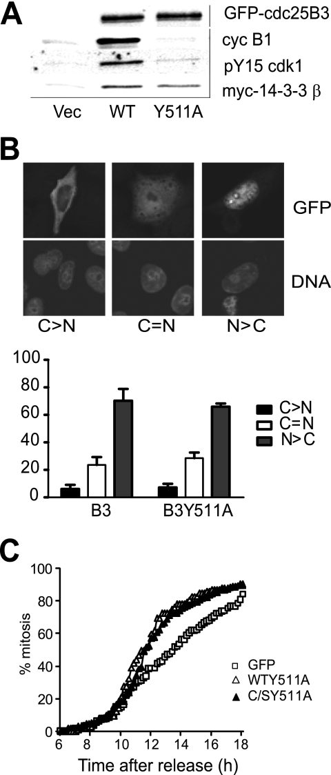
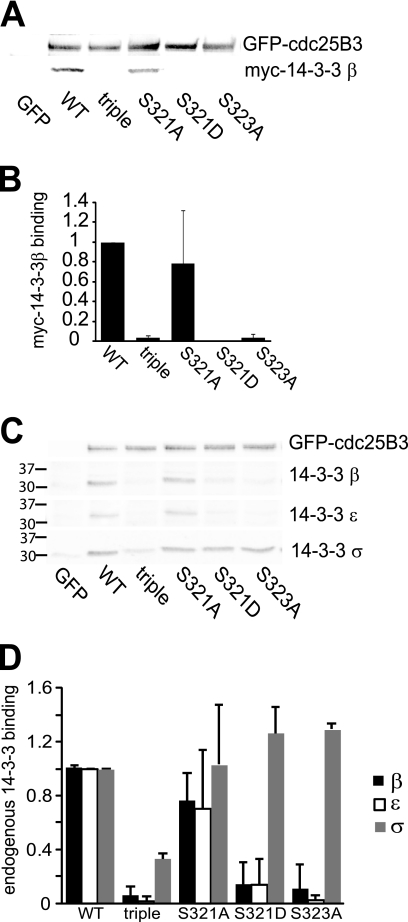
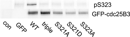
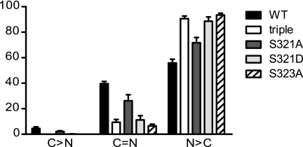
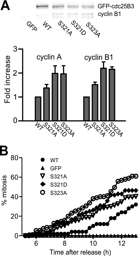
Cdc25B is a key regulator of entry into mitosis, and its activity and localization are regulated by binding of the 14-3-3 dimer. There are three 14-3-3 binding sites on Cdc25B, with Ser(323) being the highest affinity binding and is highly homologous to the Ser(216) 14-3-3 binding site on Cdc25C. Loss of 14-3-3 binding to Ser(323) increases cyclin/Cdk substrate access to the catalytic site, thereby increasing its activity. It also affects the localization of Cdc25B. Thus, phosphorylation and 14-3-3 binding to this site is essential for down-regulating Cdc25B activity, blocking its mitosis promoting function. The question of how this inhibitory signal is relieved to allow Cdc25B activation and entry into mitosis is yet to be resolved. Here, we show that Ser(323) phosphorylation is maintained into mitosis, but phosphorylation of Ser(321) disrupts 14-3-3 binding to Ser(323), mimicking the effect of inhibiting Ser(323) phosphorylation on both Cdc25B activity and localization. The unphosphorylated Ser(321) appears to have a role in stabilizing 14-3-3 binding to Ser(323), and loss of the Ser hydroxyl group appears to be sufficient to significantly reduce 14-3-3 binding. A consequence of loss of 14-3-3 binding is dephosphorylation of Ser(323). Ser(321) is phosphorylated in mitosis by Cdk1. The mitotic phosphorylation of Ser(321) acts to maintain full activation of Cdc25B by disrupting 14-3-3 binding to Ser(323) and enhancing the dephosphorylation of Ser(323) to block 14-3-3 binding to this site.


Figures

FIGURE 1.
FIGURE 1.
The substrate binding mutant of Cdc25B had no effect on 14-3-3 binding, localization, or G2/M progression. A, substrate trapping mutants of GFP-Cdc25B3 C487S/R493K, either wild type for the substrate binding site residues (WT) or Y511A mutant, were transfected into HeLa cells followed by etoposide addition overnight to arrest cells in G2 phase. The interaction with substrate cyclin B1 and the inactive pTyr15 Cdk1, and exogenously expressed Myc-14-3-3β were examined from the immunoprecipitates of GFP-Cdc25B. Immunoprecipitation from cells expressing GFP alone (Vec) was performed as a control. B, HeLa cells were transfected with Cdc25B3 wild type (WT) and the substrate binding mutant Cdc25B3 Y511A. The localization was examined 8 h post-transfection. The different localization of GFP-Cdc25B3 relative to the nucleus is shown in the inset, with predominantly cytoplasmic localization (C > N), equivalent nuclear and cytoplasmic staining (C=N) and predominantly nuclear staining (N > C). The data represent three independent experiments counting a minimum of 200 transfected cells. C, HeLa cells were either transfected with the substrate binding mutants GFP-Cdc25B3 Y511A or GFP-Cdc25B2 Y511A (C/SY511A), which also incorporated the inactivating active site mutation, or GFP alone, synchronized at G1/S transition by thymidine synchrony, and the entry into mitosis was monitored by time lapse microscopy.
FIGURE 2.
FIGURE 2.
Ser321 mutation reduces 14-3-3 binding. HeLa cells were co-transfected with the substrate binding site mutant of GFP-Cdc25B3 Y511A, either wild type for Ser321 (WT), S321A, S321D, S151A/S230A/S323A 14-3-3 binding sites triple mutants, or the S323A mutant, together with Myc-14-3-3β. The cells were synchronized and collected as they transited G2 phase, 7 h after synchrony release. The GFP-Cdc25B was immunoprecipitated and analyzed for Myc-14-3-3β binding, and the level of GFP-Cdc25B was analyzed by immunoblotting. GFP-transfected cells were used as controls. B, quantification of Myc-14-3-3β binding in A. 14-3-3 binding was normalized for GFP-Cdc25B levels in the immunoprecipitate from three independent experiments. C, similar experiment as in A but detecting endogenous 14-3-3β, -ϵ, and -σ binding. The position of the molecular weight markers for the 14-3-3 blots is shown for each isoform. D, quantification of endogenous 14-3-3 binding in C from three independent experiments.
FIGURE 3.
FIGURE 3.
Ser321 mutation reduces Ser323 phosphorylation. HeLa cells were transfected with substrate binding site mutants of GFP-Cdc25B3 as in Fig. 2 and synchronized and harvested in G2 phase. The GFP-Cdc25B was immunoprecipitated and analyzed for Ser323 phosphorylation (pS323) and GFP-Cdc25B3 levels. Controls were untransfected (con) and GFP-transfected cells.
FIGURE 4.
FIGURE 4.
Ser321 mutation increases nuclear localization of Cdc25B. HeLa cells were transfected with the indicated substrate binding site mutants of Cdc25B3, and their localization was examined by immunofluorescence 8 h post-transfection. The localization of the GFP-Cdc25B constructs expressed was quantitated as in Fig. 1B. The data are from three independent experiments counting at least 200 GFP-Cdc25B-expressing cells. C > N, predominantly cytoplasmic localization; C=N, equivalent nuclear and cytoplasmic staining, and N > C, predominantly nuclear staining.
FIGURE 5.
FIGURE 5.
Ser321 mutation increases substrate binding and activity of Cdc25B. A, HeLa cells were synchronized at G1/S and transfected with substrate trapping mutants of GFP-Cdc25B3 and the indicated phosphorylation site mutants. 7 h post-transfection, the cells were harvested and immunoprecipitated to detect the interaction between GFP-Cdc25B3 and its substrate cyclin/Cdk, cyclin A, and cyclin B1. A representative set of immunoprecipitates of the indicated GFP-Cdc25B mutants immunoblotted for GFP-Cdc25B and cyclin B1 is shown. The fold increased binding of cyclin A and cyclin B1 over the Ser321 wild type protein, normalized for GFP-Cdc25B levels from three individual experiments is shown. B, HeLa cells were synchronized at G1/S and transfected with catalytically active GFP-Cdc25B3, either wild type (WT), the indicated phosphorylation site mutants, or GFP. Etoposide (4 μm) was added 5 h post-transfection to impose a G2 phase checkpoint arrest, and the timing of cells entering mitosis was monitored by time-lapse microscopy and scored as a percent of the total population. These data are representative of three individual experiments.
FIGURE 6.
FIGURE 6.
Ser323 is phosphorylated during interphase and reduced in mitosis. A, U2OS cells expressing HA-Cdc25B were synchronized at G1/S by thymidine block and released in the absence of tetracycline to induce Cdc25B expression. The cells were harvested at indicated times and analyzed for Cdc25B Ser323 phosphorylation (pS323). The level of induced Cdc25B is shown (HA-Cdc25B), as are the levels of cyclin B1 Thr286 phosphorylated MEK1 as a marker of mitosis. PCNA was used as a loading control. The position of the 75-kDa marker is indicated by the bar on the left-hand side of the Cdc25B blots. B, the indicated substrate binding mutants of GFP-Cdc25B3and Myc-14-3-3β were transfected into HeLa cells, which were then synchronized by arrest in G2 phase with overnight of etoposide (G2), and mitotic sample produced by caffeine addition to the etoposide arrested cells for 5 h (M). Cells also were arrested in mitosis by overnight treatment with nocodazole (No). The mitotic cells from both the caffeine-promoted mitosis and nocodazole treatment were collected by mitotic shake-off. Lysates or immunoprecipitates of GFP-Cdc25B (GFP IP) were immunoblotted for the phosphorylation of Ser323 (pS323) of the overexpressed protein, the GFP-Cdc25B3, phosphorylated MEK1 Thr286 (pMEK T286) as a marker of cyclin B/Cdk1 activity and mitosis, and α-tubulin (α-tub) as a loading control. C, substrate binding site mutant of GFP-Cdc25B3 was transfected into HeLa cells that were then synchronized with thymidine. After release from the synchrony arrest, cells were treated without or with 250 nm okadaic acid (OA) 6 h after release and harvested 2 h later. Cells were immunoblotted for the indicated proteins. The position of the 100-kDa marker is indicated (bar). α-Tubulin was used as a loading control. D, substrate binding mutant of GFP-Cdc25B3 or GFP and Myc-14-3-3β were transfected in HeLa cells, synchronized, and then treated with or without okadaic acid as in C. GFP-Cdc25B was immunoprecipitated. Lysates were immunoblotted for the overexpressed protein (IB), and the immunoprecipitate for immunoblotted for the associated Myc-14-3-3β (IP) is indicated by the arrowhead. The faster migrating band was nonspecifically detected by the antibody in the immunoprecipitate.
FIGURE 7.
FIGURE 7.
Cdc25B is phosphorylated at multiple sites including Ser321 in mitosis by Cdk1. A, HeLa cells were synchronized at G1/S by thymidine block, and cells were harvested at 7 (G2) and 9.5 h post-release mitotic (M), or arrested overnight with nocodazole (No), either without or with addition of RO-3306 (Cdk1i) 30 min prior to harvesting. The mitotic cells from both the thymidine synchrony and nocodazole treatment were collected by mitotic shake-off. Lysates were immunoblotted for Cdc25B. The position of the 75-kDa marker is indicated (bar). B, HeLa cells were transfected with the substrate binding site mutant of GFP-Cdc25B3 or empty vector (Vec), synchronized with thymidine, and harvested in G2 or mitosis, or at the same time as the mitotic sample but in the presence of 9 μm Cdk1 inhibitor RO-3306 added 2 h prior to harvesting (+Cdk1i). Lysates were immunoblotted for pSer321, GFP-Cdc25B3, and pMEK Thr286 as a marker of mitosis, and MEK1 as a loading control. The position of the 100-kDa marker is indicated (bar).

References

    1. Gabrielli B. G., De Souza C. P., Tonks I. D., Clark J. M., Hayward N. K., Ellem K. A. (1996) J. Cell Sci. 109, 1081–1093 - PubMed
    1. Karlsson C., Katich S., Hagting A., Hoffmann I., Pines J. (1999) J. Cell Biol. 146, 573–584 - PMC - PubMed
    1. Lammer C., Wagerer S., Saffrich R., Mertens D., Ansorge W., Hoffmann I. (1998) J. Cell Sci. 111, 2445–2453 - PubMed
    1. Lindqvist A., Källström H., Lundgren A., Barsoum E., Rosenthal C. K. (2005) J. Cell Biol. 171, 35–45 - PMC - PubMed
    1. Goldstone S., Pavey S., Forrest A., Sinnamon J., Gabrielli B. (2001) Oncogene 20, 921–932 - PubMed

Publication types