. 2010 Nov;118(1):213-23.
doi: 10.1093/toxsci/kfq254. Epub 2010 Aug 26.

TCDD induced pericardial edema and relative COX-2 expression in medaka (Oryzias Latipes) embryos

Affiliations

TCDD induced pericardial edema and relative COX-2 expression in medaka (Oryzias Latipes) embryos

Wu Dong et al. Toxicol Sci. 2010 Nov.

Abstract

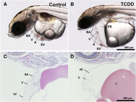
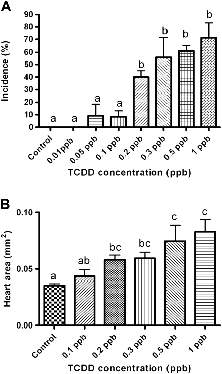
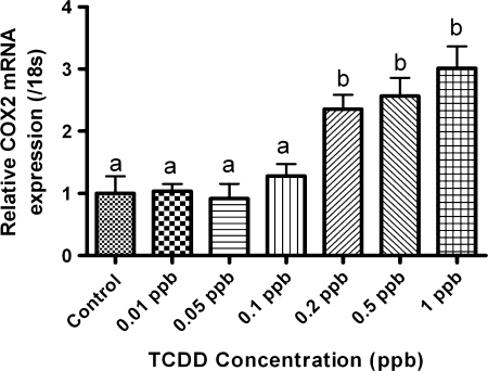
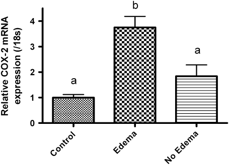
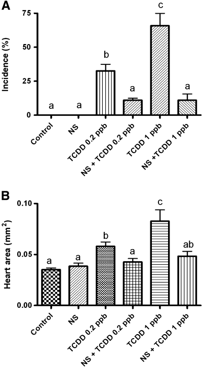
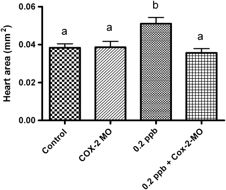
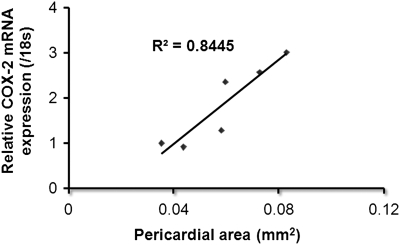
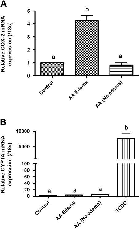
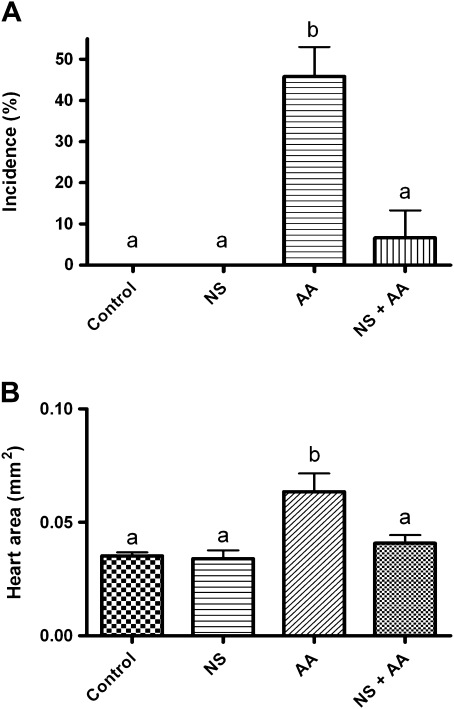
Exposure to dioxin and other aryl hydrocarbon receptor (AhR) ligands results in multiple, specific developmental cardiovascular phenotypes including pericardial edema and circulatory failure in small aquarium fish models. Although phenotypes are well described, mechanistic underpinnings for such toxicities remain elusive. Here we suggest that AhR activation results in stimulation of inflammation and "eicosanoid" pathways, which contribute to the observed developmental, cardiovascular phenotypes. We demonstrate that medaka embryos exposed to 2,3,7,8-tetrachlorodibenzo-p-dioxin (TCDD) (0.05-1 ppb) during early development result in a dose-related increase in the prevalence of pericardial edema and that this phenotype correlates with an increase in cyclooxygenase-2 (COX-2) gene expression. Those individuals exhibiting the edema phenotype had significantly greater COX-2 mRNA than their nonedematous cohort. Selective pharmacological inhibition of COX-2, with NS-398, and genetic knock down of COX-2 with a translation initiation morpholino significantly attenuated prevalence and severity of edema phenotype. Subsequently, exposures of medaka embryos to arachidonic acid (AA) resulted in recapitulation of the pericardial edema phenotype and significantly increased COX-2 expression only in those individuals exhibiting the edema phenotype compared with their nonedematous cohort. AA exposure does not result in significant induction of cytochrome P450 1A expression, suggesting that pericardial edema can be induced independent of AhR/aryl hydrocarbon receptor nuclear translocator/dioxin response element interactions. Results from this study demonstrate that developmental exposure to TCDD results in an induction of inflammatory mediators including COX-2, which contribute to the onset, and progression of heart dysmorphogenesis in the medaka model.


Figures

FIG. 1.
FIG. 1.
Cardiovascular toxicity in medaka embryos. Orientation of images is such that head is to the left, dorsal wall is at the top, and ventral wall is toward bottom of figure with tail to the right. (A) Dechorionated medaka embryo after 8 days development in 0.1% DMSO shows normal structure. Heart ventricle (V) and atrium (A) are adjacent indicating proper looping. Bulbus arteriosus (BA) is cranial to ventricle and sinus venosus (SV) is caudoventral to the heart structures. (B) 1.0 ppb TCDD (1-h exposure)-treated medaka embryo demonstrates significant heart dysmorphogenesis. Elongated heart failed to loop correctly and appears as a long linear tube stretching across the enlarged pericardial cavity. Pericardial edema is extreme and heart is visibly altered. (C and D) GMA sections of medaka hearts stained with H&E. (C) Control ventricle (V) and BA are readily visible. In this orientation, the atrium is out of the plane of section. Myocardium and endocardium appear as a thin wall at this magnification. (D) In this TCDD-treated heart significant PE is observed. Part of the elongated ventricle is in the plane of section.
FIG. 2.
FIG. 2.
Incidence and severity of PE in medaka embryos. (A) Incidence and (B) severity of PE phenotype at 6 dpe to 0.01–1 ppb TCDD (1-h exposure). Severity and incidence of PE was determined as described in the “Materials and Methods” section. Significant differences between treatment was determined using one-way ANOVA, p value < 0.05, followed by Newman-Keuls Multiple Comparison test within the Prism4 software package (GraphPad Software, Inc.). All experiments consisted of three replicate experimental wells containing 10 embryos per well and each experiment replicated at least three times.
FIG. 3.
FIG. 3.
Quantitative, real-time PCR data for COX-2 expression in medaka embryos 6 dpe to 0.01–1 ppb TCDD. Data were normalized to medaka 18s and are represented as the mean mRNA level ± SEM. A significant change in COX-2 was determined using one-way ANOVA, p value < 0.05, followed by Newman-Keuls Multiple Comparison test within the Prism4 software package (GraphPad Software Inc.). Data were logarithmically transformed as needed to improve equality of variances. All experiments consisted of three replicate experimental wells containing 10 embryos per well and each experiment replicated at least three times. All qPCR was conducted in triplicate.
FIG. 4.
FIG. 4.
Quantitative, real-time PCR data for COX-2 expression in medaka embryos 6 dpe to 1 ppb TCDD or DMSO vehicle. TCDD-treated embryos were separated into two populations “Edema” or “No-Edema” based upon presence of pericardial swelling as described in “Materials and Methods” section. Relative COX-2 mRNA transcript levels were normalized to medaka 18s and are represented as the mean mRNA level ± SEM. A significant change in COX-2 was determined using one-way ANOVA, p value < 0.05, followed by Newman-Keuls Multiple Comparison test within the Prism4 software package (GraphPad Software, Inc.). Data were logarithmically transformed as needed to improve equality of variances. All experiments consisted of three replicate experimental wells containing 10 embryos per well and each experiment replicated at least three times. All qPCR was conducted in triplicate.
FIG. 5.
FIG. 5.
Incidence and severity of PE phenotype at 6 dpe to 0.2 or 1.0 ppb TCDD with and without coexposure to 20μM NS398. (A) Incidence and (B) severity. Medaka embryos were treated to TCDD and NS398 as described in “Materials and Methods” section. Severity and incidence of PE was determined as described in “Materials and Methods” section. Significant differences between treatments were determined using one-way ANOVA, p value < 0.05, followed by Newman-Keuls Multiple Comparison test within the Prism4 software package (GraphPad Software, Inc.). All experiments consisted of three replicate experimental wells containing 10 embryos per well and each experiment replicated at least three times.
FIG. 6.
FIG. 6.
Effect of medaka COX-2 translation initiation morpholino on (A). Pericardial edema severity. Medaka embryos were microinjected with morpholinos at the one- to two-cell–stage followed by exposure to 0.2 ppb TCDD as described in “Materials and Methods” section. Severity of PE was determined by measuring total pericardial area using NIH Image J software. Significant differences between treatments were determined using one-way ANOVA, p value < 0.05, followed by Newman-Keuls Multiple Comparison test within the Prism4 software package (GraphPad Software, Inc.). All experiments consisted of three replicate experimental wells containing 10 embryos per well and each experiment replicated at least three times. All qPCR was conducted in triplicate.
FIG. 7.
FIG. 7.
Correlation analysis for relative COX-2 mRNA expression and severity of PE. Medaka embryos were treated to TCDD at a concentration of 0.05, 0.1, 0.2, 0.3, 0.5, or 1 ppb for 1 h. Severity of PE was determined by measuring total pericardial area using NIH Image J software. Following assessment of severity, embryos were harvested for mRNA and subjected to qPCR for medaka COX-2 transcript. qPCR data were normalized to medaka 18s and are represented as the mean mRNA level ± SEM. Correlation analysis was conducted in Prism4 software package (GraphPad Software, Inc.) by plotting relative medaka COX-2 expression on the y-axis and pericardial area on the x-axis. All experiments consisted of three replicate experimental wells containing 10 embryos per well and each experiment replicated at least three times. All qPCR was conducted in triplicate.
FIG. 8.
FIG. 8.
Quantitative, real-time PCR data of COX-2 and Cyp1A expression in medaka embryos following a constant 6-day exposure to 200μM AA or DMSO vehicle. AA treated embryos were separated into two populations “Edema” or “No-Edema” based upon presence of pericardial swelling as described in the “Materials and Methods” section. (A) Relative COX-2 expression and (B) relative Cyp1A expression. mRNA transcript levels were normalized to medaka 18s and are represented as the mean mRNA level ± SEM. A significant change in transcript was determined using one-way ANOVA, p value < 0.05, followed by Newman-Keuls Multiple Comparison test within the Prism4 software package (GraphPad Software, Inc.). Data were logarithmically transformed as needed to improve equality of variances. All experiments consisted of three replicate experimental wells containing 10 embryos per well, and each experiment was replicated at least three times. All qPCR was conducted in triplicate.
FIG. 9.
FIG. 9.
Incidence and severity of PE phenotype following a constant 6-day exposure to 200μM AA with and without coexposures to 20μM NS398. (A) Incidence and (B) severity. Medaka embryos were treated to AA and NS398 as described in the “Materials and Methods” section. Severity and incidence of PE was determined as described in the “Materials and Methods” section. Significant differences between treatments were determined using one-way ANOVA, p value < 0.05, followed by Newman-Keuls Multiple Comparison test within the Prism4 software package (GraphPad Software, Inc.). All experiments consisted of three replicate experimental wells containing 10 embryos per well and each experiment was replicated at least three times.

Similar articles

Cited by

References

    1. Antkiewicz DS, Burns CG, Carney SA, Peterson RE, Heideman W. Heart malformation is an early response to TCDD in embryonic zebrafish. Toxicol. Sci. 2005;84:368–377. - PubMed
    1. Antkiewicz DS, Peterson RE, Heideman W. Blocking expression of AHR2 and ARNT1 in zebrafish larvae protects against cardiac toxicity of 2,3,7,8-tetrachlorodibenzo-p-dioxin. Toxicol. Sci. 2006;94:175–182. - PubMed
    1. Aragon AC, Kopf PG, Campen MJ, Huwe JK, Walker MK. In utero and lactational 2,3,7,8-tetrachlorodibenzo-p-dioxin exposure: effects on fetal and adult cardiac gene expression and adult cardiac and renal morphology. Toxicol. Sci. 2008;101:321–330. - PMC - PubMed
    1. Arzuaga X, Reiterer G, Majkova Z, Kilgore MW, Toborek M, Hennig B. PPARalpha ligands reduce PCB-induced endothelial activation: possible interactions in inflammation and atherosclerosis. Cardiovasc. Toxicol. 2007;7:264–272. - PubMed
    1. Auman HJ, Coleman H, Riley HE, Olale F, Tsai HJ, Yelon D. Functional modulation of cardiac form through regionally confined cell shape changes. PLoS Biol. 2007;5:e53. - PMC - PubMed

Publication types

MeSH terms