Comparative Study
. 2010 Nov;192(21):5588-602.
doi: 10.1128/JB.00432-10. Epub 2010 Aug 27.

Comparative analysis of the biochemical and functional properties of C-terminal domains of autotransporters

Affiliations
Comparative Study

Comparative analysis of the biochemical and functional properties of C-terminal domains of autotransporters

Elvira Marín et al. J Bacteriol. 2010 Nov.

Abstract

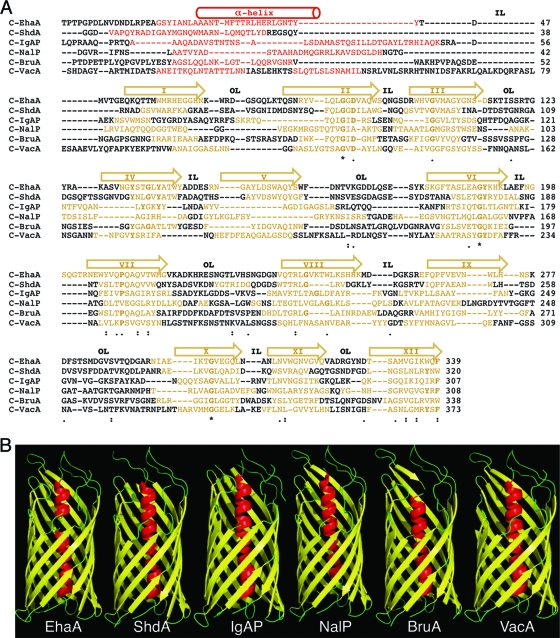
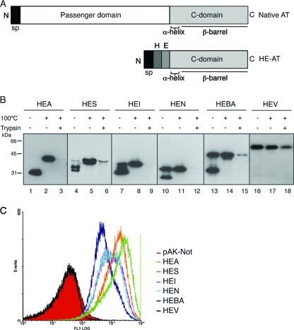
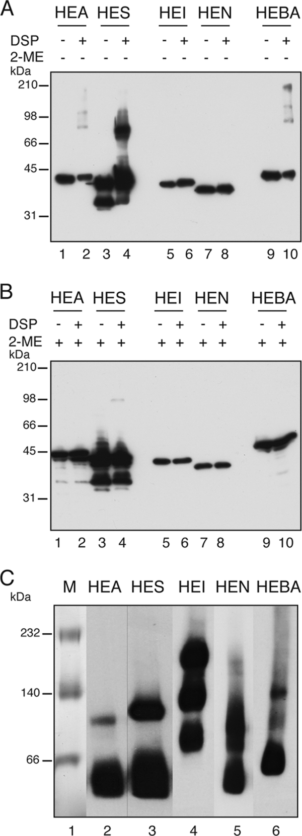
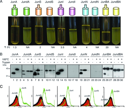
Autotransporters (ATs) are the largest group of proteins secreted by Gram-negative bacteria and include many virulence factors from human pathogens. ATs are synthesized as large precursors with a C-terminal domain that is inserted in the outer membrane (OM) and is essential for the translocation of an N-terminal passenger domain to the extracellular milieu. Several mechanisms have been proposed for AT secretion. Self-translocation models suggest transport across a hydrophilic channel formed by an internal pore of the β-barrel or by the oligomerization of C-terminal domains. Alternatively, an assisted-translocation model suggests that transport employs a conserved machinery of the bacterial OM such as the Bam complex. In this work we have investigated AT secretion by carrying out a comparative study to analyze the conserved biochemical and functional features of different C-terminal domains selected from ATs of gammaproteobacteria, betaproteobacteria, alphaproteobacteria, and epsilonproteobacteria. Our results indicate that C-terminal domains having an N-terminal α-helix and a β-barrel constitute functional transport units for the translocation of peptides and immunoglobulin domains with disulfide bonds. In vivo and in vitro analyses show that multimerization is not a conserved feature in AT C-terminal domains. Furthermore, we demonstrate that the deletion of the conserved α-helix severely impairs β-barrel folding and OM insertion and thereby blocks passenger domain secretion. These observations suggest that the AT β-barrel without its α-helix cannot form a stable hydrophilic channel in the OM for protein translocation. The implications of our data for an understanding of AT secretion are discussed.


Figures

FIG. 1.
FIG. 1.
Secondary- and tertiary-structure predictions of the selected AT C-terminal domains. (A) Alignment of the amino acid sequences of the C-terminal domains of the EhaA, ShdA, IgAP, NalP, BruA, and VacA ATs. The sequence predicted by the PSI-pred program (22) to fold as a hydrophilic α-helix is represented in red. The transmembrane amphipathic β-strands predicted with the Pred-TMBB (1) and ProfTMB (4) programs are shown in yellow. For clarity, the regions adopting α-helix or β-strand structures are also indicated with a cylinder or arrows on top, respectively. The roman numerals inside the arrows indicate the numbers of the β-strand (I to XII). Predicted inner loops (IL) and outer loops (OL) are labeled. Symbols indicating identity and similarity in the sequence alignment, according to the Tcoffe server (44), are indicated: *, 100% identity; :, >60% similarity; ·, >40% similarity. The conserved amino acid residues shown in boldface type correspond to those conserved in the alignment of the autotransporter family (Pfam accession number PF03797) from the Pfam server (12). (B) 3D structures of the C-terminal domains of selected ATs obtained with the Genesilico Metaserver-meta2 server (34). The secondary and tertiary structures of C-NalP correspond to the crystal structure data for NalP (41).
FIG. 2.
FIG. 2.
Expression of the selected AT C-terminal domains in E. coli K-12. (A) Schematic representation of a native AT, bearing the passenger domain and the C-terminal domain, and of the HE-tagged C-terminal domain AT constructs (HE-AT) containing the pelB signal peptide (sp) followed by the His tag (H) and E tag (E) epitopes and the corresponding AT C-terminal domain. (B) Western blot probed with anti-E tag MAb of whole-cell protein extracts obtained from IPTG-induced E. coli UT5600 cells expressing the HE-ATs indicated at the top. Prior to lysis, intact bacteria were incubated with (+) or without (−) trypsin (10 μg/ml), as indicated. The samples were boiled (+) or not (−) before loading onto the 10% SDS-polyacrylamide gel. The protein bands with faster mobility correspond to the folded conformation of the polypeptides, and the bands with slower mobility correspond to the unfolded conformation. The mass of protein standards is shown on the left (in kDa). Images shown here were obtained with different exposure times to reveal bands of approximately similar intensities for all AT C-terminal domains, although the intensity of the protein bands of HEA and HES was found to be ∼10-fold higher than those of other HE-ATs, according to quantification with a Chemi-doc device (Bio-Rad). (C) Flow cytometry analysis of E. coli UT5600 cells expressing the indicated HE-ATs, or bearing a control plasmid (pAK-Not), with anti-E tag MAb and secondary anti-mouse IgG-Alexa 488-conjugated antibody. The fluorescence intensities of bacteria carrying the indicated plasmids are shown in the histograms.
FIG. 3.
FIG. 3.
Surface display in E. coli of functional Ig VHH domains with disulfide bonds fused to AT C-terminal domains. (A) Western blot probed with anti-E tag MAb of whole-cell protein extracts obtained from IPTG-induced E. coli UT5600 cells expressing the VHH-ATs indicated at the top. Intact bacteria were incubated with (+) or without (−) trypsin (10 μg/ml), as indicated. The samples were boiled (+) or not (−) before loading onto a 10% SDS-polyacrylamide gel. The masses of protein standards are shown on the left (in kDa). (B) Flow cytometry analysis of E. coli UT5600 cells expressing the indicated VHH-ATs or bearing a control plasmid (pAK-Not) incubated with anti-E tag MAb and secondary anti-mouse IgG-Alexa 488-conjugated antibody. (C) ELISA to determine the antigen binding activity of the exposed VHH on E. coli UT5600 cells. Induced bacteria expressing the indicated VHH-AT fusion or bearing the empty vector (pAK-Not) were bound to ELISA plates coated with human fibrinogen (black bars) or BSA as a control (white bars). Binding signals in the ELISA developed with the anti-E tag MAb are shown. (D) ELISA (as in C) in which the VHH-A fusion was induced in wild-type (wt) E. coli UT5600 or its isogenic dsbA strain. (E) Western blot developed with anti-E tag MAb of wild-type and dsbA bacteria expressing the VHH-A fusion that were incubated (+) or not (−) with mPEG-MAL-5000 and DTT (as indicated) to analyze disulfide bond formation in the translocated VHH. The band corresponding to the PEGylated VHH-A fusion is labeled with an arrow.
FIG. 4.
FIG. 4.
Analysis of the quaternary structure of the AT C-terminal domains in vivo and in vitro. (A and B) Cross-linking with DSP of HE-ATs expressed in E. coli to determine the formation of oligomeric complexes of AT C-terminal domains in vivo. DSP-treated (+) and untreated (−) samples were subjected to nonreducing SDS-PAGE, and the Western blot was probed with anti-E tag MAb. Samples incubated with the reducing agent 2-ME are indicated (+). (C) Blue native PAGE of purified HE-ATs from the outer membrane of E. coli bacteria. The polypeptides were detected by Western blotting with an anti-E tag MAb. The masses of protein standards are shown on the left (in kDa).
FIG. 5.
FIG. 5.
Expression and cell surface translocation of α-helix deletion mutants. (A) Aggregation assay of E. coli bacteria expressing Jun fusions to the C-terminal domain of the ATs, either wild-type Jun-AT polypeptides, depicted as a small cylinder (α-helix) on top of a large cylinder (β-barrel), or the α-helix-deleted Jun-dAT mutants (large cylinder). The picture was taken at 24 h after the mixing of two independent cultures of E. coli cells expressing either Fosβ or each Jun fusion. The τ parameter is indicated in hours. NA, not applicable. (B) Western blot probed with anti-E tag MAb of whole-cell protein extracts obtained from IPTG-induced E. coli cells expressing the Jun fusion constructs indicated at the top. The samples were boiled (+) or not (−) before loading onto a 10% SDS-polyacrylamide gel. The protein bands with faster mobility correspond to the folded conformation of the polypeptides, and the band with slower mobility corresponds to the unfolded conformation. Intact bacteria were incubated with (+) or without (−) trypsin (10 μg/ml). The masses of protein standards are shown on the left (in kDa). (C) Flow cytometry analysis to determine the presence of the indicated Jun-AT fusions on the surface of E. coli cells with anti-E tag MAb. The bacterial controls and secondary antibodies used are described in the legend of Fig. 2.
FIG. 6.
FIG. 6.
Expression and cell surface translocation of AT C-terminal domains with heterologous α-helices. (A) Aggregation assay of E. coli bacteria expressing the indicated Jun fusions (JunI, JunAI, and JunNI). The picture was taken at 24 h after the mixing of the indicated E. coli cultures with a culture of E. coli cells expressing Fosβ. The τ parameter is indicated in hours. NA, not applicable. (B) Flow cytometry analysis to determine the presence of the indicated Jun-AT constructs on the surface of E. coli cells with anti-E tag MAb. Bacterial controls and secondary antibodies used are described in the legends of Fig. 2 and 5. (C) Western blot probed with anti-E tag MAb of whole-cell protein extracts obtained from IPTG-induced E. coli cells expressing the indicated Jun-AT constructs. Intact bacteria were incubated with (+) or without (−) trypsin (10 μg/ml), and the samples were either boiled (+) or not (−) before loading onto a 10% SDS-polyacrylamide gel. The masses of protein standards are shown on the left (in kDa). (D) ELISA developed with anti-PG serum to test the peptidoglycan accessibility of bacteria expressing Jun-ATs or bearing the empty vector pAK-Not (black bars). EDTA-permeabilized bacteria harboring pAK-Not were used to monitor the reactivity of the anti-PG serum (white bar).
FIG. 7.
FIG. 7.
Subcellular localization of AT C-terminal domains with a deleted or heterologous α-helix. Western blots were probed with anti-E tag MAb to detect Jun-AT constructs in the indicated subcellular fractions obtained from induced E. coli cells: soluble, envelope, TX-100-solubilized, urea-solubilized, and integral OMPs (see Materials and Methods). The detection of the periplasmic MBP and OmpA with specific antibodies (indicated in the panels) was done to control the E. coli fractionation method. The masses of protein standards are shown on the left (in kDa).

Similar articles

Cited by

References

    1. Bagos, P. G., T. D. Liakopoulos, I. C. Spyropoulos, and S. J. Hamodrakas. 2004. PRED-TMBB: a Web server for predicting the topology of beta-barrel outer membrane proteins. Nucleic Acids Res. 32:W400-W404. - PMC - PubMed
    1. Barnard, T. J., N. Dautin, P. Lukacik, H. D. Bernstein, and S. K. Buchanan. 2007. Autotransporter structure reveals intra-barrel cleavage followed by conformational changes. Nat. Struct. Mol. Biol. 14:1214-1220. - PMC - PubMed
    1. Bernstein, H. D. 2007. Are bacterial ‘autotransporters’ really transporters? Trends Microbiol. 15:441-447. - PubMed
    1. Bigelow, H. R., D. S. Petrey, J. Liu, D. Przybylski, and B. Rost. 2004. Predicting transmembrane beta-barrels in proteomes. Nucleic Acids Res. 32:2566-2577. - PMC - PubMed
    1. Bitto, E., and D. B. McKay. 2003. The periplasmic molecular chaperone protein SurA binds a peptide motif that is characteristic of integral outer membrane proteins. J. Biol. Chem. 278:49316-49322. - PubMed

Publication types

MeSH terms