. 2010 Nov;192(21):5645-56.
doi: 10.1128/JB.00590-10. Epub 2010 Aug 27.

Biogenesis of Salmonella enterica serovar typhimurium membrane vesicles provoked by induction of PagC

Affiliations

Biogenesis of Salmonella enterica serovar typhimurium membrane vesicles provoked by induction of PagC

Ryo Kitagawa et al. J Bacteriol. 2010 Nov.

Abstract

Gram-negative bacteria ubiquitously release membrane vesicles (MVs) into the extracellular milieu. Although MVs are the product of growing bacteria, not of cell lysis or death, the regulatory mechanisms underlying MV formation remained unknown. We have found that MV biogenesis is provoked by the induction of PagC, a Salmonella-specific protein whose expression is activated by conditions that mimic acidified macrophage phagosomes. PagC is a major constituent of Salmonella MVs, and increased expression accelerates vesiculation. Expression of PagC is regulated at the posttranscriptional and/or posttranslational level in a sigmaS (RpoS)-dependent manner. Serial quantitative analysis has demonstrated that MV formation can accelerate when the quantity of the MV constituents, OmpX and PagC, rises. Overproduction of PagC dramatically impacts the difference in the relative amount of vesiculation, but the corresponding overproduction of OmpX was less pronounced. Quantitative examination of the ratios of PagC and OmpX in the periplasm, outer membrane, and MVs demonstrates that PagC is preferentially enriched in MVs released from Salmonella cells. This suggests that specific protein sorting mechanisms operate when MVs are formed. The possible role(s) of PagC-MV in host cells is discussed.


Figures

FIG. 1.
FIG. 1.
Accumulation of PagC in the fraction detached from Salmonella cell surface by ClpXP depletion. Bacterial cells of Salmonella strains χ3306 (clpXP+ flhD+), CS2007 (ΔclpXP flhD+), CS2609 (clpXP+ ΔflhD), and CS2610 (ΔclpXP ΔflhD) were grown in N minimal medium. Proteins in the fraction detached from the bacterial cell surfaces by mechanical shearing were separated on an SDS-polyacrylamide gel, followed by staining with Coomassie blue. Protein bands indicated with arrowheads were excised, analyzed by mass spectrometry, and specified as SseD, PagL, and PagC.
FIG. 2.
FIG. 2.
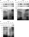
Cellular contents of PagC and expression of the pagC promoter in Salmonella strains χ3306 (clpXP+ phoP+ rpoS+), CS2007 (ΔclpXP phoP+ rpoS+), CS2011 (clpXP+ ΔphoP rpoS+), CS2045 (clpXP+ phoP+ ΔrpoS), CS2831 (ΔclpXP ΔphoP rpoS+), and CS2832 (ΔclpXP phoP+ ΔrpoS). (A) Immunoblotting of whole-cell lysates using anti-PagC and anti-sigmaE antibodies. Bacterial cells were grown in N minimal medium. (B) Coomassie blue-stained SDS-PAGE patterns of the same samples used for immunoblotting. (C) Expression of the pagC promoter from transcriptional pagC-lacZ fusion in bacterial strains with different backgrounds, as in panel A. The plasmid containing the pagC-lacZ fusion, pTKY620, was introduced into the same Salmonella strains as used in panel A. The resultant transformants were cultured in N minimal medium. The β-galactosidase activity was assayed. The values represent the means and standard deviations of samples tested in triplicate.
FIG. 3.
FIG. 3.
Elution pattern of size exclusion chromatography and protein profiles of MV preparations from Salmonella strains CS2609 (clpXP+) and CS2610 (ΔclpXP). Both strains have the flhD::Tn10 mutation expressing no flagella in the genetic background. Cells were grown in N-minimal medium and the MV preparations were isolated and subjected to gel chromatography using Superose 12. The elution patterns of the chromatography are shown in panel A. Peak a, strain CS2610 (ΔclpXP); peak b, strain CS2609 (clpXP+). The inset is an electron micrograph showing macromolecules of the highest peak. Bar, 100 nm. (B) The fractions were subjected to SDS-PAGE, followed by immunoblotting. (C and D) A portion of the biggest peak of each chromatography was subjected to SDS-PAGE, followed by immunoblotting (C) and silver staining (D). (E) Immunoblotting was performed to detect HtrA in the culture supernatants (Sup) prepared from Salmonella strains, CS2609 (clpXP+ htrA+) and CS2610 (ΔclpXP htrA+). The lysates (Lys) were prepared from CS2609 and CS2019 (clpXP+ ΔhtrA). Coomassie blue-stained SDS-PAGE patterns of the same samples used for immunoblotting are shown in panel F. Cells were grown in N minimal medium and removed by centrifugation at 13,000 × g for 30 min. The supernatant was filtrated and then concentrated as previously described (52).
FIG. 4.
FIG. 4.
Transmission electron microscopy of Salmonella cells producing MVs. Cells were grown on an N minimal agar plate at 37°C. The strains are CS2609 (clpXP+ [A]), CS2610 (ΔclpXP [B]), and CS3000 (ΔclpXP ΔpagC [C]). MVs were purified from culture supernatants of CS2610 (D) and CS3000 (E) as described in Materials and Methods. All bacterial strains used here have the flhD::Tn10 mutation expressing no flagella in the genetic background. Bars, 100 nm.
FIG. 5.
FIG. 5.
Heightened formation of MVs by cells grown under conditions that physiologically increase PagC production. Bacterial cells of strain χ3306 were grown in L broth (a) or N minimal medium (b) for 16 h. MV preparation was performed as described in Materials and Methods. A portion of MVs (MV) or whole-cell lysates (WC) was subjected to SDS-PAGE, followed by immunoblotting with antiserum against PagC, OmpA, or RpoS (A). Silver-stained SDS-PAGE patterns of the samples used for immunoblotting are presented in panel B.
FIG. 6.
FIG. 6.
Quantitative analysis of MV production by Salmonella cells expressing pagC or ompX controlled by IPTG concentration. The bacterial strains used are CS2992 (ppagC) and CS3746 (pompX). These strains do not produce flagella due to the flhD::Tn10 mutation. Bacterial cells were grown in L broth to an OD600 of 0.5, followed by the induction of pagC or ompX at different concentrations of IPTG, as indicated for 16 h. MV preparations isolated from equal amounts of culture supernatant equivalents were loaded on to a size exclusion column of Superose 12. The elution patterns of the chromatography are shown in panel A. Integral volumes of the peaks corresponding MVs are indicated in panel B. Coomassie blue-stained SDS-15% PAGE pattern of cell lysates are shown in panel C. ppagC, pTKY698; pompX, pTKY896.
FIG. 7.
FIG. 7.
pagC overexpression and ompX overexpression controlled by IPTG do not affect the cellular levels of sigmaE in Salmonella cells. Cells of Salmonella strains CS2992 (ppagC) and CS3746 (pompX) were grown in L broth to OD600 of 0.5, followed by the induction of pagC or ompX by different concentrations of IPTG. (A) Immunoblotting of whole-cell lysates with anti-sigmaE serum. Lysates of bacterial cells of strain CS2020 (ΔrpoE) were a negative control for sigmaE. (B) Coomassie blue-stained SDS-PAGE patterns of the samples used for immunoblotting. ppagC, pTKY698; pompX, pTKY896.
FIG. 8.
FIG. 8.
Specific sorting of PagC into MVs. Bacterial cells of strains CS2992 (ppagC [A]), CS3746 (pompX [B]), and CS3747 (pompA [C]) were grown in L broth to an OD600 of 0.5, followed by the induction of pagC, ompX, or ompA expression by 50 μM IPTG for 16 h. MV preparation and cell fractionation were performed as described in Materials and Methods. A portion of MVs (MV), outer membrane (OM), periplasmic space (PE), cytoplasm (CY), or whole-cell lysates (WC) was subjected to SDS-PAGE, followed by immunoblotting with antiserum against PagC, OmpX, or OmpA. Silver-stained SDS-PAGE patterns of the samples used for immunoblotting are also presented. PagC/OmpA ratio of densitometry values is indicated in panel A and B. ppagC, pTKY698; pompX, pTKY896; pompA, pTKY897.

Similar articles

Cited by

References

    1. Alix, E., T. Miki, C. Felix, C. Rang, N. Figueroa-Bossi, E. Demettre, and A. B. Blanc-Potard. 2008. Interplay between MgtC and PagC in Salmonella enterica serovar Typhimurium. Microb. Pathog. 45:236-240. - PubMed
    1. Alpuche-Aranda, C. M., J. A. Swanson, W. P. Loomis, and S. I. Miller. 1992. Salmonella typhimurium activates virulence gene transcription within acidified macrophage phagosomes. Proc. Natl. Acad. Sci. U. S. A. 89:10079-10083. - PMC - PubMed
    1. Arnold, T., M. Poynor, S. Nussberger, A. N. Lupas, and D. Linke. 2007. Gene duplication of the eight-stranded β-barrel OmpX produces a functional pore: a scenario for the evolution of transmembrane β-barrels. J. Mol. Biol. 366:1174-1184. - PubMed
    1. Balsalobre, C., J. M. Silvan, S. Berglund, Y. Mizunoe, B. E. Uhlin, and S. N. Wai. 2006. Release of the type I secreted alpha-haemolysin via outer membrane vesicles from Escherichia coli. Mol. Microbiol. 59:99-112. - PubMed
    1. Bearson, B. L., L. Wilson, and J. W. Foster. 1998. A low pH-inducible, PhoPQ-dependent acid tolerance response protects Salmonella typhimurium against inorganic acid stress. J. Bacteriol. 180:2409-2417. - PMC - PubMed

Publication types

MeSH terms

LinkOut - more resources