. 2010 Dec;9(12):2654-65.
doi: 10.1074/mcp.M110.004119. Epub 2010 Aug 27.

Construction of a large extracellular protein interaction network and its resolution by spatiotemporal expression profiling

Affiliations

Construction of a large extracellular protein interaction network and its resolution by spatiotemporal expression profiling

Stephen Martin et al. Mol Cell Proteomics. 2010 Dec.

Abstract

Extracellular interactions involving both secreted and membrane-tethered receptor proteins are essential to initiate signaling pathways that orchestrate cellular behaviors within biological systems. Because of the biochemical properties of these proteins and their interactions, identifying novel extracellular interactions remains experimentally challenging. To address this, we have recently developed an assay, AVEXIS (avidity-based extracellular interaction screen) to detect low affinity extracellular interactions on a large scale and have begun to construct interaction networks between zebrafish receptors belonging to the immunoglobulin and leucine-rich repeat protein families to identify novel signaling pathways important for early development. Here, we expanded our zebrafish protein library to include other domain families and many more secreted proteins and performed our largest screen to date totaling 16,544 potential unique interactions. We report 111 interactions of which 96 are novel and include the first documented extracellular ligands for 15 proteins. By including 77 interactions from previous screens, we assembled an expanded network of 188 extracellular interactions between 92 proteins and used it to show that secreted proteins have twice as many interaction partners as membrane-tethered receptors and that the connectivity of the extracellular network behaves as a power law. To try to understand the functional role of these interactions, we determined new expression patterns for 164 genes within our clone library by using whole embryo in situ hybridization at five key stages of zebrafish embryonic development. These expression data were integrated with the binding network to reveal where each interaction was likely to function within the embryo and were used to resolve the static interaction network into dynamic tissue- and stage-specific subnetworks within the developing zebrafish embryo. All these data were organized into a freely accessible on-line database called ARNIE (AVEXIS Receptor Network with Integrated Expression; www.sanger.ac.uk/arnie) and provide a valuable resource of new extracellular signaling interactions for developmental biology.


Figures
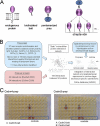
Fig. 1.
Fig. 1.
Summary of AVEXIS method and overall approach to construct and resolve extracellular protein interaction networks. A, the entire ectodomains of endogenous cell surface receptors (pink) are expressed as both monomeric biotinylated baits (B) and pentamerized β-lactamase-tagged (β) preys (P). Bait proteins are immobilized in individual wells of streptavidin-coated microtiter plates and probed with a normalized prey, which, if the two proteins physically interact, is captured within the well. B, a flowchart presenting an overall summary of the work described here, including how interactions from two previous screens (Refs. (Bushell 2008) and (Söllner 2009)) were integrated into the larger network of interactions. C, positive interactions are detected by adding a colorimetric β-lactamase substrate, nitrocefin, which is converted from a yellow to red product. Two typical screening plates are shown illustrating a heterophilic interaction between Cadm3 and Cadm4 that is detected in both bait-prey orientations and a homophilic interaction involving Cadm3. The controls for each prey included a negative bait, the rat Cd4d3+4 protein tag alone (well G6), and a biotinylated anti-rat Cd4 monoclonal antibody (OX68), which captured the Cd4-tagged preys (well G7). Each plate also contained positive control interactions: the rat Cd200R prey was probed against rat Cd200 baits immobilized at the normalized screening threshold and at 1:500 and 1:1,000 dilutions (wells G8–G10, respectively) and against the negative Cd4d3+4 bait (well G12). An additional negative bait (Fgfr1b) was included for both preys (wells G5 and G11). SLRPs, small leucine-rich proteoglycans.
Fig. 2.
Fig. 2.
Large extracellular protein interaction network systematically determined by AVEXIS. The interaction network consists of 188 interactions between 92 proteins. The family to which each protein belongs and its predicted subcellular localization are indicated: red, secreted; blue, membrane-tethered; circle, IgSF; triangle, LRR; diamond, IgSF + LRR; square, other domains.
Fig. 3.
Fig. 3.
Genes encoding proteins within extracellular interaction network are expressed in tissue-restricted manner during early development. A, the development of the zebrafish embryo. Drawings of representative stages within each of the five main periods of zebrafish development are shown above a brief description of the main morphogenetic landmarks within each period. Drawings are taken with permission from Kimmel et al. (45). B, a summary of the tissue expression patterns of the genes encoding proteins from the interaction network. The expression patterns were annotated and placed into all the appropriate non-exclusive tissue categories. C, a summary of the temporal expression patterns of the genes encoding proteins from the interaction network. The number of genes expressed at each period of development is plotted, indicating those genes whose expression was ubiquitous (green) or restricted to particular tissues (blue).
Fig. 4.
Fig. 4.
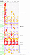
Integration of spatiotemporal gene expression profiles with extracellular protein interaction network. All 188 gene pairs that encode interacting proteins are listed vertically; those encoding a secreted protein are highlighted in green. Each gene of a pair is arbitrarily assigned 1 (left column, purple) or 2 (right column, gold), and their spatiotemporal expression patterns are indicated by shading an appropriate box in the matrix, which is organized into 10 organ systems (nervous and sensory to immune), each of which is subdivided in up to five developmental stages as appropriate (G, gastrula; S, segmentation; P, pharyngula; H, hatching; L, larval). Where both genes are compatibly expressed, the box is shaded red. Interactions are first organized into those whose gene pairs are compatibly or incompatibly expressed and then divided further into functional subcategories as indicated.
Fig. 5.
Fig. 5.
Resolution of extracellular interaction network into stage- and tissue-specific signaling networks. A, graphs show time-resolved extracellular interaction networks at each period of development within the nervous and sensory system. The node key is as follows: blue, gene whose expression is restricted to nervous system; green, ubiquitously expressed gene at the stage considered. B, heat map showing the percentage of extracellular interaction network edges at a given developmental period relative to the others within each tissue. For comparison, the percentage of genes expressed at each period is also shown. G, gastrula; S, segmentation; P, pharyngula; H, hatching; L, larval.

Similar articles

Cited by

References

    1. Wright G. J. (2009) Signal initiation in biological systems: the properties and detection of transient extracellular protein interactions. Mol. Biosyst. 5, 1405–1412 - PMC - PubMed
    1. van der Merwe P. A., Barclay A. N. (1994) Transient intercellular adhesion: the importance of weak protein-protein interactions. Trends Biochem. Sci. 19, 354–358 - PubMed
    1. Urech D. M., Lichtlen P., Barberis A. (2003) Cell growth selection system to detect extracellular and transmembrane protein interactions. Biochim. Biophys. Acta 1622, 117–127 - PubMed
    1. de Wildt R. M., Tomlinson I. M., Ong J. L., Holliger P. (2002) Isolation of receptor-ligand pairs by capture of long-lived multivalent interaction complexes. Proc. Natl. Acad. Sci. U.S.A. 99, 8530–8535 - PMC - PubMed
    1. Cain S. A., McGovern A., Small E., Ward L. J., Baldock C., Shuttleworth A., Kielty C. M. (2009) Defining elastic fiber interactions by molecular fishing: an affinity purification and mass spectrometry approach. Mol. Cell. Proteomics 8, 2715–2732 - PMC - PubMed

Publication types