. 2011 Jul;45(1):40-52.
doi: 10.1165/rcmb.2010-0097OC. Epub 2010 Aug 27.

Effect of cocaine on human immunodeficiency virus-mediated pulmonary endothelial and smooth muscle dysfunction

Affiliations

Effect of cocaine on human immunodeficiency virus-mediated pulmonary endothelial and smooth muscle dysfunction

Navneet K Dhillon et al. Am J Respir Cell Mol Biol. 2011 Jul.

Abstract

Human immunodeficiency virus (HIV)-associated pulmonary arterial hypertension (PAH) is a devastating, noninfectious complication of acquired immune deficiency syndrome, and the majority of HIV-PAH cases occur in individuals with a history of intravenous drug use (IVDU). However, although HIV-1 and IVDU have been associated with PAH independently or in combination, the pathogenesis of the disproportionate presence of HIV-PAH in association with IVDU has yet to be characterized. The objective of this study was to obtain a better understanding of the interactions between HIV-1 and cocaine to help uncover the mechanism(s) of the development of HIV-PAH. We observed that exposure of HIV-infected macrophages or HIV-Trans-Activator of Transcription (Tat)-treated pulmonary endothelial cells to cocaine enhanced the expression of platelet-derived growth factor (PDGF)-BB. Simultaneous treatment with Tat and cocaine, on the other hand, exacerbated both the disruption of tight junction proteins (TJPs), with enhanced permeability in pulmonary endothelial cells, and the proliferation of pulmonary smooth muscle cells (pSMCs) compared with either treatment alone. Histological examination of HIV plus IVDU human lung sections showed signs of early pulmonary arteriopathy, severe down-modulation of TJPs, and increased expression of PDGF-BB compared with the lung sections from individuals who are infected with HIV and without history of IVDU. Interestingly, blocking of PDGF receptor signaling with the receptor antagonist or small interfering RNA has been shown to inhibit the increase in proliferation of pSMCs on Tat and cocaine exposure. Our results, therefore, support an additive effect of cocaine to HIV infection in the development of pulmonary arteriopathy through enhancement of endothelial dysfunction and proliferation of pSMCs, while also suggesting PDGF-PDGF receptor axis as a potential target for use in clinical intervention.


Figures

Figure 1.
Figure 1.
Cocaine potentiates the expression of platelet-derived growth factor (PDGF)–BB in human immunodeficiency virus (HIV)–infected human macrophages and Trans-Activator of Transcription (Tat)-treated pulmonary endothelial cells (pECs), but not in Tat-treated pulmonary smooth muscle cells (SMCs). Real-Time RT-PCR analysis of PDGF-B chain in (A) HIVBal-infected monocyte-derived macrophages (MDMs), (C) Tat (25ng/ml)-treated primary human pulmonary arterial endothelial cells (HPAECs), and (E) Tat-treated human pulmonary arterial SMCs (HPASMCs), in the absence or presence of cocaine (1 μM). The data represent means (±SD) of three or more independent experiments done in triplicates. Western blot analysis of PDGF-BB in cellular extracts from (B) HIVBal-infected MDMs in the absence or presence of cocaine, (D) HPAECs, and (F) HPASMCs treated with or without cocaine and/or Tat. The blots were reprobed with human β-actin antibodies. Representative Western blot images (upper panel) are shown with histograms (lower panel), showing the average densitometric analysis of the PDGF-BB band normalized to corresponding β-actin band from three experiments. Statistically significant differences are shown as *P ≤ 0.05, **P ≤ 0.01, ***P ≤ 0.001, treatment versus control, and #P < 0.05, ##P < 0.01, HIV+cocaine versus cocaine or HIV alone; Tat+cocaine versus cocaine or Tat alone.
Figure 2.
Figure 2.
Down-regulation in the expression of tight junction proteins (TJPs) in pulmonary arterial endothelial cells on exposure to HIV-Tat and/or cocaine. HPAECs grown in six-well plates were serum starved (0.5% FBS) overnight, followed by treatment with Tat (25 ng/ml) and/or cocaine (1 μM) for mRNA analysis by real-time RT-PCR at 24 hours after treatment. Values represent fold decrease of (A) claudin (CLDN)-4, (B) TJP-1, (C) TJP-3, and (D) occludin (OCLN) mRNA on treatment with Tat and/or cocaine compared with untreated cells. Means (±SD) are shown; *P < 0.05, **P ≤ 0.01, ***P ≤ 0.001, compared with untreated cells. (E) HPAECs grown on coverslips, treated with cocaine (1 μM) and/or Tat (25 ng/ml) for 24 hours, respectively, were immunostained for TJP-1. Control represents untreated cells. (F) Quantitative analysis of TJP-1 immunofluorescence staining. The TJP-1 staining images were acquired from three independent experiments performed on endothelial monolayers stimulated with cocaine and/or Tat. Graph represents the average fluorescent units present as a percentage of untreated control cells (*P ≤ 0.001, treatment versus control; #P ≤ 0.01, cocaine +Tat versus cocaine or Tat alone.). (G) Effect of cocaine (1 μM) and Tat (25 ng/ml) on barrier function of HPAECs as assessed by FITC-dextran. Cells were grown on collagen-coated Transwell filters. Confluent monolayers were incubated for 6 hours with cocaine and Tat, followed by treatment with the FITC-dextran for 5 minutes. The fluorescence in the lower compartment was then measured and expressed as percentage of basal fluorescence. The values shown are means (±SD) of three independent experiments (*P ≤ 0.05, **P < 0.01 treatment versus control; #P < 0.05 Cocaine +Tat versus Tat, ##P < 0.01 Cocaine +Tat versus cocaine).
Figure 3.
Figure 3.
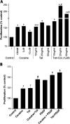
Increased proliferation of SMCs with Tat, cocaine, or PDGF-BB. The SMCs grown in 96 wells were starved in 0.1% serum–containing media for 48 hours before treatment. (A) Exposure of quiescent human aorta–vascular SMCs (T/G HA-VSMC) to Tat and cocaine results in synergistic or additive increase in proliferation of cells compared with cells treated with Tat or cocaine alone. Means (±SD) are shown, and are representative of three independent experiments; *P < 0.05, **P ≤ 0.01, ***P ≤ 0.001, treated versus untreated cells; #P ≤ 0.05, compared with Tat (25 ng/ml) treatment alone; and $P ≤ 0.05, compared with Tat (50 ng/ml) treatment alone. (B) Increased proliferation of primary HPASMCs with Tat (25 ng/ml), cocaine (1 μM), or PDGF-BB (100 ng/ml) treatment with further enhancement on simultaneous treatment with cocaine and Tat or either cocaine or Tat with PDGF-BB. The data shown are means (±SD), and are representative of three independent experiments, in triplicates. *P < 0.05, compared with untreated cells; #P ≤ 0.01, compared with cocaine treatment alone; $P ≤ 0.01, compared with PDGF-BB treatment alone.
Figure 4.
Figure 4.
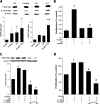
Involvement of PDGF signaling in HIV-Tat– and cocaine-induced proliferation of SMCs. (A) HIV-Tat and cocaine induces expression and activation of PDGF-β receptor (PDGF-βR) in pulmonary SMCs (pSMCs) as shown in the representative Western blot image and by densitometric analysis of the signal intensity. Quiescent HPASMCs were incubated with Tat (25 ng/ml) and cocaine (1 μM) for 24 hours, followed by extraction of protein and sequential immunoblotting with antibodies specifically directed to the PDGF-βR, phosphorylated active form of PDGF-βR, and finally reprobed with β-actin antibody for normalization. *P ≤ 0.05 versus control; #P ≤ 0.05 versus cocaine or Tat treatment. (B) Imatinib mesylate reverses the effect of Tat and cocaine on the proliferation of SMCs. Quiescent HPASMCs were pretreated with 1 μM imatinib mesylate, followed by treatment with Tat (25 ng/ml) and cocaine (1 μM) for 48 hours. Means (±SD) are shown, and are representative of three independent experiments. *P ≤ 0.01, treated versus untreated cells; #P ≤ 0.001 versus cocaine plus Tat treatment. (C) Evaluation of PDGF-βR knockdown by Western blot analysis of whole-cell lysates from HPASMCs transfected with either PDGF-βR small interfering (si) RNA (5 nM and 10 nM) or negative siRNA control (10 nM) in the absence or presence of cocaine and Tat treatment. Blot is representative of three independent experiments, with histogram (lower panel) showing the average densitometric analysis of PDGF-βR normalized to β-actin; *P < 0.01, treatment versus control; #P < 0.01, treatment versus cocaine plus Tat treatment. (D) PDGF-βR siRNA inhibited cocaine- and Tat-mediated induction of smooth muscle proliferation. Cells were transfected, treated, and analyzed for proliferation by 3-(4,5-dimethylthiazol-2-yl)-5-(3-carboxymethoxyphenyl)-2-(4-sulfophenyl)-2H-tetrazolium (MTS) assay, as described in Materials and Methods. All values are means (±SD). *P ≤ 0.001, treatment versus control; #P ≤ 0.001, treatment versus cocaine plus Tat–treated, untransfected cells.
Figure 5.
Figure 5.
Down-modulation of TJPs in the lungs from HIV-infected individuals with a history of intravenous drug use (IVDU). Expression of TJPs was analyzed by QuantiGene multiplex analysis of RNA extracted from frozen lung tissues of uninfected normal (n = 3), uninfected individual with IVDU (n = 3), HIV-1–infected individual without IVDU (n = 4), and HIV-1–infected individual with IVDU (HIV+IVDU; n = 5). Values represent the levels of (A) CLDN4, (B) TJP-1, (C) TJP-3, and (D) OCLN in various groups. Mean fluorescence intensity (MFI) obtained for each target was normalized with MFI of β-actin from the same sample. (E) Immunofluorescence demonstrated decreased expression of TJP-1 in HIV+IVDU compared with uninfected control. Paraffin-embedded lung sections were stained using human anti–TJP-1 primary antibody. Images were captured using confocal microscopy. Original magnification, 40×.
Figure 6.
Figure 6.
Representative images showing pulmonary vasculature from each individual within normal, IVDU, HIV+, and HIV+IVDU groups. Lungs were characterized for endothelial and SMC markers by immunohistochemistry for factor VIII (red color), and α–smooth muscle actin (SMA; brown color) on paraffin-embedded lung sections. Scale bars, 100 μm.
Figure 7.
Figure 7.
Expression of PDGF-BB in lungs of individuals from normal, IVDU, HIV1, and HIV+IVDU groups. (A) Representative photomicrographs of immunohistochemistry on paraffin-embedded lung sections from each group are shown. Original magnification, 20×; scale bar, 100 μm. (B and C) Quantitative analysis of PDGF-BB–stained lung sections from HIV-infected individuals with or without history of IVDU compared to normal controls. Mean staining intensity in the entire lung parenchyma (A) and in the arterial wall (B) obtained from three different lung sections within each group is shown. Data represent means (±SD); *P < 0.001, treatment versus normal; #P < 0.05, ###P < 0.001, HIV+IVDU versus IVDU or HIV alone.
Figure 8.
Figure 8.
Model showing possible changes in the pulmonary arterial walls of HIV-infected cocaine users.

Similar articles

Cited by

References

    1. Barbaro G. Cardiovascular manifestations of HIV infection. J R Soc Med 2001;94:384–390. - PMC - PubMed
    1. Barbaro G, Lipshultz SE. Pathogenesis of HIV-associated cardiomyopathy. Ann N Y Acad Sci 2001;946:57–81. - PubMed
    1. Bauersachs J, Bouloumie A, Fraccarollo D, Hu K, Busse R, Ertl G. Endothelial dysfunction in chronic myocardial infarction despite increased vascular endothelial nitric oxide synthase and soluble guanylate cyclase expression: role of enhanced vascular superoxide production. Circulation 1999;100:292–298. - PubMed
    1. Kitayama J, Faraci FM, Lentz SR, Heistad DD. Cerebral vascular dysfunction during hypercholesterolemia. Stroke 2007;38:2136–2141. - PubMed
    1. Dobbs MR, Berger JR. Stroke in HIV infection and AIDS. Expert Rev Cardiovasc Ther 2009;7:1263–1271. - PubMed

Publication types

MeSH terms