

Complex structure of electrophysiological gradients emerging during long-duration ventricular fibrillation in the canine heart
- PMID: 20802138
- PMCID: PMC2993199
- DOI: 10.1152/ajpheart.00419.2010
Complex structure of electrophysiological gradients emerging during long-duration ventricular fibrillation in the canine heart
Abstract
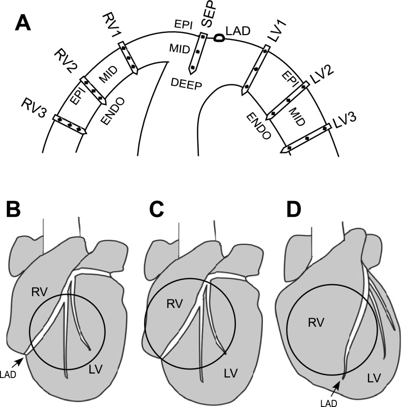
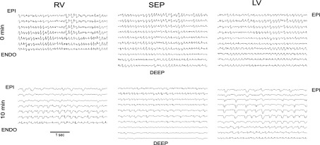
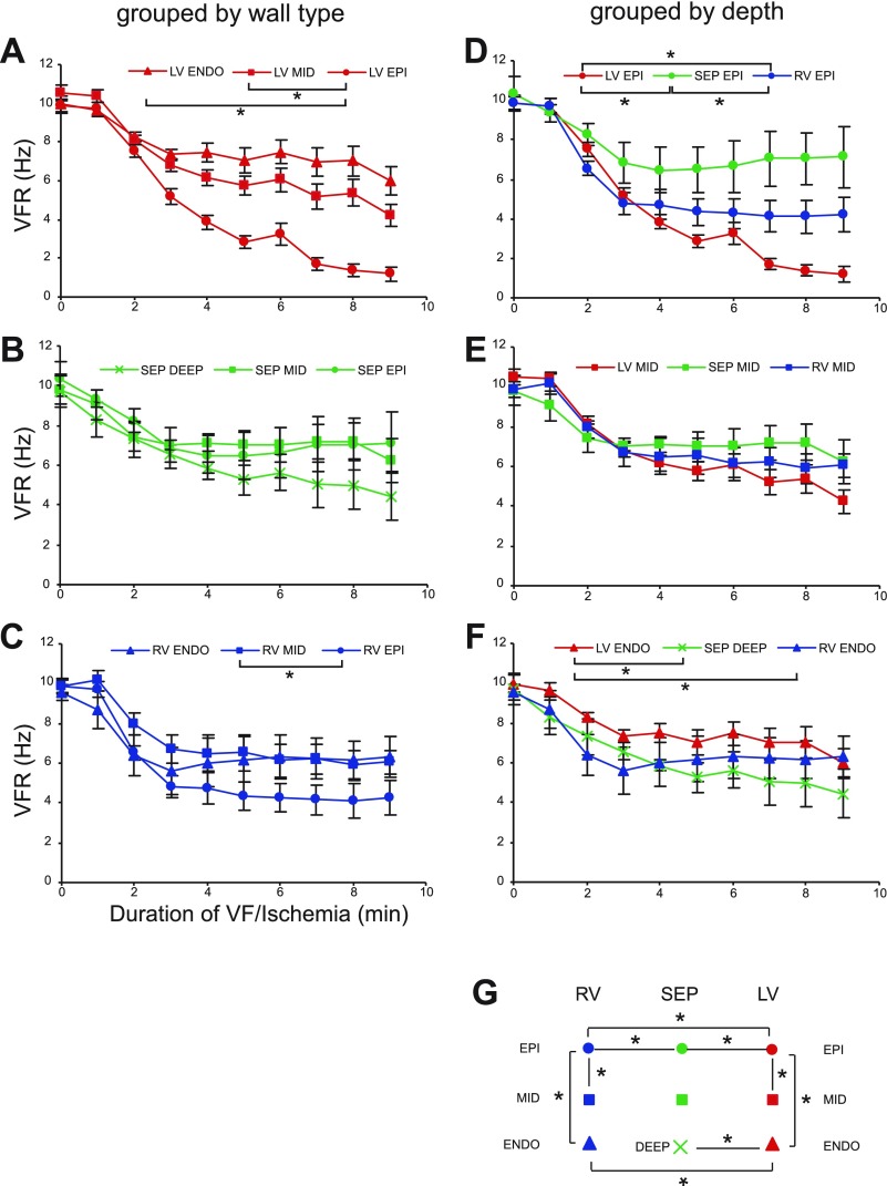
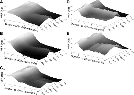
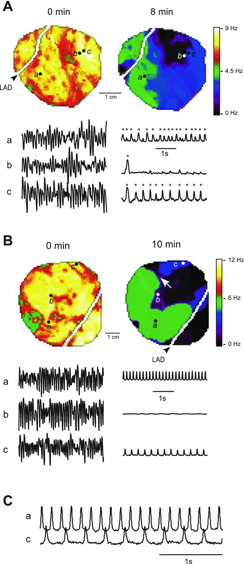
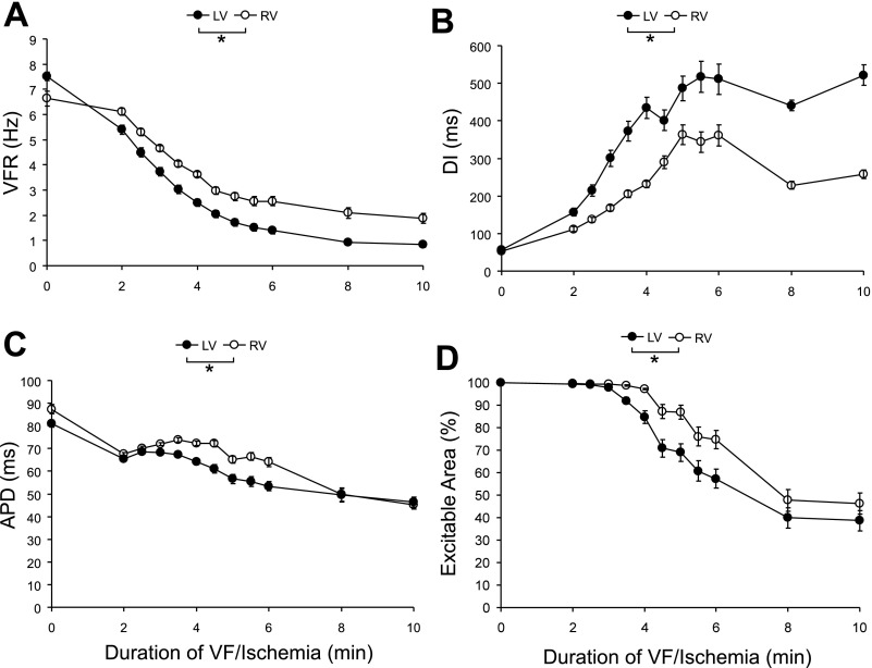
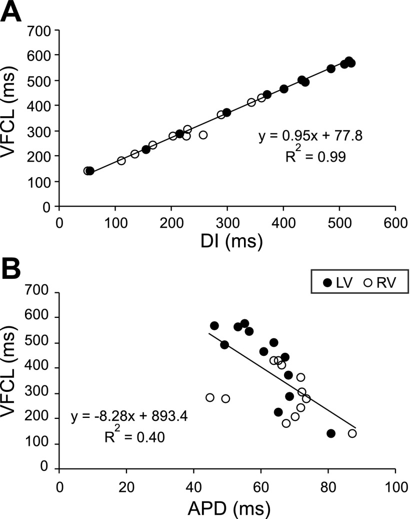
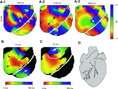
Long-duration ventricular fibrillation (LDVF) in the globally ischemic heart is a common setting of cardiac arrest. Electrical heterogeneities during LDVF may affect outcomes of defibrillation and resuscitation. Previous studies in large mammalian hearts have investigated the role of Purkinje fibers and electrophysiological gradients between the endocardium (Endo) and epicardium (Epi). Much less is known about gradients between the right ventricle (RV) and left ventricle (LV) and within each chamber during LDVF. We studied the transmural distribution of the VF activation rate (VFR) in the RV and LV and at the junction of RV, LV, and septum (Sep) during LDVF using plunge needle electrodes in opened-chest dogs. We also used optical mapping to analyze the Epi distribution of VFR, action potential duration (APD), and diastolic interval (DI) during LDVF in the RV and LV of isolated hearts. Transmural VFR gradients developed in both the RV and LV, with a faster VFR in Endo. Concurrently, large VFR gradients developed in Epi, with the fastest VFR in the RV-Sep junction, intermediate in the RV, and slowest in the LV. Optical mapping revealed a progressively increasing VFR dispersion within both the LV and RV, with a mosaic presence of fully inexcitable areas after 4-8 min of LDVF. The transmural, interchamber, and intrachamber VFR heterogeneities were of similar magnitude. In both chambers, the inverse of VFR was highly correlated with DI, but not APD, at all time points of LDVF. We conclude that the complex VFR gradients during LDVF in the canine heart cannot be explained solely by the distribution of Purkinje fibers and are related to regional differences in the electrical depression secondary to LDVF.
Figures
References
-
- Allison JS, Qin H, Dosdall DJ, Huang J, Newton JC, Allred JD, Smith WM, Ideker RE. The transmural activation sequence in porcine and canine left ventricle is markedly different during long-duration ventricular fibrillation. J Cardiovasc Electrophysiol 18: 1306–1312, 2007. - PubMed
-
- Antzelevitch C, Sicouri S, Litovsky SH, Lukas A, Krishnan SC, Di Diego JM, Gintant GA, Liu DW. Heterogeneity within the ventricular wall. Electrophysiology and pharmacology of epicardial, endocardial, and M cells. Circ Res 69: 1427–1449, 1991. - PubMed
-
- Caldwell J, Burton FL, Smith GL, Cobbe SM. Heterogeneity of ventricular fibrillation dominant frequency during global ischemia in isolated rabbit hearts. J Cardiovasc Electrophysiol 18: 854–861, 2007. - PubMed
-
- Cha YM, Uchida T, Wolf PL, Peters BB, Fishbein MC, Karagueuzian HS, Chen PS. Effects of chemical subendocardial ablation on activation rate gradient during ventricular fibrillation. Am J Physiol Heart Circ Physiol 269: H1998–H2009, 1995. - PubMed
Publication types
MeSH terms
Grants and funding
LinkOut - more resources
Full Text Sources