. 2010 Oct 1;185(7):4385-92.
doi: 10.4049/jimmunol.1000803. Epub 2010 Aug 27.

Inflammasome-dependent release of the alarmin HMGB1 in endotoxemia

Affiliations

Inflammasome-dependent release of the alarmin HMGB1 in endotoxemia

Mohamed Lamkanfi et al. J Immunol. .

Abstract

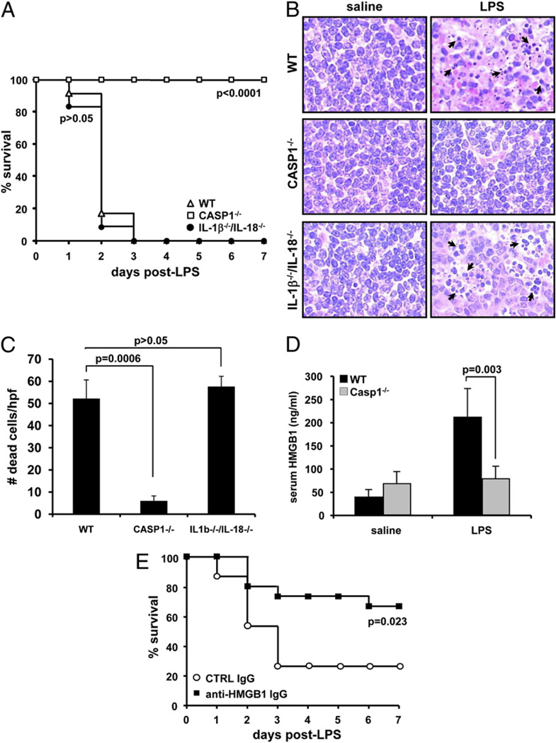
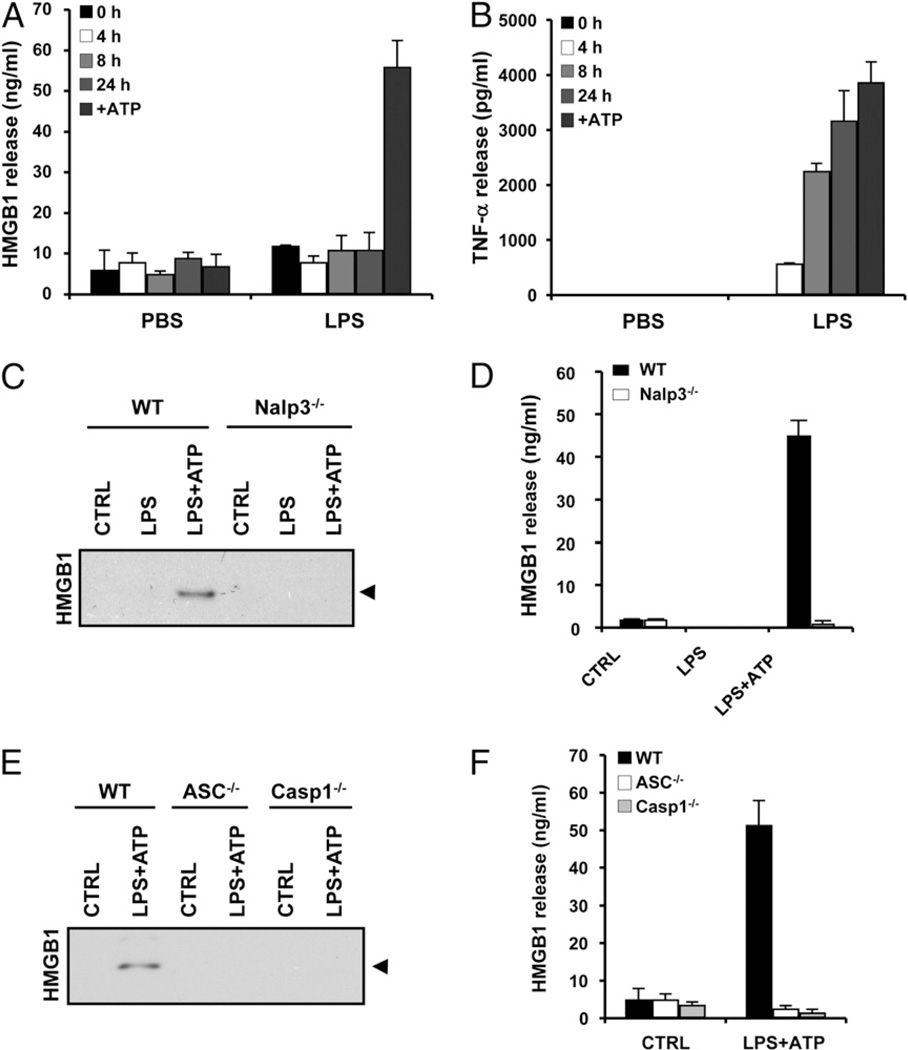
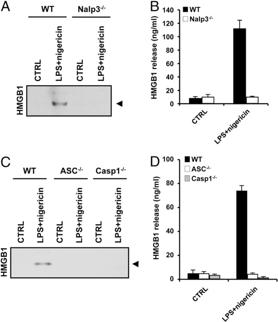
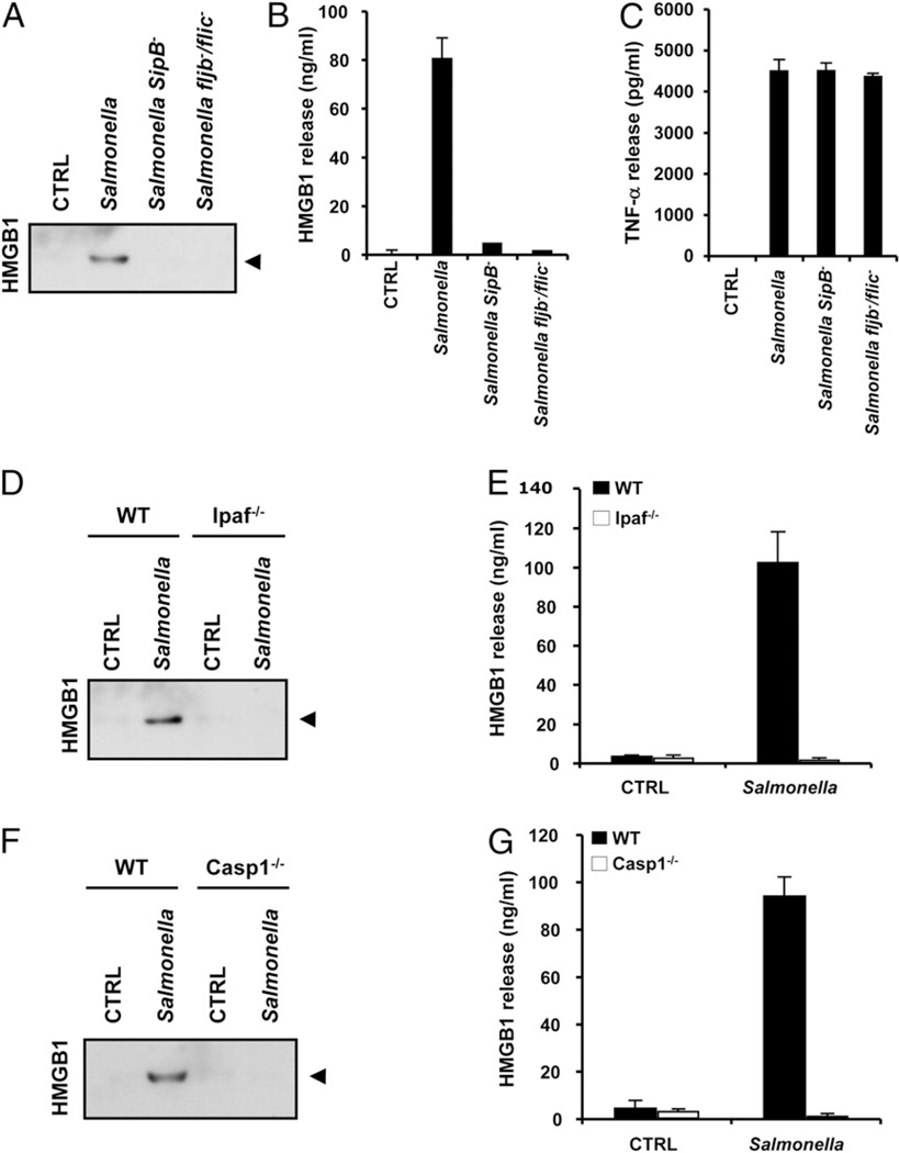
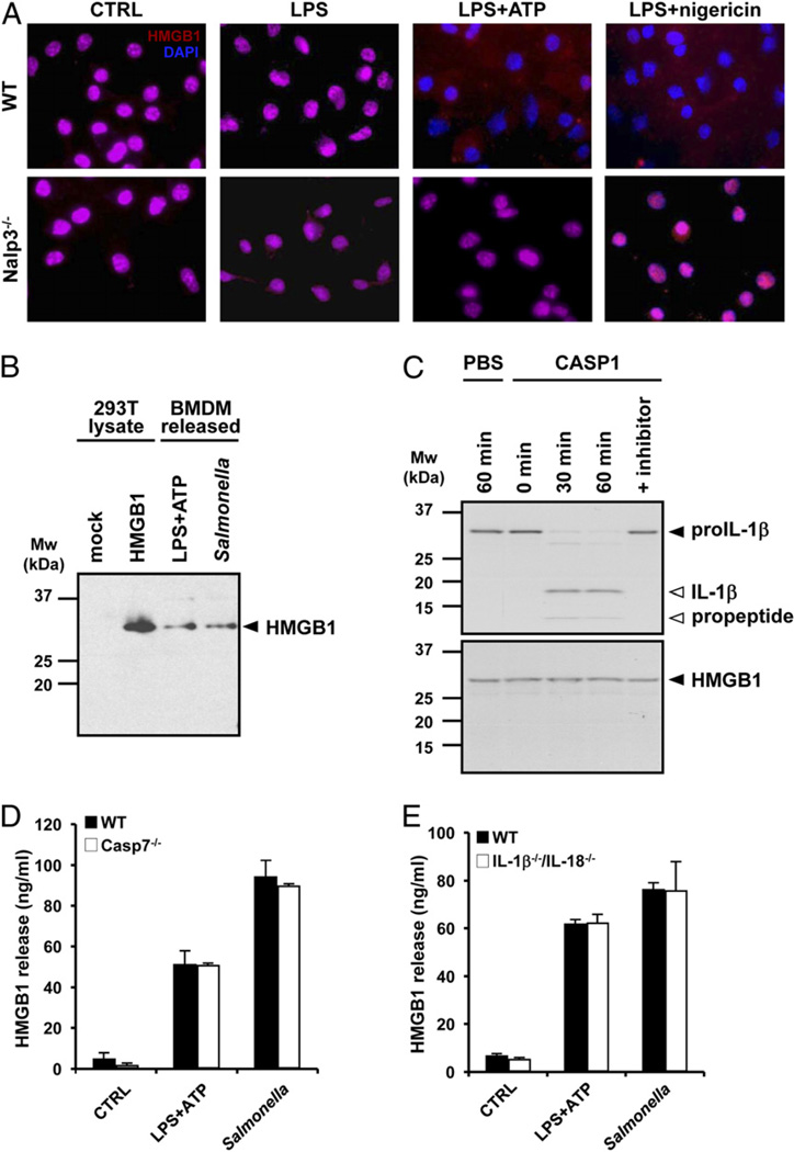
Endotoxin administration recapitulates many of the host responses to sepsis. Inhibitors of the cysteine protease caspase 1 have long been sought as a therapeutic because mice lacking caspase 1 are resistant to LPS-induced endotoxic shock. According to current thinking, caspase 1-mediated shock requires the proinflammatory caspase 1 substrates IL-1β and IL-18. We show, however, that mice lacking both IL-1β and IL-18 are normally susceptible to LPS-induced splenocyte apoptosis and endotoxic shock. This finding indicates the existence of another caspase 1-dependent mediator of endotoxemia. Reduced serum high mobility group box 1 (HMGB1) levels in caspase 1-deficient mice correlated with their resistance to LPS. A critical role for HMGB1 in endotoxemia was confirmed when mice deficient for IL-1β and IL-18 were protected from a lethal dose of LPS by pretreatment with HMGB1-neutralizing Abs. We found that HMGB1 secretion from LPS-primed macrophages required the inflammasome components apoptotic speck protein containing a caspase activation and recruitment domain (ASC), caspase 1 and Nalp3, whereas HMGB1 secretion from macrophages infected in vitro with Salmonella typhimurium was dependent on caspase 1 and Ipaf. Thus, HMGB1 secretion, which is critical for endotoxemia, occurs downstream of inflammasome assembly and caspase 1 activation.


Figures

Figure 1
Figure 1
LPS-induced endotoxemia requires caspase 1-dependent HMGB1 release, but not the caspase 1 substrates IL-1β and IL-18. A, WT (n = 12), caspase 1−/− (n = 10), and IL-1β−/−/IL-18−/− (n = 12) mice were injected i.p. with 40 mg/kg LPS, and their survival was monitored. Results shown are from a single experiment and representative for two independent experiments. Data were compared with WT mice and analyzed with the Kaplan-Meier test. B, WT (n = 4), caspase 1−/− (n = 4), and IL-1β−/−/IL-18−/− (n = 4) mice were injected i.p. with either saline or 40 mg/kg LPS for 24 h before spleens were collected, and sections were stained with H&E. Arrows indicate selected apoptotic splenocytes in the white pulp of the spleen. Original magnification ×400. C, The number of apoptotic cells in five high-power fields (×400) from the white pulp of the spleen of each animal was quantified. Results represent mean ± SD for each genotype. Data were analyzed by Student t test. D, WT and caspase 1−/− mice were injected i.p. (n = 4/group) with either saline or 40 mg/kg LPS for 24 h before serum was collected to measure secreted HMGB1. Results are expressed in ng/ml and represent mean ± SD. Data were analyzed with Student t test. E, One hour before endotoxemia was induced by i.p. injection of 20 mg/kg LPS, IL-1β−/−/IL-18−/− mice (n = 15/group) received an i.p. injection of 100 µg/kg control IgG or HMGB1-neutralizing IgG. Survival was monitored daily for 7 d. Results shown are from a single experiment and representative for two independent experiments. Data were analyzed with the Kaplan-Meier test.
Figure 2
Figure 2
HMGB1 release from LPS and ATP-stimulated BMDMs requires the Nalp3 inflammasome. A and B, BMDMs were stimulated with PBS or LPS (100 ng/ml) for the indicated durations. In one setup, BMDMs were stimulated with 100 ng/ml LPS for 24 h, the last 30 min of which in the presence of 5 mM ATP (+ATP). Culture medium was collected and analyzed for secreted HMGB1 (A) and TNF-α (B). C and D, BMDMs from WT and Nalp3−/− mice were left untreated (CTRL), stimulated with 10 µg/ml LPS for 3 h (LPS), or treated with 10 µg/ml LPS for 3 h followed by 5 mM ATP for 30 min (LPS+ATP). Culture supernatants were collected and analyzed for secreted HMGB1 by Western blotting (C) and ELISA (D). E and F, BMDMs from WT, ASC−/−, and caspase 1−/− mice were left untreated (CTRL) or stimulated with 10 µg/ml LPS for 3 h and 5 mM ATP for 30 min (LPS+ATP). Culture supernatants were collected and analyzed for secreted HMGB1 by Western blotting (E) and ELISA (F). Black arrowhead on Western blots marks HMGB1 (29 kDa). ELISA and Luminex data represent the mean ± SD of triplicate samples from a single experiment, and all results are representative of at least three independent experiments.
Figure 3
Figure 3
Nalp3 inflammasome-dependent release of HMGB1 from LPS plus nigericin-stimulated macrophages. A and B, BMDMs from WT and Nalp3−/− mice were left untreated (CTRL) or stimulated with 10 µg/ml LPS for 3 h and 20 µM nigericin for 30 min (LPS+nigericin). Culture supernatants were collected and analyzed for secreted HMGB1 by Western blotting (A) and ELISA (B). C and D, BMDMs from WT, ASC−/−, and caspase 1−/− mice were left untreated (CTRL) or stimulated with 10 µg/ml LPS for 3 h and 20 µM nigericin for 30 min (LPS+nigericin). Culture supernatants were collected and analyzed for secreted HMGB1 by Western blotting (C) and ELISA (D). Black arrowhead on Western blots marks HMGB1 (29 kDa). ELISA data represent the mean ± SD of triplicate samples from a single experiment, and all results are representative of three independent experiments.
Figure 4
Figure 4
HMGB1 release from Salmonella-infected macrophages requires bacterial flagellin, a functional type III secretion and the Ipaf inflammasome. AC, BMDMs were left untreated (CTRL), infected with WT Salmonella (MOI 10), or with the type III secretion system-deficient (SipB) or flagellin-deficient (fljB/fliC) mutants for 1 h. Culture supernatants were collected and analyzed for secreted HMGB1 by Western blotting (A) and ELISA (B) and for secreted TNF-α by Luminex assay (C). D and E, BMDMs from WT and Ipaf−/− mice were left untreated (CTRL) or infected with WT Salmonella (MOI 10) for 1 h. Culture supernatants were collected and analyzed for secreted HMGB1 by Western blotting (D) and ELISA (E). F and G, BMDMs from WT and caspase 1−/− mice were left untreated (CTRL) or infected with WT Salmonella (MOI 10) for 1 h. Culture supernatants were analyzed for secreted HMGB1 by Western blotting (F) and ELISA (G). Black arrowhead on Western blots marks HMGB1 (29 kDa). ELISA and Luminex data represent the mean ± SD of triplicate samples from a single experiment, and all results are representative of three independent experiments.
Figure 5
Figure 5
The inflammasome is essential for nuclear translocation and secretion of full-length HMGB1 independently of caspase 1 processing and substrates. A, BMDMs from WT and Nalp3−/− mice were plated on cover slips and left untreated (CTRL), stimulated with 10 µg/ml LPS for 3 h (LPS), or treated with 10 µg/ml LPS for 3 h followed by 5 mM ATP (LPS+ATP) or 20 µM nigericin for 30 min. Cells were immunostained for HMGB1, and nuclei were counterstained with DAPI. Original magnification ×20. B, Cell extracts of mock-transfected and HMGB1-overexpressing 293T cells were immunoblotted for HMGB1 together with culture supernatants of BMDMs treated with 10 µg/ml LPS and 5 mM ATP or infected with Salmonella (MOI 10). Black arrowhead on the Western blot marks full-length HMGB1 (29 kDa). C, [35S]-labeled pro-IL-1βand HMGB1 were incubated with PBS or 1 U recombinant caspase 1 for the indicated durations, and cleavage fragments were analyzed by SDS-PAGE and autoradiography. In one setup, recombinant caspase 1 was pretreated with 1 µM YVAD-cmk preincubation with pro-IL-1βand HMGB1 for 60 min. Black arrowhead marks full-length pro-IL-1β and HMGB1, and white arrowheads mark IL-1β cleavage fragments. BMDMs from WT and caspase 7−/− mice (D) and IL-1β−/−/IL-18−/− mice (E) were left untreated (CTRL), stimulated with 10 µg/ml LPS for 3 h followed by 5 mM ATP for 30 min (LPS+ATP), or infected with Salmonella (MOI 10) for 1 h. Culture supernatants were collected and analyzed for secreted HMGB1 by ELISA. Data represent the mean ± SD of triplicate samples from a single experiment, and all results are representative of three independent experiments.

References

    1. Gautier EL, Huby T, Saint-Charles F, Ouzilleau B, Chapman MJ, Lesnik P. Enhanced dendritic cell survival attenuates lipopolysaccharide-induced immunosuppression and increases resistance to lethal endotoxic shock. J. Immunol. 2008;180:6941–6946. - PubMed
    1. Wang H, Bloom O, Zhang M, Vishnubhakat JM, Ombrellino M, Che J, Frazier A, Yang H, Ivanova S, Borovikova L, et al. HMG-1 as a late mediator of endotoxin lethality in mice. Science. 1999;285:248–251. - PubMed
    1. Yang H, Ochani M, Li J, Qiang X, Tanovic M, Harris HE, Susarla SM, Ulloa L, Wang H, DiRaimo R, et al. Reversing established sepsis with antagonists of endogenous high-mobility group box 1. Proc. Natl. Acad. Sci. USA. 2004;101:296–301. - PMC - PubMed
    1. Abraham E, Arcaroli J, Carmody A, Wang H, Tracey KJ. HMG-1 as a mediator of acute lung inflammation. J. Immunol. 2000;165:2950–2954. - PubMed
    1. Bustin M. Regulation of DNA-dependent activities by the functional motifs of the high-mobility-group chromosomal proteins. Mol. Cell. Biol. 1999;19:5237–5246. - PMC - PubMed

Publication types

MeSH terms