. 2010 Nov;299(5):L694-710.
doi: 10.1152/ajplung.00140.2010. Epub 2010 Aug 27.

Sox17 modulates Wnt3A/beta-catenin-mediated transcriptional activation of the Lef-1 promoter

Affiliations

Sox17 modulates Wnt3A/beta-catenin-mediated transcriptional activation of the Lef-1 promoter

Xiaoming Liu et al. Am J Physiol Lung Cell Mol Physiol. 2010 Nov.

Abstract

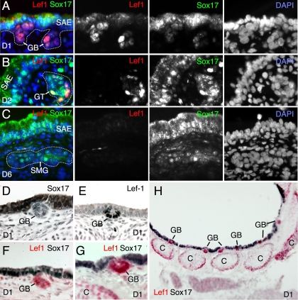
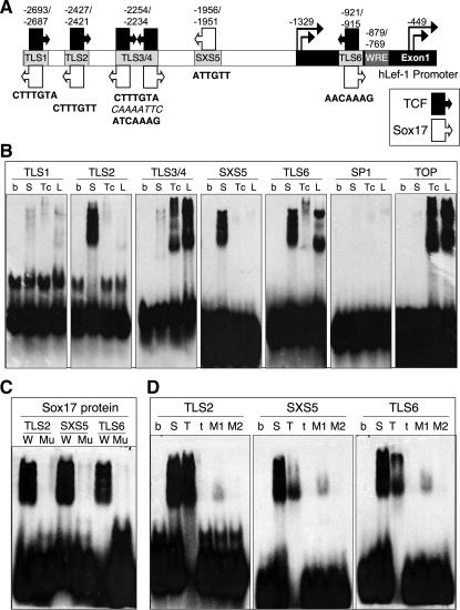
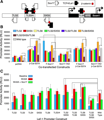
Wnt/β-catenin-dependent activation of lymphoid enhancer factor 1 (Lef-1) plays an important role in numerous developmental processes. In this context, transcription of the Lef-1 gene is increased by Wnt-mediated TCF4/β-catenin activation on the Lef-1 promoter through mechanisms that remain poorly defined. In mouse airway submucosal gland progenitor cells, Wnt3A transiently induces Lef-1 gene expression, and this process is required for epithelial cell proliferation and glandular morphogenesis. In the present study, we sought to identify additional candidate transcriptional regulators of the Lef-1 gene during glandular morphogenesis. To this end, we found that Sox17 expression is dramatically downregulated in early glandular progenitor cells that induce Lef-1 expression. Wnt stimulation of undifferentiated primary airway epithelial cells induced similar changes in Sox17 and Lef-1 expression. Reporter assays revealed that ectopic expression of Sox17 suppresses Wnt3A/β-catenin activation of the Lef-1 promoter in cell lines. EMSA and ChIP analyses defined several Sox17- and TCF4-binding sites that collaborate in transcriptional control of the Lef-1 promoter. More specifically, Sox17 bound to four sites in the Lef-1 promoter, either directly or indirectly through TCF complexes. The DNA- or β-catenin-binding domains of Sox17 controlled context-specific binding of Sox17/TCF complexes on the Lef-1 promoter. Combinatorial site-directed mutagenesis of Sox17- or TCF-binding sites in the Lef-1 promoter demonstrated that these sites control Wnt/β-catenin-mediated induction and/or repression. These findings demonstrate for the first time that Sox17 can directly regulate Wnt/β-catenin-dependent transcription of the Lef-1 promoter and reveal new context-dependent binding sites in the Lef-1 promoter that facilitate protein-protein interactions between Sox17 and TCF4.


Figures

Fig. 1.
Fig. 1.
Lymphoid enhancer factor 1 (Lef-1) and Sox17 expression changes dynamically during the development of airway submucosal glands (SMGs). A–C: frozen sections of ferret trachea from postnatal day 1 (D1), day 2 (D2), and day 6 (D6) were stained with antibodies specific to Lef-1 (red) and Sox-17 (green). Combined and individual immunofluorescence channels are shown. DAPI was used to mark nuclei, and dashed lines indicate the basal lamina of SMG buds and tubules. D–H: using a different set of Sox17 and Lef-1 antibodies, paraffin sections of day 1 ferret tracheas were histochemically stained for Sox17 and Lef-1. Two methods of histochemical staining included staining for Sox17 and Lef-1 individually using DAB peroxidase (black) histochemistry (D, E) or costaining for Sox17 and Lef-1 using DAB peroxidase (black for Sox17) and alkaline phosphatase (red for Lef-1) histochemistry. As previously reported, Lef-1 is expressed in both the newly forming gland buds (GB) and growing edges of the cartilage (C). SAE, surface airway epithelium; GT, gland tubule.
Fig. 2.
Fig. 2.
Wnt proteins stimulate Lef-1 expression in undifferentiated primary airway epithelial cells. A: HCT-116 cells were infected with the indicated recombinant adenoviral vectors expressing Wnt1 or Wnt3A (Ad.Wnt1 and Ad.Wnt3A) or with a control adenoviral vector (Ad.BglII) lacking a transgene. Conditioned HCT-116 medium was absorbed to Blue Sepharose beads, and 20 μl of the 50-ml conditioned medium (m) or 20 μl of the 500 μl of absorbed beads (b) were loaded in the indicated lanes and subjected to Western blotting with anti-Wnt3A or anti-Wnt1 antibodies as indicated. B: undifferentiated primary human bronchial airway epithelial cells (AECs) grown on plastic were treated with the indicated Wnts absorbed to beads. At 36 h following treatment, whole cell lysates were harvested for Western blotting with anti-Lef-1, anti-TCF-4, and anti-β-catenin antibodies (noted at right). C: conditioned medium harvested from Wnt3A-expressing transgenic L cells or from control L cells lacking the Wnt3A transgene was applied to undifferentiated primary human (left) or ferret (right) AECs for 72 h. Nuclear extracts were used for Western blotting with anti-Lef-1, anti-β-catenin, anti-TCF4, anti-cyclin D1, and anti-β-actin antibodies. D: undifferentiated primary AECs were treated with Wnt-conjugated beads. At 36 h following treatment, postnuclear supernatants (PNS) and nuclear extracts (NE) were generated and evaluated by Western blotting with anti-Lef-1, anti-Sox17, and anti-β-catenin antibodies. F-Sox17 (long isoform) and t-Sox17 (truncated short-form missing the HMG binding domain) are shown, at long and short exposure times, respectively. E: a recombinant adenoviral vector encoding the long form of Sox17 (F-Sox17) was used to demonstrate isoform migration. Lane 1, nuclear extract from mock-treated undifferentiated primary AECs (from D) for comparison of F-Sox17 and t-Sox17 expression (long exposure time); lanes 2 and 3, Ad.BglII-infected AECs; lanes 4 and 5, Ad.F-Sox17-infected AECs.
Fig. 3.
Fig. 3.
Sox17 dynamically regulates the Lef-1 promoter in the presence and absence of Wnt3A. A: a 2.5-kb Lef-1 promoter-luciferase reporter (Lef-1-Luciferase) construct was used to evaluate the kinetics of promoter activation in the presence of Wnt3A and/or Sox17 expression. 293 Cells were cotransfected with 0.2 μg of Lef-1-luciferase reporter and 0.5 μg of Wnt3A and/or 0.2 μg of Sox17 expression plasmids using Lipofectamine LTX reagent. For all transfections, the total DNA concentration was identical and normalized using the empty-vector control plasmid pcDNA. All transfections also contained 0.01 μg of a pCMV-Renilla luciferase plasmid for normalization of transfection efficiency. Relative activity of the Lef-1 promoter is plotted as relative light units (RLU). B: Western blot analysis of changes in total β-catenin, active β-catenin, Sox17, and phosphorylated GSK3β at 24 h posttransfection, with the transfected plasmids indicated above the blot. C and D: comparison of Lef-1-luciferase reporter (0.2 μg) (C) and TOP-flash TCF-responsive reporter (0.05 μg) (D) activities in cells transfected with the indicated reporter and Wnt3A (0.5 μg) and/or Sox17 (0.2 μg) expression constructs. E and F: comparison of Lef-1-luciferase reporter (E) and TOP-flash TCF-responsive reporter (F) activities in cell transfected with the indicated reporters and dominant-active β-catenin-S37A (0.5 μg) and/or Sox17 (0.2 μg) expression constructs. Values in A and C–F represent the mean (± SE, n = 9) RLU normalized for Renilla luciferase expression.
Fig. 4.
Fig. 4.
Sox17 transcriptionally regulates Wnt3A-mediated activation of both the Lef-1 promoter and TCF-responsive reporter TOP-flash. MCF-7, SW480, A549, or L cells were cotransfected with Wnt3A- (0.5 μg) and/or Sox17- (0.2 μg) expressing plasmid(s), together with the Lef-1 promoter luciferase (0.2 μg) (left) or TOP-flash luciferase (0.05 μg) (right) reporter. The pcDNA empty plasmid was used as a negative control and for normalizing total DNA content in each transfection. Each transfection cocktail also contained 0.01 μg of CMV-Renilla luciferase as an internal control plasmid for the normalization of transfection efficiency. At 24 h posttransfection, cell lysates were prepared, and both firefly and renilla luciferase activities were determined. Bars depict the mean (± SE, n = 3) RLU normalized for Renilla luciferase expression.
Fig. 5.
Fig. 5.
Structural analysis of Sox17-mediated regulation of the Lef-1 promoter by Wnt3A. Various deletion and point mutants of mouse Sox17 were used to determine which domains of Sox17 are required to repress Wnt3A-mediated transcriptional induction of the Lef-1 promoter. A: schematic representation of constructs encoding the full-length Sox17 protein and various Sox17 mutants (see materials and methods). All of the constructs contained a V5-tag at the COOH terminus. B: 293 cells were transfected with each of these constructs, and expression was evaluated by Western blotting with anti-V5 antibody at 24 h posttransfection. C: anti-V5 antibody was used to immunoprecipitate Sox17 from 293 cells transfected with the expression constructs shown in A, followed by Western blotting with anti-Sox17, anti-Lef-1, anti-TCF4, and anti-β-catenin antibodies. Nuclear extracts (500 μg) were used for IP reactions at 48 h posttransfection. The input lane represents direct loading of nuclear extract from full-length Sox17-transfected cells, whereas the No V5 lane omitted the capture V5 antibody from the IP reaction for full-length Sox17-transfected cells. D: analysis of the functions of different Sox17 domains in Wnt3A-mediated activation of Lef-1 promoter (top) and TCF-responsive reporter TOP-flash (bottom). 293 Cells were cotransfected with the Wnt3A (0.5 μg) expression plasmid and either the Lef-1 promoter (0.2 μg) or TOP-flash (0.05 μg) reporter plasmid, together with one of the indicated Sox17 (0.2 μg) expression plasmids. All transfections also contained 0.01 μg of a pCMV-Renilla luciferase plasmid for normalization of transfection efficiency. Bars depict the mean (± SE, n = 4) RLU at 24 h posttransfection and were normalized for Renilla luciferase expression.
Fig. 6.
Fig. 6.
ChIP survey of Sox17 and TCF4 binding sites in the endogenous Lef-1 promoter of A549 cells. A: schematic representation of the 10 PCR fragments used to survey the 2.5-kb human Lef-1 promoter (Table 3). Each PCR fragment covered 250–400 bp of the promoter. B and C: A549 cells were transfected with mouse Sox17- or TCF4-expressing plasmid, and genomic DNA sheared to an average size of 400–600 bp before ChIP was performed with anti-Sox17 and TCF4 antibodies. The recovered DNA was then evaluated by either standard PCR (B) or real-time PCR (C). B: agarose gel images show PCR products following 30 cycles of amplification from DNA immunoprecipitated with anti-Sox17 antibody (top), anti-TCF4 antibody (middle), or nonimmune total IgG from the same species as the primary capture antibody (bottom; used as a negative control). C: quantitative PCR results from the ChIP assays. Values depict the fold increase in copies of each target sequence detected with the specific capture antibody over the number detected with the species-matched IgG control antibody alone. Data are representative of 3 independent experiments.
Fig. 7.
Fig. 7.
Evaluation of Sox17 and TCF binding sites in the Lef-1 promoter by EMSA. A: schematic representation of the 2.5-kb human Lef-1 promoter, with the positions and sequences of candidate TCF and Sox17 protein binding sites indicated (as determined based on the consensus sequences described in Table 4). The direction of arrows marked TCF and Sox17 indicate whether the consensus conforms to the forward or reverse stand of the DNA sequence. B: EMSA gels depicting protein/DNA complexes formed on the indicated biotinylated probes (Table 2) following binding by purified BSA (b), Sox17 (S), TCF4 (Tc), and Lef-1 (L). SP1 and TOP are negative and positive control biotinylated oligonucleotides for SP1 and TCF/Lef-1 (same consensus in TOP-flash) binding, respectively. C: EMSA gels depicting protein/DNA complexes formed when purified Sox17 protein was incubated with the indicated wild-type (W) or mutant (Mu) biotinylated probes (Table 2) for the 3 primary Sox17 binding sites. D: EMSA gels depicting protein/DNA complexes formed on the indicated biotinylated probes following binding of purified BSA (b), full-length Sox17 (S), Sox17-TAD (T), t-Sox17 (t), Sox17-mut1 (M1), or Sox17-mut2 (M2).
Fig. 8.
Fig. 8.
Sox17, TCF4, Lef-1, and β-catenin form intermolecular complexes on sequences within the Lef-1 promoter. A: the indicated cell lines were transfected with V5-tagged Sox17-, TCF4-, and β-catenin-expressing plasmids, and nuclear extracts were prepared at 48 h posttransfection. These extracts were then immunoprecipitated with antibodies against V5 (for Sox17), TCF4, or β-catenin (Bc) before being immunoblotted against the indicated antibodies. B: schematic representation of the strategy used in the experiments represented in C and D to detect protein/DNA complexes bound to biotinylated oligonucleotide sequences from the Lef-1 promoter. Purified recombinant proteins were prebound to the biotinylated oligonucleotides before they were captured on Avidin dynabeads and Western blotted with the appropriate antibodies. C: association of β-catenin, Sox17, TCF4, and Lef-1 with the indicated Lef-1 promoter sequences. An SP1 oligonucleotide served as negative control. D: association of β-catenin, TCF4, Lef-1, and Sox17 mutant (Sox17-TAD or t-Sox17) with the indicated Lef-1 promoter sequences.
Fig. 9.
Fig. 9.
Transcriptional analysis of the Lef-1 promoter with mutated Sox17 and TCF4/Lef-1 binding sites. A: schematic representation of TCF/Lef-1 and Sox17 binding interactions on the Lef-1 promoter as determined by ChIP, EMSA, and DNA-affinity capture assays. The size of each arrowed box denotes the level of binding (i.e., smallest represents weakest binding). The presence of β-catenin (red) indicates that a particular factor is able to bind β-catenin and/or that β-catenin may have to interact with the BBD of Sox17 to mediate an intermolecular interaction (i.e., at SXS5). B: promoter activity when 293 cells were transfected with the indicated mutant or wild-type Lef-1 promoter-luciferase constructs (as indicated by color), together with plasmid expressing the protein(s) indicated on the x-axis. C: promoter activity when A549 cells were transfected with the indicated mutant or wild-type Lef-1 promoter-luciferase constructs on the x-axis, together with plasmid expressing Wnt3A and/or Sox17 (as indicated by color). Significant differences in expression levels as determined by Student's t-test (P < 0.05) are indicated by the connecting lines.
Fig. 10.
Fig. 10.
Context-dependent interactions between Sox17 and TCF4/Lef-1 at specific sites on the Lef-1 promoter and working models for Lef-1 promoter regulation. A: a summary of Sox17 and TCF4/Lef-1 binding to sites on the Lef-1 promoter, and of the context-dependent interactions that depend on the BBD and HMG domains of Sox17. Based on consensus sequences, 6 TCF/Sox sites exist in the Lef-1 promoter (sites 1–6). Site 2 (TLS2) binds only Sox17. Sites 3/4 (TLS3/4) primarily bind TCF4/Lef-1, but can also recruit Sox17 in the presence of both β-catenin and the Sox17 HMG domain. Site 5 (SXS5) primarily binds Sox17, but can recruit TCF4/Lef-1 in the presence of both β-catenin and the Sox17 β-catenin binding domain (BBD). Site 6 (TLS6) can directly bind to both Sox17 and TCF4/Lef-1. Whether context-dependent interactions between Sox17 and TCF4/Lef-1 occur at this site could not be determined from this study. However, we hypothesize that binding of Sox17 and TCFs to this site may be competitive. B: working models for Lef-1 promoter regulation. In the presence of a Wnt signal where β-catenin rises and Sox17 falls (as in submucosal gland progenitor cells), we hypothesize that sites within the Lef-1 promoter are predominantly bound by TCF4/β-catenin, and this leads to increased transcription. In the presence of a sustained Wnt signal, we hypothesize that Sox17 expression is reactivated, leading to competitive occupancy of Sox17/TCF4 sites and complex formation. This may function to repress the promoter. Alternative transcriptional states, such as the cell-specific induction of the Lef-1 promoter by Sox17 overexpression in 293 cells, may be explained by intermolecular complex formation between Sox17 and TCFs at distant sites in the promoter. These interactions may also explain the complex transcriptional consequences of mutating one or more TCF/Sox17 sites within the promoter that are inconsistent with assigning a strictly inhibitor function to Sox17 binding.

Similar articles

Cited by

References

    1. Amen M, Liu X, Vadlamudi U, Elizondo G, Diamond E, Engelhardt JF, Amendt BA. PITX2 and beta-catenin interactions regulate Lef-1 isoform expression. Mol Cell Biol 27: 7560–7573, 2007 - PMC - PubMed
    1. Anderson RD, Haskell RE, Xia H, Roessler BJ, Davidson BL. A simple method for the rapid generation of recombinant adenovirus vectors. Gene Ther 7: 1034–1038, 2000 - PubMed
    1. Arce L, Pate KT, Waterman ML. Groucho binds two conserved regions of LEF-1 for HDAC-dependent repression. BMC Cancer 9: 159, 2009 - PMC - PubMed
    1. Arce L, Yokoyama NN, Waterman ML. Diversity of LEF/TCF action in development and disease. Oncogene 25: 7492–7504, 2006 - PubMed
    1. Atcha FA, Munguia JE, Li TW, Hovanes K, Waterman ML. A new beta-catenin-dependent activation domain in T cell factor. J Biol Chem 278: 16169–16175, 2003 - PubMed

Publication types

MeSH terms