. 2010 Oct;177(4):2091-102.
doi: 10.2353/ajpath.2010.090878. Epub 2010 Aug 27.

The role of vascular endothelial growth factor-induced activation of NADPH oxidase in choroidal endothelial cells and choroidal neovascularization

Affiliations

The role of vascular endothelial growth factor-induced activation of NADPH oxidase in choroidal endothelial cells and choroidal neovascularization

Elizabeth Monaghan-Benson et al. Am J Pathol. 2010 Oct.

Abstract

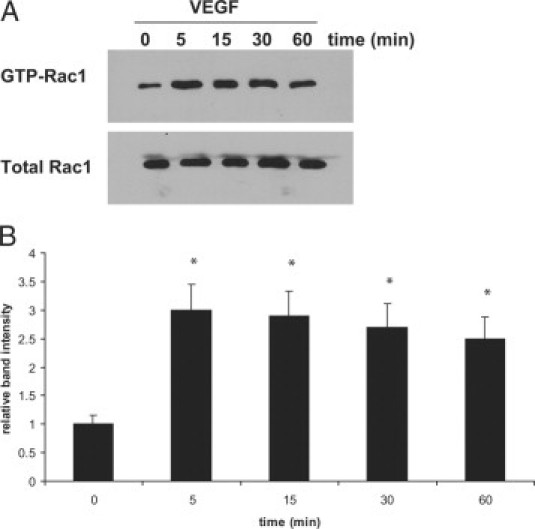
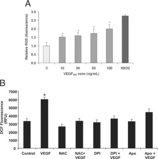
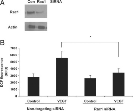
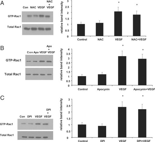
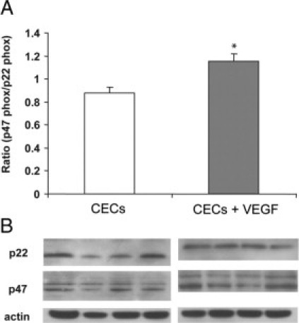
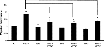
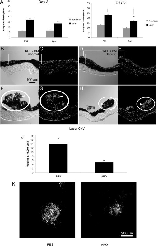
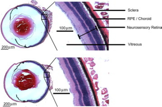
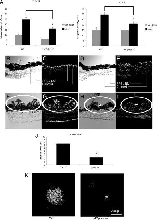
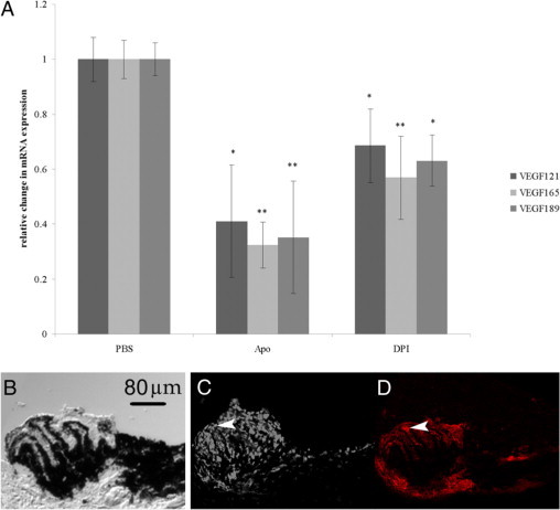
Rac1, a subunit of NADPH oxidase, plays an important role in directed endothelial cell motility. We reported previously that Rac1 activation was necessary for choroidal endothelial cell migration across the retinal pigment epithelium, a critical step in the development of vision-threatening neovascular age-related macular degeneration. Here we explored the roles of Rac1 and NADPH oxidase activation in response to vascular endothelial growth factor treatment in vitro and in a model of laser-induced choroidal neovascularization. We found that vascular endothelial growth factor induced the activation of Rac1 and of NADPH oxidase in cultured human choroidal endothelial cells. Further, vascular endothelial growth factor led to heightened generation of reactive oxygen species from cultured human choroidal endothelial cells, which was prevented by the NADPH oxidase inhibitors, apocynin and diphenyleneiodonium, or the antioxidant, N-acetyl-L-cysteine. In a model of laser-induced injury, inhibition of NADPH oxidase with apocynin significantly reduced reactive oxygen species levels as measured by dihydroethidium fluorescence and the volume of laser-induced choroidal neovascularization. Mice lacking functional p47phox, a subunit of NADPH oxidase, had reduced dihydroethidium fluorescence and choroidal neovascularization compared with wild-type controls. Taken together, these results indicate that vascular endothelial growth factor activates Rac1 upstream from NADPH oxidase in human choroidal endothelial cells and increases generation of reactive oxygen species, contributing to choroidal neovascularization. These steps may contributed to the pathology of neovascular age-related macular degeneration.


Figures

Figure 1
Figure 1
VEGF treatment activates Rac1 in choroidal ECs. Choroidal ECs were treated with 10 ng/ml VEGF for the indicated times, and the activation of Rac1 was measured using a GST-PBD pulldown assay as described in Materials and Methods. Total Rac1 was used as a loading control. A: Representative blot showing increased active Rac1 within five minutes of treatment with VEGF. B: Densitometry analysis of blots performed as described in Materials and Methods. Band intensity is expressed for each time point as a relative number compared with the 0-minute time point. Rac1 activation at the zero time point is statistically different from that of cells treated with VEGF for 5, 15, 30, or 60 minutes (*P < 0.05).
Figure 2
Figure 2
VEGF-induced ROS generation in choroidal ECs. A: Cells were incubated with different concentrations of VEGF165 for 15 minutes and then ROS generation was determined by DCF fluorescence measurement (*P < 0.05); 200 μmol/L H2O2 was used as a positive control. B: Choroidal ECs were pretreated with 1 mmol/L NAC, 500 μmol/L apocynin (Apo), or 1 μmol/L DPI for two hours. The choroidal ECs were loaded with H2DCF-DA and then treated with 10 ng/ml VEGF165 for ten minutes. The generation of ROS was measured by the fluorescence intensity of DCF. Data are expressed in relative fluorescent units (RFU) as given by the plate reader. *P < 0.05 when VEGF is compared with control choroidal ECs. Treatment with NAC, DPI, or Apo abrogated VEGF-induced generation of ROS.
Figure 3
Figure 3
Rac1-dependent VEGF-induced ROS in choroidal ECs. A: Choroidal ECs were transfected with Rac1 siRNA or nontargeting siRNA for 72 hours. Cells were then lysed in sample buffer, electrophoresed, and immunoblotted with an antibody against Rac1 or actin. B: Choroidal ECs were transfected with Rac1 siRNA or a nontargeting siRNA for 72 hours. Cells were then treated with 10 ng/ml VEGF, and ROS generation was monitored by DCF fluorescence. Data are expressed as relative fluorescent units (RFU) as given by the plate reader. ROS generation in the nontargeting siRNA cells treated with VEGF is statistically different from that in the Rac1 siRNA cells treated with VEGF at 15 minutes (*P < 0.05). Con, control.
Figure 4
Figure 4
Rac1 activated upstream of NADPH oxidase in VEGF-stimulated choroidal ECs. A: Choroidal ECs were pretreated with 1 mmol/L NAC for two hours before treatment with VEGF165 (10 ng/ml). The activation of Rac1 was measured through a GST-PBD pulldown assay. Representative blots are shown for each. B: Choroidal ECs were pretreated with 500 μmol/L apocynin for 2 hours before treatment with VEGF165 (10 ng/ml). The activation of Rac1 was measured through a GST-PBD pulldown assay. C: Choroidal ECs were pretreated with 1 μmol/L DPI for two hours before treatment with VEGF165 (10 ng/ml). The activation of Rac1 was measured through a GST-PBD pulldown assay. Representative blots are shown for each. Densitometry analysis of blots was performed as described in Materials and Methods. Band intensity is expressed for each treatment as a relative number compared with the control. Rac1 activation in the control cells is statistically different from that for cells treated with VEGF or VEGF + drug. (*P < 0.05). Con, control.
Figure 5
Figure 5
p47phox/p22phox increased in VEGF-stimulated choroidal ECs. A: Choroidal ECs (CECs) were incubated with 30 ng/ml VEGF165 for 15 minutes, and isolated membrane protein was separated by SDS-polyacrylamide gel electrophoresis. Membranes were then blotted for p47phox or p22phox. B: Figure is representative of densitometry analyses of Western blot of membrane protein probed with anti-p47phox and anti-p22phox antibodies. β-Actin was used as a loading control. The ratio in control CECs is statistically different from that in CECs treated with VEGF (*P < 0.05).
Figure 6
Figure 6
NADPH oxidase required for VEGF-induced migration of choroidal ECs. Endothelial cells were seeded onto collagen-coated transwell tissue culture inserts in the presence or absence of NAC, DPI, or apocynin (Apo) for two hours. After this initial pretreatment with inhibitors, 10 ng/ml VEGF was added to the outer chamber, and cells were allowed to migrate for two hours. After two hours, cells remaining on the top of the chamber were removed, and cells that had migrated to the bottom chamber were trypsinized and counted. Migration of the VEGF-treated choroidal ECs is statistically different from that of the choroidal ECs treated with VEGF + drug (*P < 0.05). All values are expressed as relative to the control, which has been set equal to 1. C, control choroidal ECs.
Figure 7
Figure 7
Inhibition of NADPH oxidase decreases laser-induced CNV in mice. A: ROS levels in lasered and unlasered sections of RPE-choroid in three month-old C57BL/6 mice. ROS levels were determined by measuring DHE fluorescence. Data are presented as integrated density per area (mean ± SEM [10 μmol/L]) using ImageJ 1.43 for analysis. *P < 0.05, laser-treated, PBS versus apocynin (Apo), day five. B–I: Representative images of retinal/choroidal cross sections from mice unlasered (B–E) or lasered (F–I) and administered apocynin (D, E, H and I) or PBS (B, C, F and G). Sections are oriented with CNV and/or RPE-choroid at the top and the sclera below. Phase images (B, D, F, and H) were taken of corresponding DHE-stained images (C, E, G, and I). Note the qualitatively increased number of DHE-stained cells in lasered sections (bottom row) compared with respective images (top row) and qualitatively reduced number of DHE-stained cells in lasered sections from apocynin-treated (I) compared with PBS-treated (G) mice. DHE fluorescence is found in mainly nonpigmented cells in PBS-treated lasered mice (compare arrowheads in phase [F] and DHE fluorescence [G]) and in both pigmented and nonpigmented cells in both PBS-treated (F) and apocynin-treated lasered mice (compare arrowheads in phase [H] and DHE fluorescence [I]). J: C57BL/6 mice (three months old) were treated with apocynin (10 mg/kg/day) or PBS for five days preceding and five days after photocoagulation by laser treatment. Choroidal flat mounts of lasered areas were stained with lectin and 1-μm horizontal sections were captured with confocal microscopy, measured, and analyzed as described (*P = 0.007, analysis of variance). K: Representative images of maximum projections of CNV from choroidal flat mounts are shown for each condition: left, PBS-treated; right, apocynin-treated. Scale bar = 200 μm. RPE/BM, retinal pigment epithelium/Bruch's membrane; laser CNV, choroidal neovascular lesion induced with laser injury.
Figure 8
Figure 8
Ocular histology of p47phox−/− compared with wild-type mice. Representative images of H&E stains of wild-type (top) and p47phox−/− whole eyes (bottom). Inset: magnified retinal layers. Scale bar = 200 μm.
Figure 9
Figure 9
Deficiency in the p47phox subunit of NADPH oxidase decreases laser-induced CNV in mice. A: Three-month-old wild-type and p47phox−/− mice received photocoagulation by laser treatment. ROS levels in lasered and unlasered sections of RPE-choroid were determined by measuring DHE fluorescence. Data are presented as integrated density per area (mean ± SEM, DHE [10 μmol/L] using ImageJ for analysis). *Analysis of variance, P < 0.05, day three and day five, laser-treated, wild-type versus p47phox−/− mice. B–I: Representative images of retinal-choroidal cross sections from mice untreated (top row) or laser-treated (bottom row). Phase images (B, D, F, and H) were taken of corresponding DHE-stained images (C, E, G, and I). Sections from wild-type mice (B, C, F and G) or p47phox−/− mice (D, E, H and I). Sections are oriented with CNV and/or the RPE-choroid at the top and sclera (nonpigmented layer) at the bottom. An edge artifact is noted at the inferior boundary of the sclera in G. Note the qualitatively increased number of DHE-stained cells in lasered sections (F–I) compared with unlasered respective comparisons (B–E) and qualitatively fewer DHE-stained cells in lasered sections from p47phox−/− mice (I) compared with wild-type mice (G). DHE fluorescence is found in mainly nonpigmented cells in wild-type lasered mice (compare arrowheads in phase [F] and DHE fluorescence [G]) and in both pigmented and nonpigmented cells in p47phox−/− lasered mice (compare phase [H] and DHE fluorescence [I]). J: Wild-type and p47phox−/− mice received photocoagulation by laser treatment 5 days earlier. Choroidal flat mounts of lasered areas were stained with lectin and 1-μm horizontal sections were captured with confocal microscopy, measured, and analyzed as described (analysis of variance, *P = 0.036, laser treated wild-type versus p47phox−/− mice). K: Representative images of maximum projections of lectin-stained CNV in choroidal flat mounts shown below: left, wild-type; right, p47phox−/−. Scale bar = 200 μm. WT, wild-type; RPE/BM, retinal pigment epithelium/Bruch's membrane; laser CNV, choroidal neovascular lesion induced with laser injury.
Figure 10
Figure 10
A: Inhibition of ROS generation decreases expression of VEGF splice variants in human RPE. Total RNA was extracted from human ARPE treated with PBS, apocynin (Apo), or DPI. Changes in gene expression were detected by real-time PCR. Data are presented as mean ± SEM change in gene expression normalized to 18s RNA levels and relative to PBS control normalized to 18s levels. *P < 0.01; **P < 0.001 versus PBS control, n = 10 (analyzed with REST software). Apo, apocynin. B–D: Panels of cross sections through the CNV lesion 5 days after laser treatment in wild-type mice. Sections taken in 10-μm increments are shown. B: Phase image showing pigmented and nonpigmented cells (neurosensory retina is removed and the sclera is toward the bottom of the image) of a fresh tissue section that had been stained for DHE fluorescence and imaged previously (C). D: Adjacent section through the same CNV lesion fixed and stained with lectin to visualize vascular cells. Arrowheads indicate lectin-stained tissue in D corresponding to DHE-positive cells in C. Scale bar = 80 μm.

Similar articles

Cited by

References

    1. Tielsch JA. Vision Problems in the U.S.: A Report on Blindness and Vision Impairment in Adults Age 40 and Older. Prevent Blindness America; Schaumburg, IL: 1994. pp. 1–20.
    1. Augood CA, Vingerling JR, de Jong PTVM, Chakravarthy U, Seland J, Soubrane G, Tomazzoli L, Topouzis F, Bentham G, Rahu M, Vioque J, Young IS, Fletcher AE. Prevalence of age-related maculopathy in older Europeans: the European Eye Study (EUREYE) Arch Ophthalmol. 2006;124:529–535. - PubMed
    1. Mitchell P, Smith W, Attebo K, Wang JJ. Prevalence of age-related maculopathy in Australia. The Blue Mountains Eye Study. Ophthalmology. 1995;102:1450–1460. - PubMed
    1. Friedman DS, O'Colmain BJ, Muñoz B, Tomany SC, McCarty C, de Jong PT, Nemesure B, Mitchell P, Kempen J, Eye Diseases Prevalence Research Group Prevalence of age-related macular degeneration in the United States. Arch Ophthalmol. 2004;122:564–572. - PubMed
    1. Gehrs KM, Anderson DH, Johnson LV, Hageman GS. Age-related macular degeneration—emerging pathogenetic and therapeutic concepts. Ann Med. 2006;38:450–471. - PMC - PubMed

Publication types

MeSH terms