. 2010 Nov 5;285(45):34469-76.
doi: 10.1074/jbc.M110.157552. Epub 2010 Aug 27.

Heat shock transcription factor 1 localizes to sex chromatin during meiotic repression

Affiliations

Heat shock transcription factor 1 localizes to sex chromatin during meiotic repression

Malin Akerfelt et al. J Biol Chem. .

Abstract

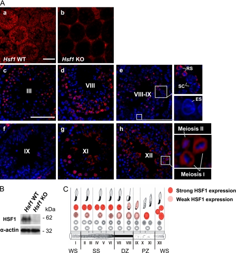
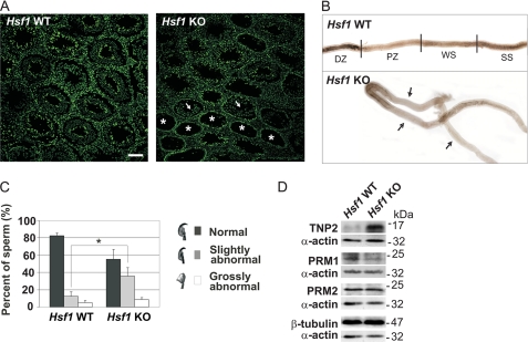
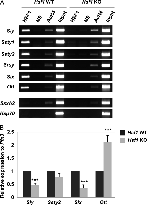
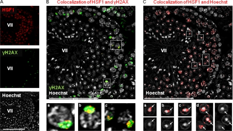
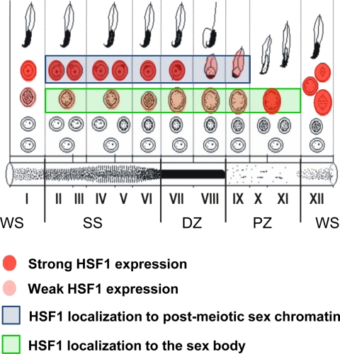
Heat shock factor 1 (HSF1) is an important transcription factor in cellular stress responses, cancer, aging, and developmental processes including gametogenesis. Disruption of Hsf1, together with another HSF family member, Hsf2, causes male sterility and complete lack of mature sperm in mice, but the specific role of HSF1 in spermatogenesis has remained unclear. Here, we show that HSF1 is transiently expressed in meiotic spermatocytes and haploid round spermatids in mouse testis. The Hsf1(-/-) male mice displayed regions of seminiferous tubules containing only spermatogonia and increased morphological abnormalities in sperm heads. In search for HSF1 target genes, we identified 742 putative promoters in mouse testis. Among them, the sex chromosomal multicopy genes that are expressed in postmeiotic cells were occupied by HSF1. Given that the sex chromatin mostly is repressed during and after meiosis, it is remarkable that HSF1 directly regulates the transcription of sex-linked multicopy genes during postmeiotic repression. In addition, our results show that HSF1 localizes to the sex body prior to the meiotic divisions and to the sex chromocenter after completed meiosis. To the best of our knowledge, HSF1 is the first known transcription factor found at the repressed sex chromatin during meiosis.


Figures

FIGURE 1.
FIGURE 1.
HSF1 is transiently expressed during mouse spermatogenesis. A, the localization and expression of HSF1 in specific cell types were examined by immunohistochemistry. Panels a and b, HSF1 immunostaining of wild-type (Hsf1 WT) and Hsf1 knock-out (Hsf1 KO) testis. No signal in the seminiferous epithelium of Hsf1 KO testis indicates specificity of the antibody. Panels c–h, HSF1 immunostaining (red) of WT testis, counterstained with Hoechst (blue). Representative stages are denoted by Roman numerals. Insets show blowups of the selected regions. SC, spermatocyte; RS, round spermatid; ES, elongating spermatid. Scale bars, 100 μm. B, Western blot analysis of HSF1 levels in Hsf1 WT and KO testis. Equal protein loading was assessed by β-tubulin and α-actin. C, a schematic drawing displaying the cell type-specific expression of HSF1 throughout the seminiferous epithelial cycle. Each vertical column, designated by a Roman numeral, depicts a stage of the seminiferous epithelial cycle and is comprised of a defined set of developing germ cells. In the schematic (C), the developmental progression of a cell is followed from the bottom row, left to right. The cycle ends with the release of morphologically mature sperm to the lumen of the tubule. The light absorption patterns of corresponding stages of the seminiferous tubule are shown at the bottom. Stages IX–XI, pale zone (PZ); stages XII–I, weak spot (WS); stages II–VI, strong spot (SS); and stages VII–VIII, dark zone (DZ).
FIGURE 2.
FIGURE 2.
Characterization of the phenotype of Hsf1−/− testis and mature sperm. A, SYBR green staining of testis cross sections of wild-type (Hsf1 WT) and Hsf1 knock-out (Hsf1 KO) mice, was used to determine the morphology of the seminiferous tubules, in addition to characterization of missing cell types. Asterisks indicate tubules containing only spermatogonia, and arrows indicate tubules containing spermatogonia and spermatocytes. Scale bar, 100 μm. B, isolated seminiferous tubules from Hsf1 WT and KO males. Arrows indicate abnormally long pale zones. To visualize long stretches of tubules, several images were merged into one using Adobe Photoshop. Stages IX–XI, pale zone (PZ); stages XII–I, weak spot (WS); stages II–VI, strong spot (SS); and stages VII–VIII, dark zone (DZ). C, analysis of hematoxylin-stained sperm smears from WT and KO males. The sperm heads were classified into categories of normal, slightly abnormal and grossly abnormal. 400–500 sperm in each category were counted in a blind experiment. Mean ± S.D. (n = 3); *, p < 0.05. D, Western blot analysis of transition protein 2 (TNP2), protamine 1 (PRM1), and protamine 2 (PRM2) levels in cauda epididymis isolated from WT and KO mice. The blots are representative of three biological repeats. Equal protein loading was assessed by β-tubulin and α-actin.
FIGURE 3.
FIGURE 3.
HSF1 is a transcriptional regulator of sex chromosomal multicopy genes in testis. A, ChIP analysis of HSF1 binding to several sex chromosomal multicopy genes in wild-type (Hsf1 WT) and Hsf1 knock-out (Hsf1 KO) testis. In addition, examples of an X-chromosomal multicopy gene, Ssxb2, and a single-copy gene, Hsp70, not bound by HSF1 are shown. NS, nonspecific antibody; AcH4, acetylated histone H4. Input represents 1% of the total material used in the ChIP assay. B, real-time RT-PCR analysis of gene expression in whole wild-type (Hsf1 WT) and knock-out (Hsf1 KO) testes. The mRNA levels were normalized to the round spermatid-specific Pfn3. The relative expression was calculated from the Hsf1 WT sample, which arbitrarily was set to 1. Mean ± S.D. (n = 3); ***, p < 0.005.
FIGURE 4.
FIGURE 4.
HSF1 localizes to meiotic and postmeiotic sex chromatin. A, detection of HSF1 (upper panel) and γH2AX (middle panel) by immunofluorescence and detection of DNA by Hoechst (lower panel) in a single confocal section of stage VII seminiferous tubule. B, colocalization of HSF1 with γH2AX (red) is superimposed on γH2AX-stained sex bodies (green), designating HSF1 localization to meiotic sex chromatin. Insets a, b, and c show HSF1 localization to sex bodies in stage VII pachytene spermatocytes. C, postmeiotic sex chromatin (white arrows in panels a–d) can be observed as a cloud-like structure peripherally to the Hoechst-dense chromocenter. The HSF1 Hoechst colocalization (red) reveals HSF1 localization to the postmeiotic sex chromatin of round spermatids. Compare HSF1 Hoechst colocalization in insets a–d (upper panels) with Hoechst-only staining (lower panels), where the sex chromocenter is clearly visible. Note that the same confocal section of the stage VII seminiferous tubule is shown in A, B, and C. The specificity of secondary antibodies that were used in the colocalization analyses was confirmed (supplemental Fig. S3). Colocalization was analyzed using ImageJ MacBiophotonics colocalization finder tool. Representative stages are shown in the figure, and the blowup is shown in the insets. Stages are denoted by Roman numerals. Scale bars, 100 μm.
FIGURE 5.
FIGURE 5.
Schematic presentation of HSF1 localization to the meiotic sex chromatin. HSF1 localizes to the meiotic sex chromatin in spermatocytes of stages II–XI, where the sex chromosomes are retained silent in a γH2AX-enriched sex body (Fig. 4 and supplemental Fig. S1). After the meiotic divisions, HSF1 localizes to the postmeiotic sex chromatin in spermatids of stages II–IX, where the sex chromatin is observed as a cloud-like structure peripherally to the Hoechst-dense chromocenter (Fig. 4 and supplemental Fig. S1). Roman numerals depict the stage of the seminiferous epithelial cycle, each of which is comprised of a defined set of developing germ cells. The light absorption patterns of corresponding stages of the seminiferous tubule are shown at the bottom. Pale zone (PZ), stages IX–XI; weak spot (WS), stages XII–I; strong spot (SS), stages II–VI, and dark zone (DZ), stages VII–VIII.

References

    1. Ellis P. J., Affara N. A. (2006) Hum. Fertil. (Camb.) 9, 1–7 - PubMed
    1. Hoyer-Fender S. (2003) Cytogenet. Genome Res. 103, 245–255 - PubMed
    1. Handel M. A. (2004) Exp. Cell. Res. 296, 57–63 - PubMed
    1. Turner J. M. (2007) Development 134, 1823–1831 - PubMed
    1. Solari A. J. (1974) Int. Rev. Cytol. 38, 273–317 - PubMed

Publication types

MeSH terms