. 2011 Jan;52(1):37-48.
doi: 10.1093/pcp/pcq129. Epub 2010 Aug 27.

CLE peptides can negatively regulate protoxylem vessel formation via cytokinin signaling

Affiliations

CLE peptides can negatively regulate protoxylem vessel formation via cytokinin signaling

Yuki Kondo et al. Plant Cell Physiol. 2011 Jan.

Abstract

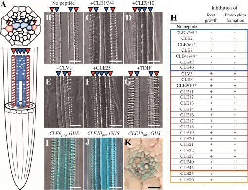
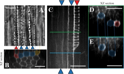
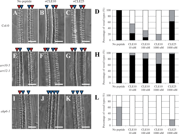
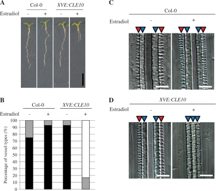
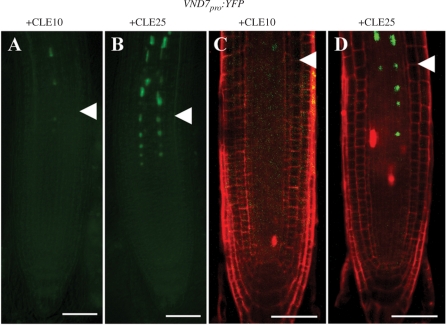
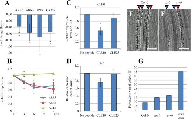
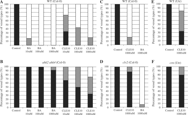
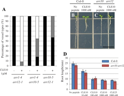
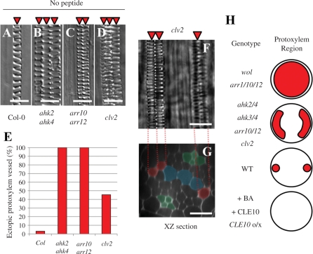
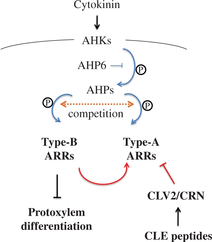
Cell-cell communication is critical for tissue and organ development. In plants, secretory CLAVATA3/EMBRYO SURROUNDING REGION-related (CLE) peptides function as intercellular signaling molecules in various aspects of tissue development including vascular development. However, little is known about intracellular signaling pathways functioning in vascular development downstream of the CLE ligands. We show that CLE peptides including CLE10, which is preferentially expressed in the root vascular system, inhibit protoxylem vessel formation in Arabidopsis roots. GeneChip analysis displayed that CLE10 peptides repressed specifically the expression of two type-A Arabidopsis Response Regulators (ARRs), ARR5 and ARR6, whose products act as negative regulators of cytokinin signaling. The arr5 arr6 roots exhibited defective protoxylem vessel formation. These results indicate that CLE10 inhibits protoxylem vessel formation by suppressing the expression of type-A ARR genes including ARR5 and ARR6. This was supported by the finding that CLE10 did not suppress protoxylem vessel formation in a background of arr10 arr12, a double mutant of type-B ARR genes. Thus, our results revealed cross-talk between CLE signaling and cytokinin signaling in protoxylem vessel formation in roots. Taken together with the indication that cytokinin signaling functions downstream of the CLV3/WUS signaling pathway in the shoot apical meristem, the cross-talk between CLE and cytokinin signaling pathways may be a common feature in plant development.


Figures

Fig. 1
Fig. 1
Effects of CLE peptides on protoxylem vessel formation and growth in the primary root. (A) A schematic vascular structure in Arabidopsis roots. Red and blue arrowheads represent protoxylem and metaxylem cell files, respectively. (B–H) Arabidopsis seeds were germinated in liquid medium with one of 26 dodeca-amino acid CLE peptides at 1 μM. Seven-day-old seedlings were collected to observe root growth and protoxylem vessel formation in roots. (B–G) Effects on protoxylem vessel formation of no peptide (B), CLE1/3/4 (C), CLE9/10 (D), CLV3 (E), CLE25 (F) and TDIF (G). (H) Twenty-six CLE peptides were classified into three groups (blue, red and orange frames) depending on their effects on the suppression of root growth and protoxylem vessel formation. Asterisks indicate synonymous names for the same peptide. For example, CLE1/3/4 means the same peptide derived from three different CLE1, CLE3 and CLE4 genes. Root growth and protoxylem formation were represented as inhibited (+) or not inhibited (−). (I–K) Vascular-specific expression of CLE9 and CLE10 in the primary root. GUS activity was observed in the root of Arabidopsis seedlings harboring a chimeric gene of the promoter of a CLE gene and the GUS gene. (I) CLE9pro:GUS. (J) CLE10pro:GUS. (K) is a cross-section of (J). Scale bars are 25 μm.
Fig. 2
Fig. 2
Confocal sections of the root vasculature. Confocal images of the protoxylem vessels and metaxylem vessels were taken from the mPS–PI-stained primary root of seedlings treated with 1 μM CLE10. (A) A bright field image. (B) A radial optical section of (A), which was constructed from Z-stack images by using image J (http://rsbweb.nih.gov/ij/). (C) A longitudinal image of a partially disrupted protoxlem vessel. (D, E) Radial optical sections of (C) at the green line position (D) and at the blue line position (E). Red and blue indicate protoxylem and metaxylem vessels, respectively. Orange indicates a cell where differentiation into protoxylem vessels has been inhibited. Scale bars are 25 μm.
Fig. 3
Fig. 3
CLE10 peptide-dependent suppression of protoxylem vessel formation in mutants defective in cytokinin signaling. (A–C, E–G, I–K) The WT, arr10-5 arr12-1 and ahp6-1 seedlings were grown for 7 d on agar plates (with 0.8% agar) containing no peptide, 1 μM CLE10 and 1 μM CLE25. (D, H, L) Dose-dependent suppression of protoxylem vessel formation by CLE10. The WT (D), arr10-5 arr12-1 (H) and ahp6-1 (L) seedlings were grown for 7 d on plates with no peptide, 10–1000 nM CLE10 and 1000 nM CLE25, and classified into three types depending on the phenotype of protoxylem vessels in the primary root. Black represents normal protoxylem vessel formation where two protoxylem cell files differentiated correctly. Gray represents a weak abnormal phenotype showing that one of the two protoxylem cell files was missing or interrupted. White represents a severe abnormal phenotype showing that both protoxylem cell files were missing or interrupted. (N = 19–30). Scale bars are 25 μm.
Fig. 4
Fig. 4
Effects of CLE10 overexpression on root growth and protoxylem vessel formation. The WT (Col-0) and XVE:CLE10-harboring seedlings were grown on agar plates (with 1.5% agar) for 7 d with (+) or without (−) 5 μM estradiol. (A) Inhibition of root growth by overexpression of CLE10. Scale bar is 5 mm. (B) Inhibition of protoxylem vessel formation by overexpression of CLE10. Black, gray and white bars are explained in Fig. 3 (N = 12–14). (C, D) Images of root vessels in the WT (C) and XVE:CLE10-harboring (D) seedlings, when treated with and without estradiol. Scale bars are 25 μm.
Fig. 5
Fig. 5
Inhibition of VND7pro:YFP expression by CLE10 peptide. (A–D) Seedlings were grown for 5 d in liquid medium with 1 μM CLE10 (A, C) or CLE25 (B, D). Fluorescence from VND7pro:YFP was detected in the non-stained primary root under a fluorescence microscope (A, B), and in the PI-stained primary root under a confocal microscope (C, D). White arrowheads represent the boundary between the RBM and the EZ. Scale bars are 50 μm.
Fig. 6
Fig. 6
Cross-talk between type-A ARRs and CLE10 signaling in protoxylem vessel formation. (A, B) Reduction of transcript levels of cytokinin-related genes including type-A ARRs by CLE10 peptide. Arabidopsis seedlings were grown in liquid medium containing 1 μM CLE10 or 1 μM CLE25 for 3–12 h, and transcript levels of cytokinin-related genes were measured by GeneChip (A) or with quantitative PCR (B). (A) Expression levels of four genes (ARR5, ARR6, IPT7 and CKX3) were significantly decreased by 12 h treatment of CLE10. The fold change was calculated as the ratio of transcript levels of plants treated with CLE10 compared with CLE25 (in log2 scale). Asterisks indicate statistically significant differences (P < 0.1). (B) Changes in two type-A ARR transcripts by 3, 6 and 12 h treatment of 1 μM CLE10 or CLE25. ACT2 was used as a control. The relative expression was calculated as the ratio of transcript levels of plants treated with CLE10 when compared with CLE25. Asterisks indicate statistically significant differences (P < 0.01). (C, D) ARR5 expression levels of plants treated with no peptide, 1 μM CLE10 or 1 μM CLE25 for 6 h in the WT (C) or clv2 (D). The relative expression was calculated as the ratio of transcript levels of plants treated with CLE peptides when compared with no peptide. Asterisks indicate statistically significant differences when compared with no peptide treatment (P < 0.01). (A–D) Error bars represent SD (N = 3). (E–G) Defects in protoxylem vessel formation in lateral roots. (E, F) Protoxylem vessel formation in lateral roots of 8-day-old WT and arr5 arr6 seedlings. (G) The frequency of suppressed protoxylem vessel formation in lateral roots of WT, arr5, arr6, arr5 arr6 (N = 32–53). One to four lateral roots located next to the hypocotyl, per seedling, were used for measurement. Scale bars are 25 μm.
Fig. 7
Fig. 7
Effects of BA and CLE10 peptide on protoxylem vessel formation in ahk2 ahk4, clv2 and crn. (A, B) Seedlings of WT (A) and ahk2 ahk4 (B) were grown in liquid medium containing BA and CLE10 at 10, 100 or 1000 nM for 7 d. (C–F) Seedlings of WT (Col-0) (C), clv2 (D), WT (Utr) (E) and crn (F) were treated with no peptide, BA or CLE10 at 1000 nM for 7 d. Black, gray and white bars are explained in Fig. 3 (N = 12–18).
Fig. 8
Fig. 8
Different sensitivities to CLE10 among three type-B ARR double mutants. (A) Seedlings of arr1 arr12, arr1 arr10 and arr10 arr12 were grown in liquid medium with or without 1 μM CLE10 for 7 d, and the numbers of primary roots with defects in protoxylem vessels was counted. Black, gray and white bars are explained in Fig. 3 (N = 7–12). (B–D) CLE10-induced root growth inhibition in arr10 arr12. The WT and arr10 arr12 seedlings were grown for 7 d on agar plates (with 1.5% agar) containing no peptide and 10–1000 nM CLE10. (A, B) Seedlings of the WT (A) and arr10 arr12 (B) grown with no peptide (left) and with 1 μM CLE10 (right). (C) Root length in WT and arr10 arr12 seedlings. Error bars represent SD (N = 7–11).
Fig. 9
Fig. 9
Ectopic protoxylem vessel formation in the primary root of mutants. (A–D) Protoxylem vessel formation in the WT (A), ahk2 ahk4 (B), arr10 arr12 (C) and clv2 (D). Arabidopsis roots exhibit diarch symmetry of the vascular bundle in which a protoxylem vessel file per arch is formed. However, ahk2 ahk4, arr10 arr12 and clv2 roots often have two or three protoxylem vessel files per arch. (E) The frequency of the primary root with ectopic protoxylem vessel files in the WT, ahk2 ahk4, arr10 arr12 and clv2 (N = 32–33). (F, G) Confocal images of the primary root of clv2. (F) A bright field image of the mPS–PI-stained stele. (G) An optical section of (F). Red and green indicate protoxylem vessels and phloem cells, respectively. Blue indicates undifferentiated metaxylem vessel cells. (H) Protoxylem vessel phenotypes in the primary root caused by loss-of-function and gain-of-function of CLE and cytokinin signaling. Circles show the root stele. Red indicates the area that protoxylem vessel cells occupy. CLE10o/x shows plants in which CLE10 is overexpressed. Scale bars are 25 μm.
Fig. 10
Fig. 10
A model illustrating the cross-talk between cytokinin and CLE peptide signaling pathways to suppress protoxylem vessel formation. CLE peptides positively regulate cytokinin signaling via type-A ARRs in protoxylem vessel formation. Blue and red lines indicate phosphorylational and transcriptional regulation, respectively.

Similar articles

Cited by

References

    1. Argyros R.D., Mathews D.E., Chiang Y.H., Palmer C.M., Thibault D.M., Etheridge N., et al. Type B response regulators of Arabidopsis play key roles in cytokinin signaling and plant development. Plant Cell. 2008;20:2102–2116. - PMC - PubMed
    1. Bleckmann A., Weidtkamp-Peters S., Seidel C.A., Simon R. Stem cell signaling in Arabidopsis requires CRN to localize CLV2 to the plasma membrane. Plant Physiol. 2010;152:166–176. - PMC - PubMed
    1. Brand U., Grünewald M., Hobe M., Simon R. Regulation of CLV3 expression by two homeobox genes in Arabidopsis. Plant Physiol. 2002;129:565–575. - PMC - PubMed
    1. Butenko M.A., Vie A.K., Brembu T., Aalen R.B., Bones A.M. Plant peptides in signalling: looking for new partners. Trends Plant Sci. 2009;14:255–263. - PubMed
    1. Carlsbecker A., Lee J.Y., Roberts C.J., Dettmer J., Lehesranta S., Zhou J., et al. Cell signalling by microRNA165/6 directs gene dose-dependent root cell fate. Nature. 2010;465:316–321. - PMC - PubMed

Publication types

MeSH terms