. 2010 Dec;59(12):3090-8.
doi: 10.2337/db10-0624. Epub 2010 Aug 29.

Conditional gene targeting in mouse pancreatic ß-Cells: analysis of ectopic Cre transgene expression in the brain

Affiliations

Conditional gene targeting in mouse pancreatic ß-Cells: analysis of ectopic Cre transgene expression in the brain

Barton Wicksteed et al. Diabetes. 2010 Dec.

Abstract

Objective: Conditional gene targeting has been extensively used for in vivo analysis of gene function in β-cell biology. The objective of this study was to examine whether mouse transgenic Cre lines, used to mediate β-cell- or pancreas-specific recombination, also drive Cre expression in the brain.

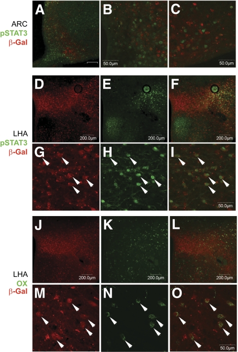
Research design and methods: Transgenic Cre lines driven by Ins1, Ins2, and Pdx1 promoters were bred to R26R reporter strains. Cre activity was assessed by β-galactosidase or yellow fluorescent protein expression in the pancreas and the brain. Endogenous Pdx1 gene expression was monitored using Pdx1(tm1Cvw) lacZ knock-in mice. Cre expression in β-cells and co-localization of Cre activity with orexin-expressing and leptin-responsive neurons within the brain was assessed by immunohistochemistry.

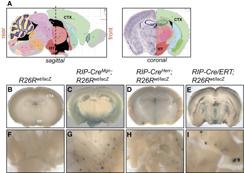
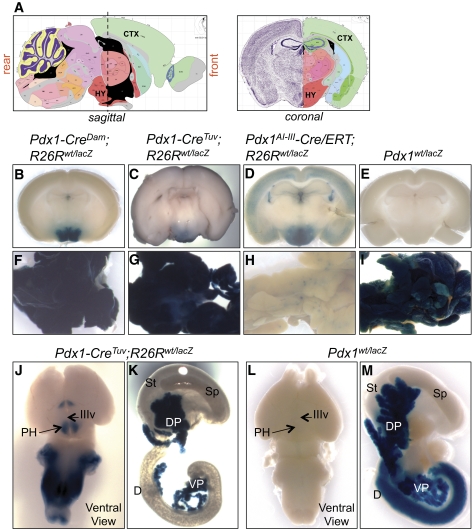
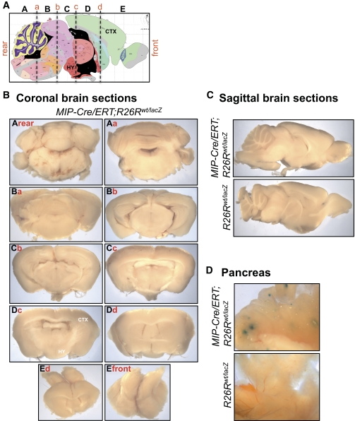
Results: All transgenic Cre lines examined that used the Ins2 promoter to drive Cre expression showed widespread Cre activity in the brain, whereas Cre lines that used Pdx1 promoter fragments showed more restricted Cre activity primarily within the hypothalamus. Immunohistochemical analysis of the hypothalamus from Tg(Pdx1-cre)(89.1Dam) mice revealed Cre activity in neurons expressing orexin and in neurons activated by leptin. Tg(Ins1-Cre/ERT)(1Lphi) mice were the only line that lacked Cre activity in the brain.

Conclusions: Cre-mediated gene manipulation using transgenic lines that express Cre under the control of the Ins2 and Pdx1 promoters are likely to alter gene expression in nutrient-sensing neurons. Therefore, data arising from the use of these transgenic Cre lines must be interpreted carefully to assess whether the resultant phenotype is solely attributable to alterations in the islet β-cells.


Figures

FIG. 1.
FIG. 1.
RIP-Cre transgenic lines display Cre-mediated recombination in multiple regions of the brain. Adult brains were sliced into four or five coronal sections and subjected to whole mount X-gal staining. Images of individual brain slices from each sectioning plane are available in supplementary Figs. 1–4. A: Sagittal and coronal views of mouse brain (adapted from Allen Mouse Brain Atlas, http://www.brain-map.org/) (29). A dashed vertical line marks coronal sectioning plane spanning the hypothalamic region of the brain. B–E: Images of coronal brain slices located on the left side of the sectioning plane in the sagittal view in A. B: R26Rwt/lacZ littermate control mice (n = 17) lacked X-gal staining in the brain. The cortex (CTX) and hypothalamus (HY) are labeled and correspond to regions marked on the coronal view in A. C: RIP-CreMgn;R26Rwt/lacZ mice (n = 8) showed X-gal staining throughout the brain with high signal intensity in the mid-brain and ventral regions. D: RIP-CreHerr;R26Rwt/lacZ mice (n = 14) showed weaker, punctate X-gal staining throughout the brain without obvious regionalization. E: RIP-Cre/ERT;R26Rwt/lacZ mice (n = 4) injected intraperitoneally with three 2-mg doses of tamoxifen over a 5-day period displayed strong, punctate X-gal staining throughout the brain with expression pattern more restricted than in RIP-CreMgn; R26Rwt/lacZ mice. Brains from littermate controls injected with corn oil vehicle were negative for X-gal staining (data not shown). F–I: Whole-mount X-gal staining of pancreas from R26Rwt/lacZ in F, RIP-CreMgn; R26Rwt/lacZ in H, RIP-CreHerr;R26Rwt/lacZ in G, and RIP-Cre/ERT;R26Rwt/lacZ mice in I. (A high-quality digital representation of this figure is available in the online issue.)
FIG. 2.
FIG. 2.
Pdx1-Cre transgenic lines show localized Cre-mediated recombination within specific regions of the brain including the hypothalamus. Adult brains were sliced, labeled, and imaged as described in Fig. 1. Images of individual brain slices from each sectioning plane are available in supplementary Figs. 5–7 and supplementary Fig. 9. A: Sagittal and coronal views of mouse brain. A dashed vertical line marks coronal sectioning plane spanning hypothalamic region of the brain. B–D: Images of coronal brain slices located on the left side of sectioning plane in the sagittal view in A. The schematics of the mouse brain are from the Allen Mouse Brain Atlas (http://www.brain-map.org/) (29). B: X-gal staining in Pdx1-CreDam;R26Rwt/lacZ brain (n = 7) was localized to the brain stem and hypothalamus. C: X-gal positive cells in Pdx1-CreTuv;R26Rwt/lacZ brain (n = 4) were localized to hypothalamic region. D: Adult Pdx1AI-III-Cre/ERT;R26Rwt/lacZ mice (n = 4) were injected subcutaneously with three 8-mg doses of tamoxifen (right panel) and analyzed for lacZ expression. X-gal staining had a broader punctate pattern with high-intensity signal localized to the hypothalamic region. Brains from littermate controls injected with corn oil vehicle (n = 2) were negative for X-gal staining (data not shown). E: Brains from adult Pdx1lacZ/wt mice (n = 4) were negative for X-gal staining. F–I: Whole-mount X-gal staining of pancreas from Pdx1-CreDam;R26Rwt/lacZ in F, Pdx1-CreTuv;R26Rwt/lacZ in G, Pdx1AI-III-Cre/ERT;R26Rwt/lacZ mice in H, and Pdx1lacZ/wt mice in I. J and K: Brains from Pdx1-CreTuv;R26Rwt/lacZ embryos at e15.5 (n = 6) in J were analyzed for LacZ expression. X-gal staining indicated expression of Pdx1-CreTuv transgene in the brain stem and ventral region of the brain that gives rise to the hypothalamus (arrows). Pancreas in K had expected X-gal staining. Similar results were obtained using the R26RYFP reporter strain in supplementary Fig. 8. Brain and pancreas from R26Rwt/lacZ (n = 5) and R26Rwt/YFP (n = 6) e15.5 controls were negative for X-gal staining and YFP fluorescence, respectively (supplementary Fig. 8). L and M: Brains from e15.5 Pdx1lacZ/wt embryos (n = 7) in L were negative for X-gal staining, while pancreas showed expected X-gal positivity in M. In e15.5 Pdx1wt/wt embryos (n = 10), both brain and pancreas were X-gal negative (supplemental Fig. 9). CTX, cortex; D, duodenum; DP, dorsal pancreas; HY, hypothalamus; IIIv, third ventricle; PH, posterior hypothalamic region; Sp, spleen; St, stomach; VP, ventral pancreas. (A high-quality digital representation of this figure is available in the online issue.)
FIG. 3.
FIG. 3.
Pdx1-CreDam;R26Rwt/lacZ mice display a complex pattern of Cre-mediated recombination that partially overlaps with orexin-positive and leptin-responsive neuronal populations. Adult Pdx1-CreDam;R26Rwt/lacZ mice were treated with leptin (5 mg/kg, intraperitoneally, 2 h), perfusion-fixed and brains isolated for immunohistochemical detection of pSTAT3, orexin, and β-gal positive. Localization of β-gal signal in brain sections of adult Pdx1-CreDam;R26Rwt/lacZ mice is available in supplementary Fig. 10. A–C: pSTAT3 (green) and β-gal (red) immunoreactivity do not co-localize efficiently in the arcuate nucleus (ARC). D–I: Co-localization of pSTAT3 (green) and β-gal (red) immunoreactivity in a subpopulation of neurons in the lateral hypothalamus (LHA). Despite extensive β-gal labeling within the preoptic area, there was essentially no co-localization with leptin-responsive neurons (data not shown). J–O: Co-localization of orexin (green) and β-gal (red) immunoreactivity within neurons in the LHA. Arrows indicate co-labeled neurons. All scale bars are either 50 μm or 200 μm (as indicated). The unlabeled scale bar in the ARC panel in A is 50 μm. (A high-quality digital representation of this figure is available in the online issue.)
FIG. 4.
FIG. 4.
Cre activity is undetectable in MIP-Cre/ERT brain. Mice were injected intraperitoneally with three 2-mg doses of tamoxifen over a 5-day period. Brains from adult MIP-Cre/ERT; R26Rwt/lacZ (n = 5) and their R26Rwt/lacZ littermates (n = 5) (24) were sliced into five coronal sections and subjected to whole-mount X-gal staining. A: Sagittal view of mouse brain. The schematic of the mouse brain is from the Allen Mouse Brain Atlas (http://www.brain-map.org/) (29). Brain slices examined are shown in capital letters (A, B, C, etc.). Vertical dashed lines mark coronal sectioning plane designated as face in lowercase letters (a, b, c, etc.). B: Images of individual brain slices from each coronal sectioning plane. C: Sagittal brain sections from MIP-Cre/ERT; R26Rwt/lacZ (top panel) and R26Rwt/lacZ littermates (bottom panel). D: Whole-mount X-gal staining of pancreas from MIP-Cre/ERT; R26Rwt/lacZ (top panel) and R26Rwt/lacZ littermates (bottom panel). Brains from MIP-Cre/ERT; R26Rwt/lacZ mice and controls in B and C were negative for X-gal staining, while MIP-Cre/ERT; R26Rwt/lacZ pancreas showed robust X-gal labeling in the islets in D. CTX, cortex; HY, hypothalamus. (A high-quality digital representation of this figure is available in the online issue.)

Comment in

Similar articles

Cited by

References

    1. Fawell SE, Lees JA, White R, Parker MG: Characterization and colocalization of steroid binding and dimerization activities in the mouse estrogen receptor. Cell 1990;60:953–962 - PubMed
    1. Hayashi S, McMahon AP: Efficient recombination in diverse tissues by a tamoxifen-inducible form of Cre: a tool for temporally regulated gene activation/inactivation in the mouse. Dev Biol 2002;244:305–318 - PubMed
    1. Gannon M, Gamer LW, Wright CV: Regulatory regions driving developmental and tissue-specific expression of the essential pancreatic gene pdx1. Dev Biol 2001;238:185–201 - PubMed
    1. Wiebe PO, Kormish JD, Roper VT, Fujitani Y, Alston NI, Zaret KS, Wright CV, Stein RW, Gannon M: Ptf1a binds to and activates area III, a highly conserved region of the Pdx1 promoter that mediates early pancreas-wide Pdx1 expression. Mol Cell Biol 2007;27:4093–4104 - PMC - PubMed
    1. Whelan J, Poon D, Weil PA, Stein R: Pancreatic beta-cell-type-specific expression of the rat insulin II gene is controlled by positive and negative cellular transcriptional elements. Mol Cell Biol 1989;9:3253–3259 - PMC - PubMed

Publication types

MeSH terms

Grants and funding