. 2010 Oct 6;29(19):3318-29.
doi: 10.1038/emboj.2010.203. Epub 2010 Aug 27.

Disulphide production by Ero1α-PDI relay is rapid and effectively regulated

Affiliations

Disulphide production by Ero1α-PDI relay is rapid and effectively regulated

Christian Appenzeller-Herzog et al. EMBO J. .

Abstract

The molecular networks that control endoplasmic reticulum (ER) redox conditions in mammalian cells are incompletely understood. Here, we show that after reductive challenge the ER steady-state disulphide content is restored on a time scale of seconds. Both the oxidase Ero1α and the oxidoreductase protein disulphide isomerase (PDI) strongly contribute to the rapid recovery kinetics, but experiments in ERO1-deficient cells indicate the existence of parallel pathways for disulphide generation. We find PDI to be the main substrate of Ero1α, and mixed-disulphide complexes of Ero1 primarily form with PDI, to a lesser extent with the PDI-family members ERp57 and ERp72, but are not detectable with another homologue TMX3. We also show for the first time that the oxidation level of PDIs and glutathione is precisely regulated. Apparently, this is achieved neither through ER import of thiols nor by transport of disulphides to the Golgi apparatus. Instead, our data suggest that a dynamic equilibrium between Ero1- and glutathione disulphide-mediated oxidation of PDIs constitutes an important element of ER redox homeostasis.


Conflict of interest statement

The authors declare that they have no conflict of interest.

Figures

Figure 1
Figure 1
Vesicular transport, glutathione concentration and protein translocation only moderately influence ER redox homeostasis. (A) HEK293 cells pre-treated with BFA/mon for 0.5 h or left untreated were incubated with 5 mM diamide (dia) for 5 min, washed twice with PBS and incubated in the same buffer for 0, 5, 10 or 15 min (wo, washout). BFA/mon was present throughout the time course. For comparison, steady-state samples±treatment with BFA/mon for 0.5 h were included. The reactions were stopped by rinsing the cells in ice-cold PBS containing 20 mM NEM, and the redox distributions of TMX3 and ERp57 visualized by western blotting (WB) after differential alkylation with NEM and AMS. The mobility of the oxidized (ox) and reduced (red) forms of TMX3 and ERp57 are indicated. (B) Recovery of the TMX3 redox ratio assessed by densitometry in BFA/mon-treated and control cells as shown in panel A (n=3, mean±s.d.). (C) The redox states of TMX3 and ERp57 in HEK293 cells depleted of glutathione (using BSO) or nascent proteins (using CHX). Treatment with BSO reduced cellular glutathione levels to 20% of control (data not shown). Oxidized fractions (%) as determined by densitometry are indicated. Lanes labelled with dia represent oxidized control lanes using diamide-treated cell lysates. The hairlines indicate where intervening lanes have been removed. The results are representative of three independent experiments.
Figure 2
Figure 2
In vivo DTT resistance of PDI, TMX3 and ERp57. (A) HEK293 cells were treated with the indicated concentrations of DTT, and the in vivo redox state of the two active sites in PDI (a and a′) determined by immunoprecipitation (IP) of 35S-labelled and mPEG-mal-modified PDI. Samples completely reduced with DTT and TCEP, or oxidized with diamide (dia) served as mobility markers. The contrast enhancement of the region marked by the rectangle more clearly shows the different behaviour of the two semi-oxidized forms of PDI (a domain oxidized/a′ domain reduced (a-ox); a domain reduced/a′ domain oxidized (a′-ox)), for which we have previously determined the relative mobility (Appenzeller-Herzog and Ellgaard, 2008a). One of two independent experiments with equal outcome is shown. Red, both active sites reduced; ox, both active sites oxidized; asterisks, reduced PDI modified with mPEG-mal on its non-catalytic cysteines (Appenzeller-Herzog and Ellgaard, 2008a). (B) After treatment of HEK293 cells with the indicated concentrations of DTT, the in vivo redox states of TMX3 and ERp57 were determined as in Figure 1A. The mobilities of the oxidized (ox) and reduced (red) species, as verified by control samples using lysates from diamide- or DTT (104 μM)-treated cells, are marked. (C) Densitometric analysis of (B) (n=3, mean±s.d.).
Figure 3
Figure 3
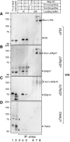
Mixed-disulphide interactions of PDIs with Ero1α/β. Doxycyclin-induced and in situ acid-trapped negative control (Neg ctrl), Ero1αmyc6his and Ero1βmyc6his cells were subjected to αmyc immunoprecipitation (IP) followed by reducing (R) or non-reducing (NR) SDS–PAGE and western blot (WB) analysis using αPDI (A), αERp57 (B), αERp72 (C) or αTMX3 (D). The 35S-signal recorded by phosphorimaging of one of the membranes is shown in Supplementary Figure S2B. As positive controls for western blotting, 1% of the Ero1αmyc6his lysate (1% of total) as well as reduced and non-labelled HEK293 lysate (cold lysate) were loaded in lanes 1 and 2. The results are representative of two independent experiments. Filled arrowheads, monomeric PDIs; open arrowheads, dimeric mixed-disulphide complexes of PDIs with Ero1 (the precise mobility of which is unclear in the case of ERp72; indicated by a vertical line); asterisks, potential mixed-disulphide complexes of PDIs with Ero1α dimers; X, background bands.
Figure 4
Figure 4
ER reoxidation after DTT treatment is fast and affected by exogenous Ero1. (A) Intracellular levels of GSSG and GStot were recorded from DTT-treated HEK293 cells after washout of the reductant for 0, 10, 30 s, 1, 3, 5 or 20 min. The GSSG:GStot ratio is expressed as percentage of the steady-state value that was independently measured (mean±s.d., n=8, for individual experiments, see Supplementary Figure S3A). (B) Negative control cells were grown on plastic coverslips, treated with or without doxycyclin (dox) for 24 h, and left untreated (−) or incubated with DTT. After 0, 5, 10 or 20 s of DTT washout (wo), the cells were processed for AMS alkylation and western blotting (WB) using αTMX3 or αERp57. Ox, oxidized species; red, reduced species; dia, oxidized control lane using diamide-treated cells. (CF) DTT washout assays followed by the determination of cellular levels of GSSG and GStot after 0, 10, 60 or 300 s using Ero1αmyc6his (C), Ero1βmyc6his (D), Ero1αmyc6his–C131A (E) and Ero1αmyc6his–C394A (F) cells cultured for 24 h with or without (control) the addition of doxycyclin (mean±s.d., two independent experiments each performed in triplet; Supplementary Figures S3D–G). *P<0.05; **P<0.01; ***P<0.001 (Student's t-test). Notice the different scaling on the y axis in the individual panels.
Figure 5
Figure 5
Rapid oxidative recovery of the ER depends on Ero1 and PDI. (A) Lysates of Ero1 wild type (+/+;+/+) or double mutant (i/i;i/i) mouse embryonic fibroblasts (MEFs) were analysed by reducing (R) or non-reducing (NR) SDS–PAGE and αEro1α western blotting after ConA precipitation. The gel mobilities of the three known redox forms of Ero1α (R, OX1, OX2) are indicated. (B) Redox state analysis of ERp57 after DTT washout using Ero1 wild-type (+/+;+/+) or double mutant (i/i;i/i) MEFs. The experiment was performed as in Figure 4B and Supplementary Figure S4B except that oxidative recovery was allowed for longer periods. Open arrowheads indicate the delayed formation of oxidized ERp57 in double mutant cells. (C) Densitometric analysis of (B) (n=3, mean±s.d.). (D) GSSG:GStot was determined in Ero1 wild-type (+/+;+/+) or double mutant (i/i;i/i) MEFs at the indicated intervals after DTT washout (mean±s.d., three independent experiments each performed in triplet; Supplementary Figure S4D). For unknown reasons, GSSG:GStot rises above the steady-state value after 300 s of oxidative recovery in wild-type cells. The GSSG:GStot ratios in Ero1 wild-type and double mutant MEFs at steady state are shown in the inset (n=12). (E) Redox state analysis of TMX3 and ERp57 after DTT washout performed as in Figure 4B, but using PDI shRNA clones 5-1 (control cells) and 4-1 (PDI knockdown (kd) cells). Open arrowheads indicate the delayed formation of oxidized TMX3/ERp57 upon knockdown of PDI. (F) Densitometric analysis of (D) (n=3, mean±s.d.). (G, H) The oxidative recovery of GSSG:GStot after DTT washout was determined as in panel D using clone 5-1 (control) and clone 4-1 (PDI kd) cells (G) or 2175+ (control) and 2175 (ERp57 ko) cells (H) (mean±s.d., two independent experiments each performed in triplet; Supplementary Figure S4E and F). For unknown reasons, GSSG:GStot rises above the steady-state value after 300 s of oxidative recovery in 2175+ and 2175 cells. *P<0.05; **P<0.01; ***P<0.001 (Student's t-test).
Figure 6
Figure 6
Upon reductive challenge, activated Ero1α rapidly reacts with PDI. (A) Co-immunoprecipitation performed in analogy to the experiment presented in Figure 3A except that, where indicated, cells were treated with DTT ahead of TCA lysis. A phosphoimager scan (IP: αmyc (Ero1)) and a western blot (WB) using αPDI are shown. The mobility differences between the Ero1–PDI mixed-disulphide complexes (Ero1+PDI) under steady-state conditions (OX.) and upon DTT-mediated reduction (RED.) are marked. For unknown reasons, the intensity of WB detection of Ero1+PDI did not reflect the relative intensities observed by phosphorimaging. Note that an NEM- and redox state-dependent mobility shift of Ero1α in reducing SDS–PAGE (compare lanes 2 and 3; see also panel B, WB: αEro1α) has been reported previously (Benham et al, 2000). The result is representative of two independent experiments. Filled arrowhead, monomeric PDI; asterisks, Ero1α/βmyc6his–ERp57 mixed-disulphide complex (inferred from Supplementary Figure S5); X, background band. (B) TCA pellets from HEK293 cells incubated with DTT or left untreated were solubilized/neutralized in the presence of NEM, and Ero1α was precipitated from the lysate using ConA-sepharose (Benham et al, 2000). The precipitate was boiled under reducing (R) or non-reducing (NR) conditions and analysed by αPDI western blotting (WB, left panel). After stripping, the membrane was probed with αEro1α (right panel). The mobilities of PDI, the known monomeric redox forms of Ero1α (R, OX1, OX2; visible upon contrast enhancement) and of the Ero1α+PDI complex (both RED. and OX.) are indicated. The results are representative of three independent experiments. Asterisk, potential mixed-disulphide complex of PDI with an Ero1α dimer; double asterisk, unidentified, DTT-resistant mixed-disulphide complex. (C) Phosphorimager scan of a co-immunoprecipitation experiment performed as in panel A. In addition to doxycyclin-induced Ero1αmyc6his cells (dox), HEK293 transiently transfected (cDNA) with pcDNA3/Ero1αmyc6his or pcDNA3/Ero1αmyc6his-C94S were used. For unknown reasons, the monomeric form of transiently transfected Ero1αmyc6his is more exposed than stably transfected Ero1αmyc6his to DTT-mediated reduction (as indicated by enhanced conversion of OX2 into more reduced, slower migrating forms). The result is representative of two independent experiments. Asterisk, Ero1αmyc6his–ERp57 mixed-disulphide complex (compare Supplementary Figure S5).
Figure 7
Figure 7
Model for glutathione-buffered ER redox homeostasis. Graphical depiction of two disulphide relay pathways that both lead to the oxidation of nascent proteins (substrate) in the ER. (A) In the Ero1α-driven oxidation pathway for de novo disulphide formation, oxidizing equivalents are transferred from O2 to Ero1α that in turn oxidizes PDI. The byproduct H2O2 (Wang et al, 2009; Enyedi et al, 2010) can also oxidize PDI yielding two molecules of H2O (Karala et al, 2009). A potential in vivo catalyst of this reaction remains to be identified (question mark). Abundant levels of reduced PDI keep Ero1α in an active state (green arrow) (Appenzeller-Herzog et al, 2008). Being the main substrate of Ero1α, disulphides are passed on primarily to PDI, but other PDI-family members (PDIs) may also participate to some extent in this pathway. GSH competes with substrate for reaction with oxidized PDI, resulting in the formation of GSSG. (B) GSSG-driven oxidation of reduced PDIs (yellow arrows) will be prominent when ER GSSG is abundant, which will also promote shutdown of Ero1α because of low availability of reduced PDI. Similar to the Ero1α-driven oxidation pathway, the PDIs will then oxidize substrate proteins (blue arrows). The interplay between the two pathways depends on the redox state of the glutathione redox couple in the ER. For instance, during oxidative recovery after DTT treatment de novo disulphide generation is dominant immediately after DTT washout. However, as GSSG levels rise, the GSSG-driven oxidation pathway will become increasingly more prominent until homeostasis is reinstalled. For simplicity, the scheme only illustrates the net flow of oxidizing equivalents onto substrate and excludes the reduction of, for example, aberrantly disulphide-bonded substrates by PDIs. Likewise, the direct reaction of GSSG with reduced substrates that results in glutathionylated substrates (Bass et al, 2004; Hansen et al, 2009) has been omitted. The model does not account for the contribution to ER thiol-disulphide homeostasis by Ero1-independent pathway(s) as the exact nature of these is not yet known. Red, reduced; ox, oxidized.

References

    1. Appenzeller-Herzog C, Ellgaard L (2008a) In vivo reduction-oxidation state of protein disulfide isomerase: the two active sites independently occur in the reduced and oxidized forms. Antioxid Redox Signal 10: 55–64 - PubMed
    1. Appenzeller-Herzog C, Ellgaard L (2008b) The human PDI family: versatility packed into a single fold. Biochim Biophys Acta 1783: 535–548 - PubMed
    1. Appenzeller-Herzog C, Riemer J, Christensen B, Sorensen ES, Ellgaard L (2008) A novel disulphide switch mechanism in Ero1alpha balances ER oxidation in human cells. EMBO J 27: 2977–2987 - PMC - PubMed
    1. Baker KM, Chakravarthi S, Langton KP, Sheppard AM, Lu H, Bulleid NJ (2008) Low reduction potential of Ero1alpha regulatory disulphides ensures tight control of substrate oxidation. EMBO J 27: 2988–2997 - PMC - PubMed
    1. Banhegyi G, Lusini L, Puskas F, Rossi R, Fulceri R, Braun L, Mile V, di Simplicio P, Mandl J, Benedetti A (1999) Preferential transport of glutathione versus glutathione disulfide in rat liver microsomal vesicles. J Biol Chem 274: 12213–12216 - PubMed

Publication types