. 2010 Sep;17(9):1144-51.
doi: 10.1038/nsmb.1899. Epub 2010 Aug 29.

Human HDAC1 and HDAC2 function in the DNA-damage response to promote DNA nonhomologous end-joining

Affiliations

Human HDAC1 and HDAC2 function in the DNA-damage response to promote DNA nonhomologous end-joining

Kyle M Miller et al. Nat Struct Mol Biol. 2010 Sep.

Abstract

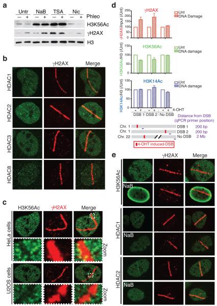
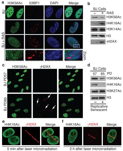
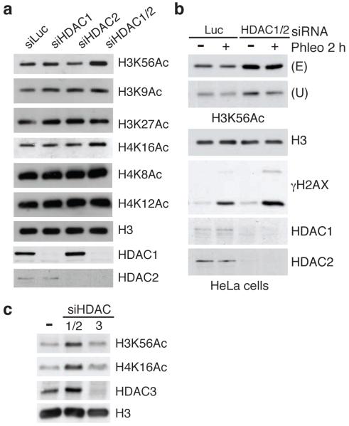
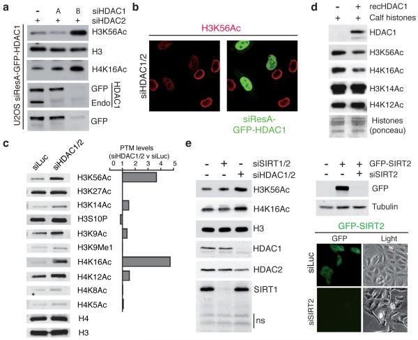
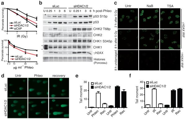
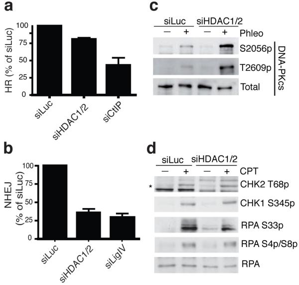
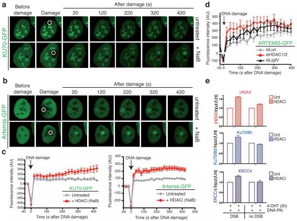
DNA double-strand break (DSB) repair occurs within chromatin and can be modulated by chromatin-modifying enzymes. Here we identify the related human histone deacetylases HDAC1 and HDAC2 as two participants in the DNA-damage response. We show that acetylation of histone H3 Lys56 (H3K56) was regulated by HDAC1 and HDAC2 and that HDAC1 and HDAC2 were rapidly recruited to DNA-damage sites to promote hypoacetylation of H3K56. Furthermore, HDAC1- and 2-depleted cells were hypersensitive to DNA-damaging agents and showed sustained DNA-damage signaling, phenotypes that reflect defective DSB repair, particularly by nonhomologous end-joining (NHEJ). Collectively, these results show that HDAC1 and HDAC2 function in the DNA-damage response by promoting DSB repair and thus provide important insights into the radio-sensitizing effects of HDAC inhibitors that are being developed as cancer therapies.


Figures

Figure 1
Figure 1
HDAC1 and HDAC2 localize to, and H3K56Ac is reduced at, sites of DNA damage. (a) Treatment with the Class I/II HDAC inhibitors sodium butyrate (NaB) or Trichostatin A (TSA) increases H3K56Ac levels and blocks damage-dependent deacetylation of H3K56. U2OS cells were untreated or treated with HDAC inhibitors followed by DNA damage induction by phleomycin. Samples were analyzed by western blotting (WB) with the indicated antibodies; γH2AX detection denotes DNA damage. (b) The Class I HDACs, HDAC1 and HDAC2, localize to sites of laser-induced DNA damage. DNA damage was generated by laser micro-irradiation followed by immunofluorescence (IF, 5 min after damage) with the indicated antibodies. γH2AX marks the “laser-lines” containing damaged DNA. (c) H3K56Ac levels are reduced at DNA-damage sites in both Hela and U2OS cells. DNA damage was generated as in b followed by IF (2 h) for H3K56Ac and γH2AX. Boxes show zoomed sections of damaged areas. (d) H3K56Ac levels are reduced at site-specific DSBs. Site-specific DSBs were induced by addition of 4-OHT into U2OS cells containing AsiSI-ER. 4-OHT treated or untreated cells were analyzed by ChIP followed by qPCR. AsiSI sites (DSB1 and DSB2) were compared to locus lacking AsiSI sites (no DSB). For H3 acetylations, qPCR values were normalized to input and total H3. All values were then normalized to untreated samples for each respective antibody and genomic locus (n=3, error bars represent S.E.M.). (e) H3K56Ac loss, and HDAC1 and HDAC2 localization, at DNA-damage sites is blocked by HDAC inhibitors. Analysis was performed as in a and b.
Figure 2
Figure 2
H3K56Ac levels decrease during oncogene-induced and replicative senescence and H4K16Ac is a DNA-damage responsive histone mark. (a) H3K56Ac levels are reduced in 53BP1-positive, RAS-overexpressing OIS cells. Human BJ primary fibroblast cells untreated or undergoing OIS were analyzed by IF with the indicated antibodies. Note that H3K56Ac is excluded from 53BP1 foci. (b) H3K56Ac is reduced and H4K16Ac is increased in OIS cells as analyzed by WB from cells in a. H3K56Ac is reduced in replicative-senescent BJ cells (arrows) by IF (c) and WB (d). PD: population doublings. (e and f) H4K16Ac levels rapidly decrease (5 min) then increase (2 h) at sites of damage following laser-microirradiation. Cells were analyzed as in Fig. 1b.
Figure 3
Figure 3
HDAC1 and HDAC2, co-regulate H3K56Ac and H4K16Ac. (a) siRNA depletion of HDAC1/2 together, but not singularly, causes H3K56 and H4K16 hyper-acetylation in U2OS cells; siLuciferase (siLuc) used as control. After siRNA transfections, cells were analyzed by WB. (b) HDAC1/2 regulates H3K56Ac in HeLa cells. Experiments were performed as in Figure 1a. An increase in H3K56Ac in siHDAC1/2-depleted cells was confirmed with two antibodies against H3K56Ac (Upstate and Epitomics). (b) The down-regulation of the Class I HDAC, HDAC3, does not affect H3K56Ac or H4K16Ac levels. Cells were analyzed as in a.
Figure 4
Figure 4
HDAC1 and HDAC2 specifically and directly target H3K56 and H4K16 (a) siResistant HDAC1 (siResA-GFP-HDAC1) rescues defects of H3K56 and H4K16 hyper-acetylation in siHDAC1/2 U2OS cells. (see methods for details; Endo = endogenous protein). (b) U2OS cells expressing siResA-GFP-HDAC1 rescues H3K56 hyper-acetylation in cells depleted for endogenous HDAC1/2. Cells were treated with the indicated siRNAs and analyzed by IF with a H3K56Ac antibody (Epitomics). (c) HDAC1/2-depleted U2OS cells exhibit H3K56 and H4K16 hyper-acetylation. Quantification of samples in a was performed with the LI-COR Odyssey infrared imaging system; ratios of signals in siHDAC1/2 versus siLuc are given and histone-modification levels were normalized to histone H3 or H4 levels. (d) Recombinant HDAC1 deacetylates H3K56 and H4K16 in vitro. (e) Depletion of SIRT1/2 does not increase H3K56Ac and H4K16Ac levels in HeLa cells. Experiments were performed as in a. siRNA sequences targeting SIRT1 and SIRT2 were obtained from a previous study. Lower right panel confirms the documented cytoplasmic localization of SIRT2 and the efficient depletion of GFP-SIRT2 by fluorescence microscopy.
Figure 5
Figure 5
HDAC1/2-depleted cells exhibit defective DNA-damage responses. (a) HDAC1/2-depleted cells exhibit hypersensitivity to IR and phleomycin; data are from triplicate experiments, and CtIP acts as a positive control; error bars, +/− S.D. (b) HDAC1/2-depleted cells exhibit increased DDR signaling as monitored by WB; samples were from untreated (U) cells or cells harvested at the indicated times after release from a 2 h phleomycin-treatment (*cross-reacting proteins). (c) HDAC inhibition following IR causes increased γH2AX. Immediately after IR exposure, cells were incubated in the absence (Untr) or presence of NaB (5 mM) or TSA (1.3 mM), and then were analyzed by IF 2 h or 8 h later. Undamaged cells were untreated or treated with HDAC inhibitors and analyzed at 8 h. (d) HDAC1/2 depletion causes DSB-repair deficiency. Neutral comet assays were performed on control (siLuc) and HDAC1/2 depleted cells; representative images are shown. Quantification of tail moments for cells untreated (Untr) or treated with phleomycin (e) or IR (f). Cells were DNA damaged (2 h Phleo, (60 μg ml−1) or IR, (20 Gy) followed by 1 h recovery (Rec).
Figure 6
Figure 6
HDAC1 and HDAC2 promote efficient DNA repair, particularly through NHEJ. HDAC1/2 depleted cells are defective in HR (a) and NHEJ (b); graphs represent three experiments +/− S.D. CtIP and Ligase IV depletions are controls. (c) DNA-PKcs is hyper-phosphorylated upon DNA damage in HDAC1/2-depleted cells. Experiments were performed as in Fig. 5b. (d) DNA-damage induction resulting from camptothecin (CPT) does not hyper-activate DDR signaling in siHDAC1/2 cells. Cells were siRNA treated and processed as in b but treated with CPT (1 μM for 1 h; * denotes CHK2 T68p).
Figure 7
Figure 7
HDAC inhibition results in NHEJ factor persistence at sites of DNA damage. (a) and (b) Persistence of KU70 and Artemis is increased after HDAC inhibition at DSBs. Accumulation of KU70-GFP or ARTEMIS-GFP to sites of laser-microirradiation (white circle) from cells either in the absence or presence of the HDAC inhibitor NaB are shown. (c) The difference in average fluorescence intensity in the damaged versus an undamaged region is plotted in relationship to time for each condition as described in methods for KU70-GFP and ARTEMIS-GFP. Error bars represent S.E.M. (d) HDAC1 and HDAC2 depletion affects ARTEMIS persistence at DSBs. siRNA experiments were performed as in b and quantifications were done as in h. (e) HDAC inhibition results in enhanced NHEJ factor binding and γH2AX production at a site-specific DSB. U2OS-AsiSI-ER cells were untreated or treated with 5 mM NaB for 16 h. Cells were pre-treated for 1 h with 10 mM DNA-PKcs inhibitor followed by 2 h with 4-OHT, then analyzed by ChIP as in Fig. 1d with the indicated antibodies (n=2, error bars= S.E.M.).
Figure 8
Figure 8
Model for the role of HDAC1/2, as well as the inhibitory effects of Class I/II HDAC inhibitors, in the DDR. HDAC1 and HDAC2 participate in the DDR through their (i) recruitment to DNA damage, (ii) regulation of H3K56 and H4K16 acetylations and (iii) requirement for DNA repair, particularly through NHEJ. Class I/II HDAC inhibitors block the activity of HDAC1 and HDAC2 resulting in defects in the DDR, including hyper-acetylation of H3K56 and H4K16 as well as an impairment of NHEJ.

References

    1. Jackson SP, Bartek J. The DNA-damage response in human biology and disease. Nature. 2009;461:1071–8. - PMC - PubMed
    1. Harper JW, Elledge SJ. The DNA damage response: ten years after. Mol Cell. 2007;28:739–45. - PubMed
    1. Huertas P. DNA resection in eukaryotes: deciding how to fix the break. Nat Struct Mol Biol. 2010;17:11–6. - PMC - PubMed
    1. Gottlieb TM, Jackson SP. The DNA-dependent protein kinase: requirement for DNA ends and association with Ku antigen. Cell. 1993;72:131–42. - PubMed
    1. Mahaney BL, Meek K, Lees-Miller SP. Repair of ionizing radiation-induced DNA double-strand breaks by non-homologous end-joining. Biochem J. 2009;417:639–50. - PMC - PubMed

Publication types