. 2010 Sep;5(9):676-82.
doi: 10.1038/nnano.2010.160. Epub 2010 Aug 29.

In vitro assembly of cubic RNA-based scaffolds designed in silico

Affiliations

In vitro assembly of cubic RNA-based scaffolds designed in silico

Kirill A Afonin et al. Nat Nanotechnol. 2010 Sep.

Abstract

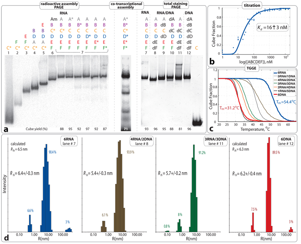
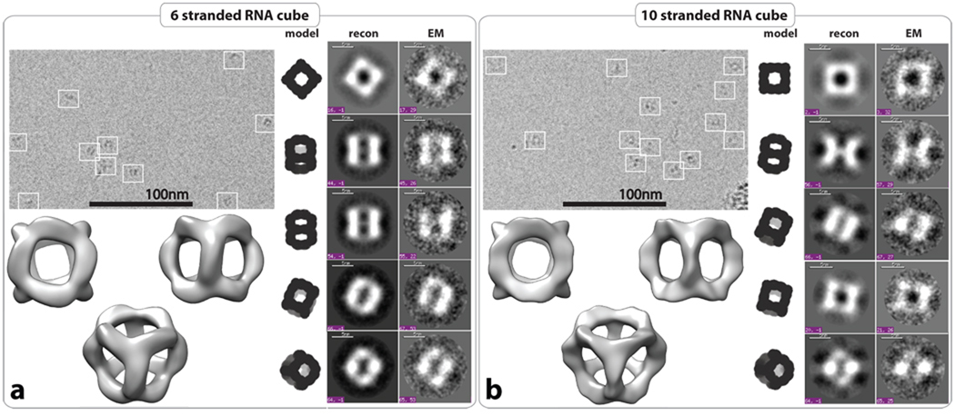
The organization of biological materials into versatile three-dimensional assemblies could be used to build multifunctional therapeutic scaffolds for use in nanomedicine. Here, we report a strategy to design three-dimensional nanoscale scaffolds that can be self-assembled from RNA with precise control over their shape, size and composition. These cubic nanoscaffolds are only approximately 13 nm in diameter and are composed of short oligonucleotides, making them amenable to chemical synthesis, point modifications and further functionalization. Nanocube assembly is verified by gel assays, dynamic light scattering and cryogenic electron microscopy. Formation of functional RNA nanocubes is also demonstrated by incorporation of a light-up fluorescent RNA aptamer that is optimally active only upon full RNA assembly. Moreover, we show that the RNA nanoscaffolds can self-assemble in isothermal conditions (37 degrees C) during in vitro transcription, which opens a route towards the construction of sensors, programmable packaging and cargo delivery systems for biomedical applications.


Figures

Figure 1
Figure 1
3D models for six and ten stranded cubes with corresponding 2D schematics of sequence interactions. Note that 5' start sequences (in black) are base paired in (a) and single-stranded in (b) and (c). The diagrams are drawn to emphasize the symmetry of 3` and 5` positions. Note that the six RNA strands for the 6-stranded cube and the 10 RNA strands for the 10-stranded cube have different length and sequences (Table S1).
Figure 2
Figure 2
Characterization of 6 stranded cube assemblies (without dangling ends). a. Native PAGE assembly experiments: (left) radioactive assemblies with 32P radiolabeled RNA molecules indicated with asterisks. Am (or A6m) was designed to assemble with B–F (or B6–F6) to form an open hexamer. (middle) Co-transcriptional self-assembly of body-labeled RNA cube strands. (right) Native PAGE assembly experiments with RNA visualization by total SYBR Green II staining. Estimated yields of the hexamers (in %) are shown at the bottom of corresponding lanes. All lanes are numbered to distinguish between twelve different compositions of RNA, RNA/DNA and DNA complexes. b. Titration curve fitting data collected from three independent experiments of RNA cube assembly. c. Thermal melting curves of RNA, DNA, and RNA/DNA hybrid cubes. Corresponding Tm`s are shown Figure S3 in SI. d. Size histograms of six stranded cubes measured by DLS. Compositions are specified for each measurement. Color code is consistent with b and c. Relative assembly yields are calculated from each histogram. All RNA complexes used in a, c, and d experiments were assembled as described in Materials and Methods at 1 µM concentrations.
Figure 3
Figure 3
Structural characterization of RNA cubes by cryo-EM with single particle image reconstruction. Panels a and b correspond to the characterization of 6 and 10 stranded RNA cubes respectively. Each panel on the top left represents typical cryo-EM images of the RNA particles. On the right side, class averages for each RNA cube as observed by cryo-EM (EM) with corresponding projections of the reconstructed 3D structure and theoretical RNA cube model. Reconstructed 3D models of the six and 10-stranded RNA cubes have been obtained at 8.9 Å and 11.7 Å resolution, respectively. All RNA complexes used in cryo-EM experiments were assembled at 1 µM of each RNA strand as described in the Materials and Methods.
Figure 4
Figure 4
Functionalization of RNA nano-cube scaffold with Malachite Green (MG) aptamer. a. Scheme showing the functionalization. b. Emission spectra representing binding of MG to RNA aptamer and native PAGE demonstrating the formation of the constructs. Monomer, dimer and nonamer samples (S1, S2, S3, S4) are unable to bring the aptamers into close enough proximity necessary for fluorescent emission in presence of MG. The functionalized cube sample (S5) shows an increase in fluorescence demonstrating correct formation of the MG binding pocket. The cube sample (S6) shows two-fold increase in fluorescence demonstrating simultaneous correct formation of its two MG binding pockets. All RNA complexes used in the fluorescent experiments were assembled at RNA strand concentration of 1 µM as described in Materials and Methods. Based on the emission signal of the control molecule, the yield of the functionally active cube (S5) was estimated to be 77.3 %. c. Comparison of co-transcriptional self-assembly of nonamers S3 and S4 with 10-stranded RNA nano-cube (S5) functionalized with one MG aptamer at 37°C. Aliquots of the transcription mixture were taken after 2, 3, 4, 5, and 7 hours, followed by the addition of DNAse to stop the reaction. MG was added just prior to fluorescent data acquisition. Note that after 5h, more T7 RNA polymerase was added to each transcription mix. Control S7 corresponds to a MG aptamer molecule.

Comment in

  • RNA nanotechnology: inspired by DNA.
    Famulok M, Ackermann D. Famulok M, et al. Nat Nanotechnol. 2010 Sep;5(9):634-5. doi: 10.1038/nnano.2010.183. Nat Nanotechnol. 2010. PMID: 20818409 No abstract available.

References

    1. Seeman NC. An overview of structural DNA nanotechnology. Mol Biotechnol. 2007;37:246–257. - PMC - PubMed
    1. Aldaye FA, Palmer AL, Sleiman HF. Assembling materials with DNA as the guide. Science. 2008;321:1795–1799. - PubMed
    1. Lin C, Liu Y, Yan H. Designer DNA Nanoarchitectures. Biochemistry. 2009;48:1663–1674. - PMC - PubMed
    1. Chen JH, Seeman NC. The electrophoretic properties of a DNA cube and its substructure catenanes. Electrophoresis. 1991;12:607–611. - PubMed
    1. Goodman RP, et al. Reconfigurable, braced, three-dimensional DNA nanostructures. Nat Nanotechnol. 2008;3:93–96. - PubMed

Publication types