Essential roles of Jab1 in cell survival, spontaneous DNA damage and DNA repair
- PMID: 20802511
- PMCID: PMC3495558
- DOI: 10.1038/onc.2010.345
Essential roles of Jab1 in cell survival, spontaneous DNA damage and DNA repair
Abstract
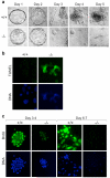
Jun activation domain-binding protein 1 (JAB1) is a multifunctional protein that participates in the control of cell proliferation and the stability of multiple proteins. JAB1 overexpression has been implicated in the pathogenesis of human cancer. JAB1 regulates several key proteins and thereby produces varied effects on cell cycle progression, genome stability and cell survival. However, the biological significance of JAB1 activity in these cellular signaling pathways is unclear. Therefore, we developed mice that were deficient in Jab1 and analyzed the null embryos and heterozygous cells. This disruption of Jab1 in mice resulted in early embryonic lethality due to accelerated apoptosis. Loss of Jab1 expression sensitized both mouse primary embryonic fibroblasts and osteosarcoma cells to γ-radiation-induced apoptosis, with an increase in spontaneous DNA damage and homologous recombination (HR) defects, both of which correlated with reduced levels of the DNA repair protein Rad51 and elevated levels of p53. Furthermore, the accumulated p53 directly binds to Rad51 promoter, inhibits its activity and represents a major mechanism underlying the HR repair defect in Jab1-deficient cells. These results indicate that Jab1 is essential for efficient DNA repair and mechanistically link Jab1 to the maintenance of genome integrity and to cell survival.
Figures


Similar articles
-
Emerging roles of Jab1/CSN5 in DNA damage response, DNA repair, and cancer.Cancer Biol Ther. 2014 Mar 1;15(3):256-62. doi: 10.4161/cbt.27823. Epub 2014 Feb 4. Cancer Biol Ther. 2014. PMID: 24495954 Free PMC article. Review.
-
Suppression of Jab1/CSN5 induces radio- and chemo-sensitivity in nasopharyngeal carcinoma through changes to the DNA damage and repair pathways.Oncogene. 2013 May 30;32(22):2756-66. doi: 10.1038/onc.2012.294. Epub 2012 Jul 16. Oncogene. 2013. PMID: 22797071 Free PMC article.
-
Multiple functions of Jab1 are required for early embryonic development and growth potential in mice.J Biol Chem. 2004 Oct 8;279(41):43013-8. doi: 10.1074/jbc.M406559200. Epub 2004 Aug 6. J Biol Chem. 2004. PMID: 15299027
-
The crucial p53-dependent oncogenic role of JAB1 in osteosarcoma in vivo.Oncogene. 2020 Jun;39(23):4581-4591. doi: 10.1038/s41388-020-1320-6. Epub 2020 May 10. Oncogene. 2020. PMID: 32390003 Free PMC article.
-
Jab1 as a mediator of nuclear export and cytoplasmic degradation of p53.Mol Cells. 2006 Oct 31;22(2):133-40. Mol Cells. 2006. PMID: 17085963 Review.
Cited by
-
Genetic controls of Tas1r3-independent sucrose consumption in mice.Mamm Genome. 2021 Apr;32(2):70-93. doi: 10.1007/s00335-021-09860-w. Epub 2021 Mar 12. Mamm Genome. 2021. PMID: 33710367
-
Emerging roles of Jab1/CSN5 in DNA damage response, DNA repair, and cancer.Cancer Biol Ther. 2014 Mar 1;15(3):256-62. doi: 10.4161/cbt.27823. Epub 2014 Feb 4. Cancer Biol Ther. 2014. PMID: 24495954 Free PMC article. Review.
-
Loss of jab1 in osteochondral progenitor cells severely impairs embryonic limb development in mice.J Cell Physiol. 2014 Nov;229(11):1607-17. doi: 10.1002/jcp.24602. J Cell Physiol. 2014. PMID: 24604556 Free PMC article.
-
THE COP9 SIGNALOSOME, A NOVEL, ESSENTIAL REGULATOR OF SKELETAL DEVELOPMENT AND TUMORIGENESIS.Case Orthop J. 2011;8(1):45-50. Case Orthop J. 2011. PMID: 26807424 Free PMC article.
-
JAB1/CSN5: a new player in cell cycle control and cancer.Cell Div. 2010 Oct 18;5:26. doi: 10.1186/1747-1028-5-26. Cell Div. 2010. PMID: 20955608 Free PMC article.
References
-
- Bae MK, Ahn MY, Jeong JW, Bae MH, Lee YM, Bae SK, et al. Jab1 interacts directly with HIF-1alpha and regulates its stability. J Biol Chem. 2002;277:9–12. - PubMed
-
- Buscemi G, Perego P, Carenini N, Nakanishi M, Chessa L, Chen J, et al. Activation of ATM and Chk2 kinases in relation to the amount of DNA strand breaks. Oncogene. 2004;23:7691–700. - PubMed
Publication types
MeSH terms
Substances
Grants and funding
LinkOut - more resources
Full Text Sources
Molecular Biology Databases
Research Materials
Miscellaneous

