. 2010 Dec 2;29(48):6331-42.
doi: 10.1038/onc.2010.362. Epub 2010 Aug 30.

DNA copy number aberrations in small-cell lung cancer reveal activation of the focal adhesion pathway

Affiliations

DNA copy number aberrations in small-cell lung cancer reveal activation of the focal adhesion pathway

S Ocak et al. Oncogene. .

Abstract

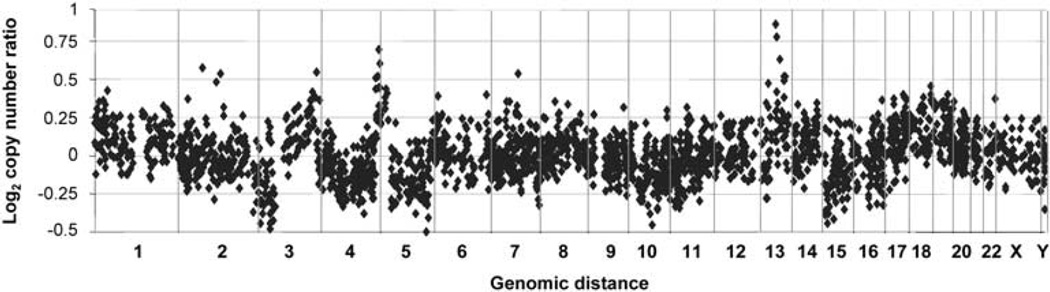
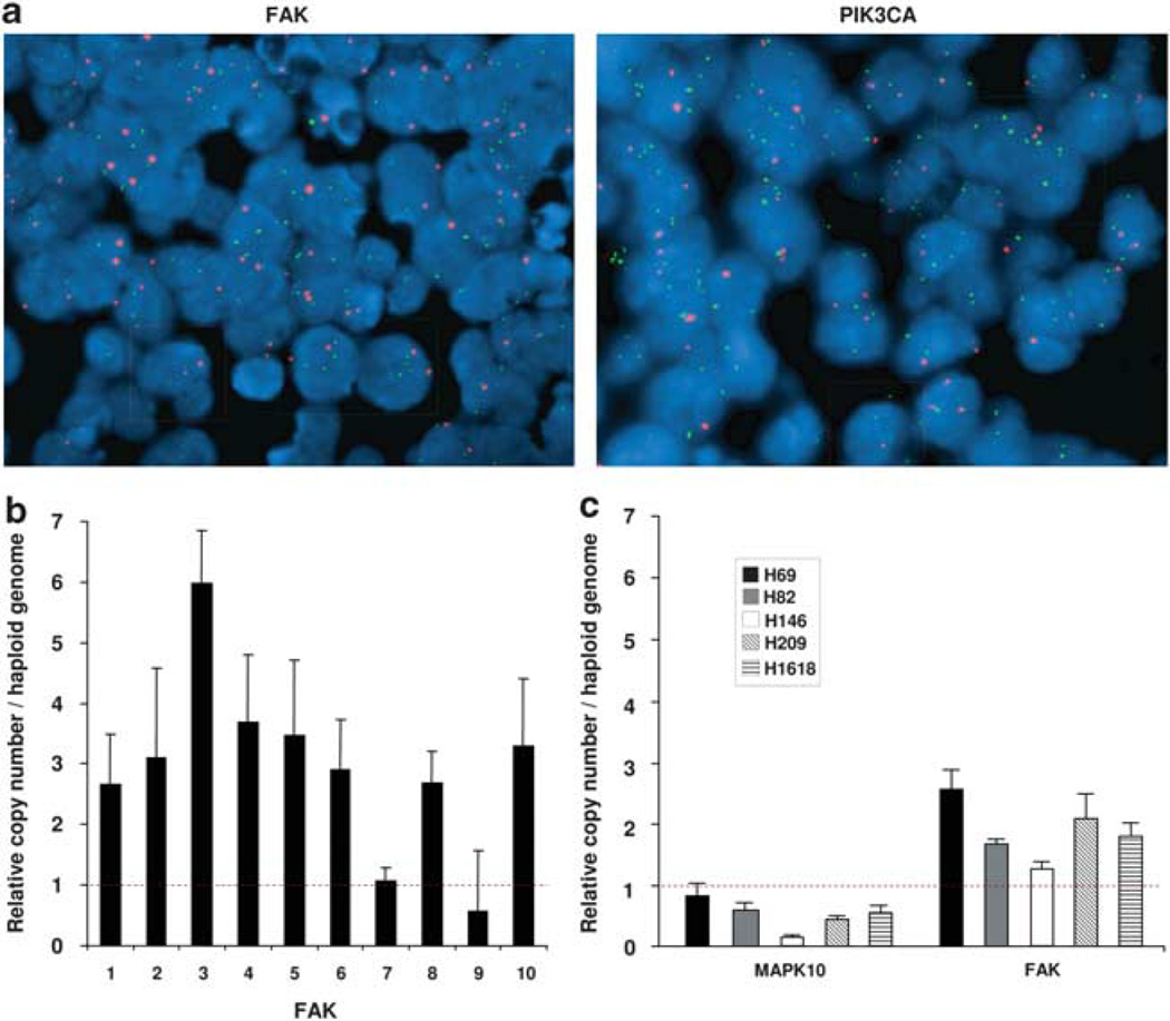
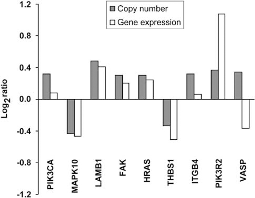
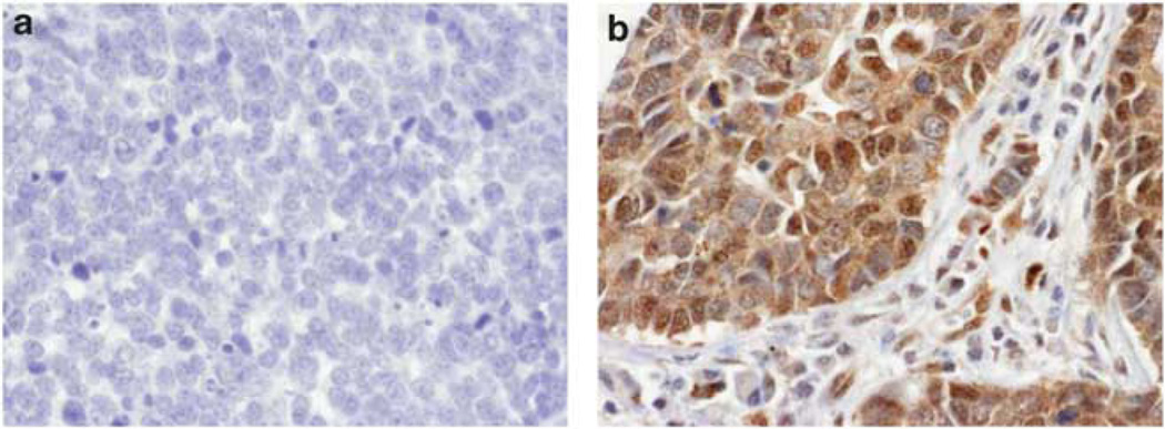
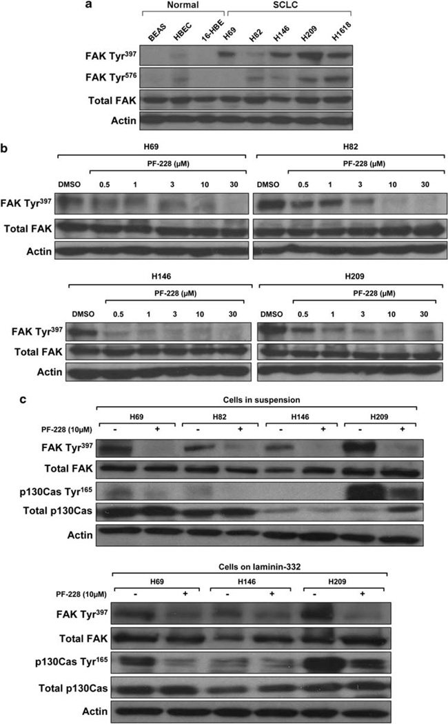
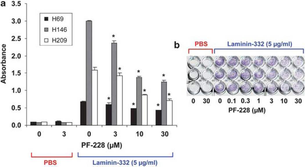
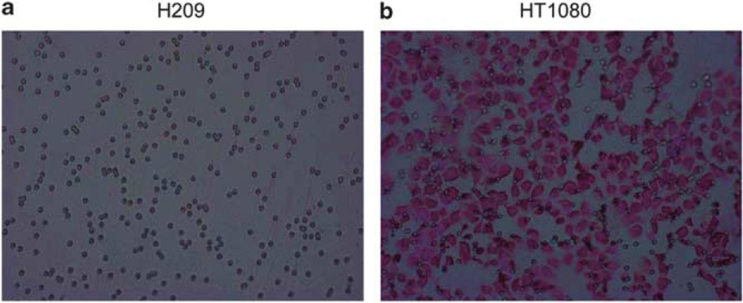
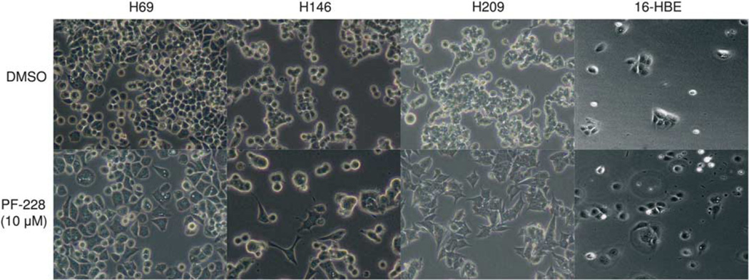
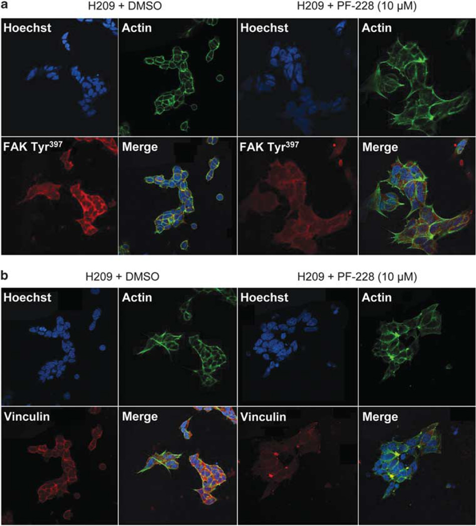
Small-cell lung cancer (SCLC) is the most aggressive subtype of lung cancer in its clinical behavior, with a 5-year overall survival as low as 5%. Despite years of research in the field, molecular determinants of SCLC behavior are still poorly understood, and this deficiency has translated into an absence of specific diagnostics and targeted therapeutics. We hypothesized that tumor DNA copy number alterations would allow the identification of molecular pathways involved in SCLC progression. Array comparative genomic hybridization was performed on DNA extracted from 46 formalin-fixed paraffin-embedded SCLC tissue specimens. Genomic profiling of tumor and sex-matched control DNA allowed the identification of 70 regions of copy number gain and 55 regions of copy number loss. Using molecular pathway analysis, we found a strong enrichment in these regions of copy number alterations for 11 genes associated with the focal adhesion pathway. We verified these findings at the genomic, gene expression and protein level. Focal Adhesion Kinase (FAK), one of the central genes represented in this pathway, was commonly expressed in SCLC tumors and constitutively phosphorylated in SCLC cell lines. Those were poorly adherent to most substrates but not to laminin-322. Inhibition of FAK phosphorylation at Tyr(397) by a small-molecule inhibitor, PF-573,228, induced a dose-dependent decrease of adhesion and an increase of spreading in SCLC cell lines on laminin-322. Cells that tended to spread also showed a decrease in focal adhesions, as demonstrated by a decreased vinculin expression. These results support the concept that pathway analysis of genes in regions of copy number alterations may uncover molecular mechanisms of disease progression and demonstrate a new role of FAK and associated adhesion pathways in SCLC. Further investigations of FAK at the functional level may lead to a better understanding of SCLC progression and may have therapeutic implications.


Conflict of interest statement

Conflict of interest

The authors declare no conflict of interest.

Figures

Figure 1
Figure 1
Gene copy number alterations in 46 primary small-cell lung cancers (SCLCs). Each dot represents the average log2 copy number ratio in 46 SCLC tumors at a given genomic locus across the genome tested by array comparative genomic hybridization (CGH). The value of 0 represents an equal fluorescence intensity ratio between SCLC and normal reference genomic DNA. Log2 ratios > 0.3 or < −0.3 are defined as copy number gain or loss. Vertical lines represent chromosomal boundaries.
Figure 2
Figure 2
Verification of FAK and PIK3CA increased gene copy number by FISH in primary SCLCs and by quantitative RT–PCR in SCLC cell lines. (a) Dual color fluorescence in situ hybridization of FAK gene (8q24.3, green spots) and CEP8 probe (red spots) or of PIK3CA gene (3q26.3, green spots) and CEP3 probe (red spots) on the centromeric region of the same chromosome in representative SCLC tissue samples. Interphase nuclei (stained in blue with DAPI) with increased copy number of FAK (left panel) and PIK3CA (right panel) are shown in green boxes. Interphase nuclei with normal gene copy number are shown in red boxes. Pictures magnification: ×100. (b) Quantitative RT–PCR of FAK in primary SCLCs. Copy number gain of FAK was confirmed in 8 of 10 tested tumors. (c) Quantitative RT–PCR of FAK and MAPK10 genes in SCLC cell lines. Copy number gain of FAK and copy number loss of MAPK10 were observed in all tested cell lines. Copy number changes per haploid genome were calculated using the formula 2(Nt–NRNase P)–(Dt–DRNase P), where Nt is the average threshold cycle number observed for an experimental primer in the normal DNA sample, NRNase P is the threshold cycle number observed for an RNase P primer in the normal DNA sample, Dt is the average threshold cycle number observed for the experimental primer in the SCLC DNA sample and DRNase P is the average threshold cycle number observed for an RNase P primer in the SCLC DNA sample. Data represent averages and error bars ± 1 s.d. (n = 3).
Figure 3
Figure 3
Positive correlation between the focal adhesion pathway gene copy number data and gene expression level. Array CGH gene copy number of 46 SCLC tumors was compared with gene expression level of the same genes represented in Garber et al.’s public gene expression data set, based on the analysis of five primary SCLC tumors. A positive correlation between the median log2 gene expression ratio (SCLC/normal) and the median log2 gene copy number ratio (SCLC/normal) was found (Pearson: 0.68) for 9 of 11 genes represented in the focal adhesion pathway.
Figure 4
Figure 4
FAK expression by immunohistochemistry: a representative example of 52 studied SCLCs. Tissue sections were incubated with an antibody against FAK (A-17). All immunostains were independently scored by two independent observers (SO and PPM) as follows: 0—no staining, 1—weak staining, 2—moderate staining and 3—strong staining. FAK displayed a cytoplasmic staining. (a) Representative image of SCLC with absence of total FAK expression. (b) Representative image of SCLC tumor with maximum staining score for total FAK. It is noteworthy that positively stained cancer cells are infiltrating the stroma. Pictures magnification: ×40.
Figure 5
Figure 5
FAK is activated in SCLC cell lines and inhibited by PF-228 in a dose-dependent manner. (a) FAK is phosphorylated in SCLC cell lines. Whole-cell lysates were resolved with SDS-PAGE and blots were incubated with antiphospho-FAK Tyr397/576, total FAK or actin antibodies. FAK Tyr397/576 and total FAK levels were normalized to actin. FAK phosphorylation at tyrosine residues was mainly observed in SCLC cell lines, demonstrating an activation of FAK in SCLC. (b) Dose-dependent inhibition of FAK Tyr397 phosphorylation by PF-228 in SCLC cell lines. SCLC cell lines in suspension were treated with the indicated concentrations of PF-228 for 90 min. Whole-cell lysates were resolved with SDS-PAGE and blots were incubated with antiphospho-FAK Tyr397, total FAK or actin antibodies. FAK Tyr397 and total FAK levels were normalized to actin. Significant decrease of FAK phosphorylation at Tyr397 was achieved with PF-228 in a dosedependent manner. (c) PF-228 decreases p130CAS Tyr165 phosphorylation, downstream substrate of FAK, in SCLC cell lines. SCLC cell lines in suspension, as well as adhering on laminin-332, were treated with 10 µm PF-228 for 90 min. Whole-cell lysates were resolved with SDS-PAGE and blots were incubated with antiphospho-FAK Tyr397, total FAK, phospho-p130CAS Tyr165, total p130CAS or actin antibodies. FAK Tyr397 phosphorylation, total FAK, phospho-p130CAS Tyr165 and total p130CAS levels were normalized to actin. Significant decrease of p130CAS phosphorylation at Tyr165 was achieved with PF-228, parallel to the decrease of FAK phosphorylation at Tyr397.
Figure 6
Figure 6
PF-228 induces a dose-dependent decrease in the adhesion of SCLC lines to laminin-332. Cells were treated with increasing concentrations of PF-228 for 30 min before seeding on laminin-332-coated plates and then during the entire experiment. Adhesion was evaluated by crystal violet staining 24 h after seeding. The extent of cell adhesion was determined by measuring the absorbance of wells at 595nm with an ELISA reader. (a) This figure, representative of one of the experiments performed in triplicate wells three independent times, shows that treatment with PF-228 induced a dose-dependent decrease of the adhesion of the three SCLC cell lines to laminin-332. Data represent averages and error bars ±1 s.d. (n = 3). P-values: absorbance of wells treated with PF-228 vs those treated with DMSO; *P<0.05. (b) This image, representative of one of the experiments performed with NCI-H209, shows the decrease of crystal violet staining with increasing concentrations of PF-228.
Figure 7
Figure 7
Absence of motility and invasion of SCLC cells in a transwell assay. Migration and invasion were assessed for up to 24 h by counting cells on the lower side of the transwell’s membrane in five microscopic fields of high magnification (40×) under a light microscope. Cells on the membrane were fixed and stained with hematoxylin and eosin. (a) This image, representative of one of the numerous experiments performed in triplicate wells to address migration and invasion under different conditions, shows the absence of SCLC cells on the lower side of the membrane. Only pores of the membrane are observed. (b) This is in sharp contrast to the large number of HT1080 (human fibrosarcoma) cells observed on the lower side of the membrane, as demonstrated in this figure representative of one of the three independent experiments performed in triplicate wells to address migration. Pictures magnification: ×40.
Figure 8
Figure 8
Treatment of SCLC cells with PF-228 increases their spreading on laminin-332. NCI-H69, NCI-H146, NCI-H209 and 16-HBE cells were plated for 12 h on laminin-332-coated dishes and then treated with 10 µm PF-228 or DMSO for 24 h. Pictures of unstained cells were taken after 24 h treatment and show that SCLC cells treated with PF-228 spread on laminin-332, losing their rounded shape and presenting extended processes. No change in morphology was observed in 16-HBE, an immortalized normal bronchial epithelial cell constitutively lacking phospho-FAK Tyr397 expression. Pictures magnification: ×20.
Figure 9
Figure 9
Treatment of SCLC cells adherent to laminin-332 with PF-228 decreases immunofluorescence staining for phospho-FAK Tyr397 and vinculin. NCI-H209 cells were plated for 12 h on laminin-332-coated dishes and then treated with 10 µm PF-228 or DMSO for 12 h. After 12 h treatment, they were fixed and immunostained using Hoechst (blue, for nuclei), Phalloidin (green, for Actin) and phospho-FAK Tyr397 (red) in a or vinculin (red) in b. (a) Cells treated with PF-228 spread on laminin-332 and showed decreased phospho-FAK Tyr397 accumulation at the spreading edges and at cell–cell contact sites compared with NCI-H209 cells treated with DMSO. (b) Immunofluorescence staining of NCI-H209 cells also showed a decrease in vinculin expression in the presence of 10 µm PF-228. Pictures magnification: ×63.

Similar articles

Cited by

References

    1. Balsara BR, Testa JR. Chromosomal imbalances in human lung cancer. Oncogene. 2002;21:6877–6883. - PubMed
    1. Bolstad BM, Irizarry RA, Astrand M, Speed TP. A comparison of normalization methods for high density oligonucleotide array data based on variance and bias. Bioinformatics. 2003;19:185–193. - PubMed
    1. Burridge K, Turner CE, Romer LH. Tyrosine phosphorylation of paxillin and pp125FAK accompanies cell adhesion to extracellular matrix: a role in cytoskeletal assembly. J Cell Biol. 1992;119:893–903. - PMC - PubMed
    1. Cappuzzo F, Hirsch FR, Rossi E, Bartolini S, Ceresoli GL, Bemis L, et al. Epidermal growth factor receptor gene and protein and gefitinib sensitivity in non-small-cell lung cancer. J Natl Cancer Inst. 2005;97:643–655. - PubMed
    1. Coe BP, Lockwood WW, Girard L, Chari R, Macaulay C, Lam S, et al. Differential disruption of cell cycle pathways in small cell and non-small cell lung cancer. Br J Cancer. 2006;94:1927–1935. - PMC - PubMed

Publication types

MeSH terms

Substances