. 2010 Nov 25;29(47):6267-79.
doi: 10.1038/onc.2010.359. Epub 2010 Aug 30.

ERK and JNK activation is essential for oncogenic transformation by v-Rel

Affiliations

ERK and JNK activation is essential for oncogenic transformation by v-Rel

J Kralova et al. Oncogene. .

Abstract

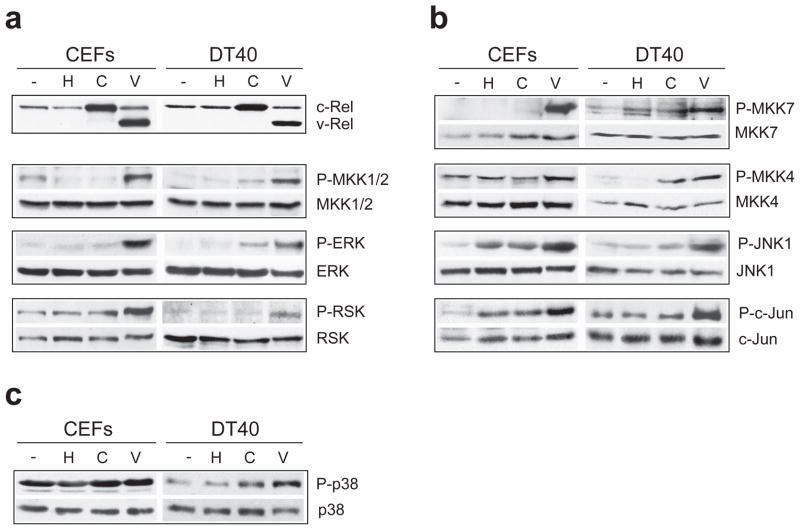
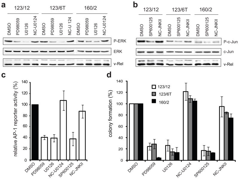
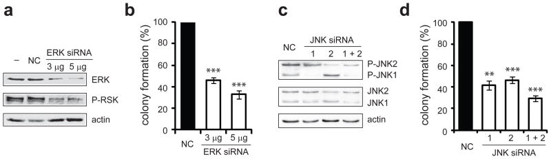
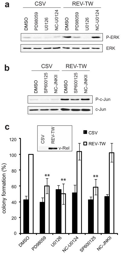
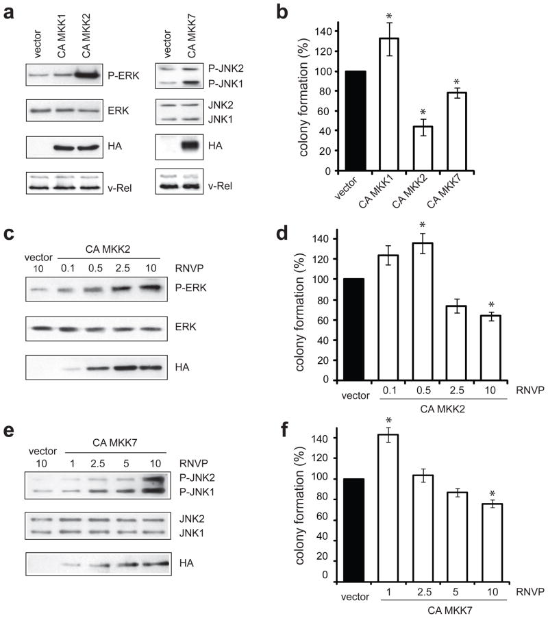
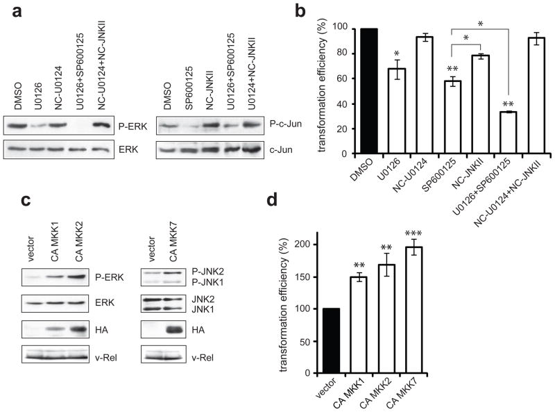
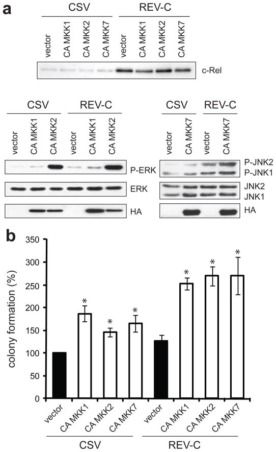
v-Rel is the acutely oncogenic member of the NF-κB family of transcription factors. Infection with retroviruses expressing v-Rel rapidly induces fatal lymphomas in birds and transforms primary lymphocytes and fibroblasts in vitro. We have previously shown that AP-1 transcriptional activity contributes to v-Rel-mediated transformation. Although v-Rel increases the expression of these factors, their activity may also be induced through phosphorylation by the mitogen-activated protein kinases (MAPKs). The expression of v-Rel results in the strong and sustained activation of the ERK and JNK MAPK pathways. This induction is critical for the v-Rel-transformed phenotype, as suppression of MAPK activity with chemical inhibitors or small interfering RNA severely impairs colony formation of v-Rel-transformed lymphoid cell lines. However, signaling must be maintained within an optimal range in these cells, as strong additional activation of either pathway beyond the levels induced by v-Rel through the expression of constitutively active MAPK proteins attenuates the transformed phenotype. MAPK signaling also has an important role in the initial transformation of primary spleen cells by v-Rel, although distinct requirements for MAPK activity at different stages of v-Rel-mediated transformation were identified. We also show that the ability of v-Rel to induce MAPK signaling more strongly than c-Rel contributes to its greater oncogenicity.


Conflict of interest statement

Conflict of Interest

The authors declare no conflict of interest.

Figures

Figure 1
Figure 1
Induction of the ERK and JNK MAPK pathways by v-Rel. Chicken embryo fibroblasts (CEFs) or DT40 cells were left uninfected (−) or were infected with helper virus alone (H) or with retroviruses expressing c-Rel (C) or v-Rel (V). Cell lysates were prepared 7–10 days later, when cells expressing v-Rel exhibited characteristic morphological changes. Western blot analysis examined the levels of total and phosphorylated protein for components of the (a) ERK, (b) JNK, and (c) p38 MAPK signaling cascades. The expression of c-Rel and v-Rel in these cells is shown in panel (a).
Figure 2
Figure 2
Inhibition of ERK and JNK pathways attenuates colony formation of v-Rel transformed cells. Three established v-Rel transformed cell lines of histologically distinct origin were utilized, including a T-cell (160/2), B-cell (123/12), and non-B/non-T (123/6T) cell line. (a) Cell lines were treated for one hour with carrier alone (DMSO), MEK inhibitors (PD98059, U0126), or negative control (U0124) at a 3 μM concentration. Cell lysates were prepared and levels of phosphorylated and total ERK were examined by Western blot. (b) Cell lines were treated for one hour with carrier alone (DMSO), JNK inhibitor (SP600125), or negative control (NC-JNK II) at a 3 μM concentration and cell lysates were examined by Western blot for phosphorylated and total c-Jun. (c) Reporter assays evaluated the effect of MAPK inhibitors on AP-1 activity in cells expressing v-Rel. CEFs were co-transfected with a pGL2 luciferase reporter (1 μg) containing multiple repeats of an AP-1 consensus site (Kralova et al., 1998), the Rc/RSV expression vector (1 μg) encoding v-Rel, and with the pRL-TK reporter vector (0.3 μg). Eight hours later, MAPK inhibitors or their respective negative controls were added to the media at a 10 μM concentration. Luciferase activity in cells treated with carrier alone (DMSO) was standardized to 100. The average and standard deviation for four independent experiments are shown. (d) Cell lines were incubated with inhibitors or negative controls (3 μM) for two days and plated into soft agar in the presence of the same concentration of inhibitors. Colonies were scored microscopically 10 days later and colony numbers for cells treated with carrier alone (DMSO) were standardized to 100. The average and standard deviation for four independent experiments are shown.
Figure 3
Figure 3
ERK and JNK knockdown inhibits colony formation of v-Rel transformed cells. (a–b) The v-Rel transformed cell line, 160/2, was electroporated with siRNA targeting ERK (3 or 5 μg) or with negative control siRNA (3 μg). Cells from the same electroporation population were plated into soft agar 16 hours after transfection and harvested at 48 hours for protein. (a) The levels of total ERK, as well as the levels of phosphorylation of a downstream target, p90-RSK were examined by Western blot. (b) Soft agar colonies were scored microscopically 7–8 days after plating. Colony numbers from cells electroporated with negative control siRNA were standardized to 100. The average for four independent experiments is shown with standard error. (c–d) 160/2 cells were electroporated with negative control siRNA or with siRNA (1 μg) targeting JNK1, JNK2, or both. Cells were plated into soft agar and harvested for protein 16 hours after transfection. (c) Western blot analysis examined the levels of phosphorylated and total JNK1 and JNK2. (d) Soft agar colonies were scored microscopically 7–8 days after plating. Colony numbers from cells electroporated with negative control siRNA were standardized to 100. The average for four independent experiments is shown with standard error. Statistically significant differences in colony formation of cells electroporated with specific siRNA relative to negative control are indicated (**P<0.01, ***P<0.001).
Figure 4
Figure 4
ERK and JNK activation is a specific requirement for v-Rel transformation. DT40 cells were infected with helper virus alone (CSV) or with retroviruses expressing v-Rel (REV-TW). Cells were grown for 7–10 days, until those expressing v-Rel exhibited characteristic morphological changes. (a) Infected cells were treated for one hour with carrier alone (DMSO), MEK inhibitors (PD95089, U0126), or negative control (U0124) at a 3 μM concentration and cell lysates were examined for phosphorylated and total ERK. (b) Infected cells were treated for one hour with carrier alone (DMSO), JNK inhibitor (SP600125), or negative control (NC-JNK II) at a 3 μM concentration and cell lysates were examined for phosphorylated and total c-Jun. (c) Infected cells were incubated for one hour with inhibitors or negative controls (3 μM) and then plated into soft agar containing the same concentration of inhibitors. Colonies were scored microscopically after 7–8 days. Colony numbers for REV-TW infected cells treated with DMSO were standardized to 100. The average and standard deviation for four independent experiments are shown. Statistically significant differences in colony formation of REV-TW-infected cells treated with various inhibitors relative to those treated with DMSO are indicated (**P<0.01). v-Rel expression in REV-TW infected cells was verified by Western blot analysis, as shown in the inset of panel (c).
Figure 5
Figure 5
Constitutive ERK and JNK activity attenuates colony formation of v-Rel transformed cells. (a) The v-Rel cell line (160/2) was infected with DS retroviruses encoding the CA MKK1, CA MKK2, and CA MKK7 mutants or with empty vector. Cells were infected with each virus with relative numbers of virus particles (RNVP) of 10. Western blot analysis demonstrated the expression of the CA MKK mutants by detection of the HA epitope located at the N-terminus of each protein and the levels of phosphorylated and total ERK in cells expressing CA MKK1 and CA MKK2 (left panels) and phosphorylated and total JNK in cells expressing CA MKK7 (right panels) relative to cells infected with empty DS virus. (b) Cells were plated into soft agar five days after infection and colonies were scored microscopically 7–9 days later. Colony numbers from cells infected with empty DS retroviruses were standardized to 100. The average of at least two independent experiments is shown with standard error. Statistically significant differences from cells infected with empty vector are indicated (*P<0.05). (c–f) Dose-dependent effect of ERK and JNK activity on colony formation of the v-Rel cell line (160/2). Cells were infected with empty DS retroviruses (RNVP of 10) or with increasing amounts of viruses encoding CA MKK2 or CA MKK7 and cell lysates were prepared. Western analysis examined (c) the levels of phosphorylated and total ERK in cells infected with virus expressing CA MKK2 (RNVP of 0.1, 0.5, 2.5, and 10) and (e) the levels of phosphorylated and total JNK in cells infected with virus expressing CA MKK7 (RNVP of 1, 2.5, 5, and 10) relative to control cells. (d,f) Infected cells were plated into soft agar and colonies were scored microscopically 10–14 days later. Colony numbers for cells infected with empty DS retroviruses were standardized to 100. In each panel, the average of at least two independent experiments is shown with standard error. Statistically significant differences from cells infected with empty vector are indicated (*P<0.05).
Figure 6
Figure 6
Role of ERK and JNK signaling in the transformation of primary splenic lymphocytes by v-Rel. (a–b) Primary splenic lymphocytes were isolated from three week-old chickens and infected with retroviruses expressing v-Rel (REV-TW). (a) The next day, cells were treated for one hour with carrier alone (DMSO), with MAPK pathway inhibitors (MEK inhibitor, U0126; JNK inhibitor, SP600125) or their respective negative controls (U0124, NC-JNK II) singly, or with MAPK inhibitors or negative controls in combination. All treatments were performed at a 3 μM concentration of inhiibitor or control. Cell lysates were analyzed for phosphorylated and total ERK and c-Jun. (b) The day following infection, cells were incubated for six hours with inhibitors or negative controls at a 3 μM concentration and then plated into soft agar containing the same concentration of inhibitors. Colonies were scored microscopically 10–14 days later. Colony numbers for cells treated with carrier alone (DMSO) were standardized to 100. The average of at least three independent experiments is shown with standard error. Statistically significant differences from cells treated with DMSO are indicated (*P<0.05, **P<0.01). (c–d) Primary splenic lymphocytes were co-infected with retroviruses expressing v-Rel (REV-TW) and DS retroviruses encoding CA MKK1, CA MKK2, or CA MKK7 or empty vector. (c) Infections were expanded in liquid culture and cell lysates were prepared 10 days after infection. Western blots examined the expression of v-Rel and the CA MKK mutants. The levels of phosphorylated and total ERK in cells expressing CA MKK1 and CA MKK2 and phosphorylated and total JNK in cells expressing CA MKK7 relative to cells infected with empty DS viruses were also determined. (d) The day following infection, an aliquot of each infection was plated into soft agar and colonies were scored microscopically 10–14 days later. Colony numbers for cells infected with empty DS viruses were standardized to 100. The average of nine independent experiments is shown with standard error (*P<0.05, **P<0.01, ***P<0.001).
Figure 7
Figure 7
CA MKK mutants enhance colony formation of DT40 cells infected with helper virus or retroviruses encoding c-Rel. DT40 cells were co-infected with CSV alone or with retroviruses expressing c-Rel (REV-C) as well as with retroviruses expressing the CA MKK mutants. Infections were expanded in liquid culture for 8–14 days and cells were harvested for protein and plated into soft agar. (a) The expression of c-Rel and the CA MKK mutants were examined by Western blot. The levels of phosphorylated and total ERK in cells expressing CA MKK1 and CA MKK2 and phosphorylated and total JNK in cells expressing CA MKK7 relative to cells infected with empty DS viruses were also determined. (b) Soft agar colonies were scored microscopically 7–9 days later. Colony numbers for cells infected with CSV and with empty DS retroviruses were standardized to 100. Statistically significant differences from cells infected with CSV and empty DS vector are indicated (*P<0.05). The average of at least three independent experiments is shown with standard error.

Similar articles

Cited by

References

    1. Basseres DS, Baldwin AS. Nuclear factor- B and inhibitor of B kinase pathways in oncogenic initiation and progression. Oncogene. 2006;25:6817–30. - PubMed
    1. Blume-Jensen P, Hunter T. Oncogenic kinase signalling. Nature. 2001;411:355–65. - PubMed
    1. Bode AM, Dong Z. The functional contrariety of JNK. Mol Carcinog. 2007;46:591–8. - PMC - PubMed
    1. Bonizzi G, Karin M. The two NF- B activation pathways and their role in innate and adaptive immunity. Trends Immunol. 2004;25:280–8. - PubMed
    1. Bost F, McKay R, Bost M, Potapova O, Dean NM, Mercola D. The Jun kinase 2 isoform is preferentially required for epidermal growth factor-induced transformation of human A549 lung carcinoma cells. Mol Cell Biol. 1999;19:1938–49. - PMC - PubMed

Publication types

MeSH terms

Substances