. 2011 Jan 6;30(1):21-31.
doi: 10.1038/onc.2010.395. Epub 2010 Aug 30.

Hypoxia and cell cycle regulation of the von Hippel-Lindau tumor suppressor

Affiliations

Hypoxia and cell cycle regulation of the von Hippel-Lindau tumor suppressor

W Liu et al. Oncogene. .

Abstract

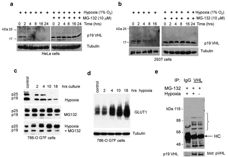
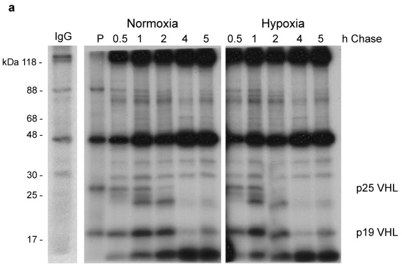
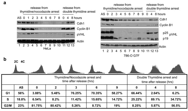
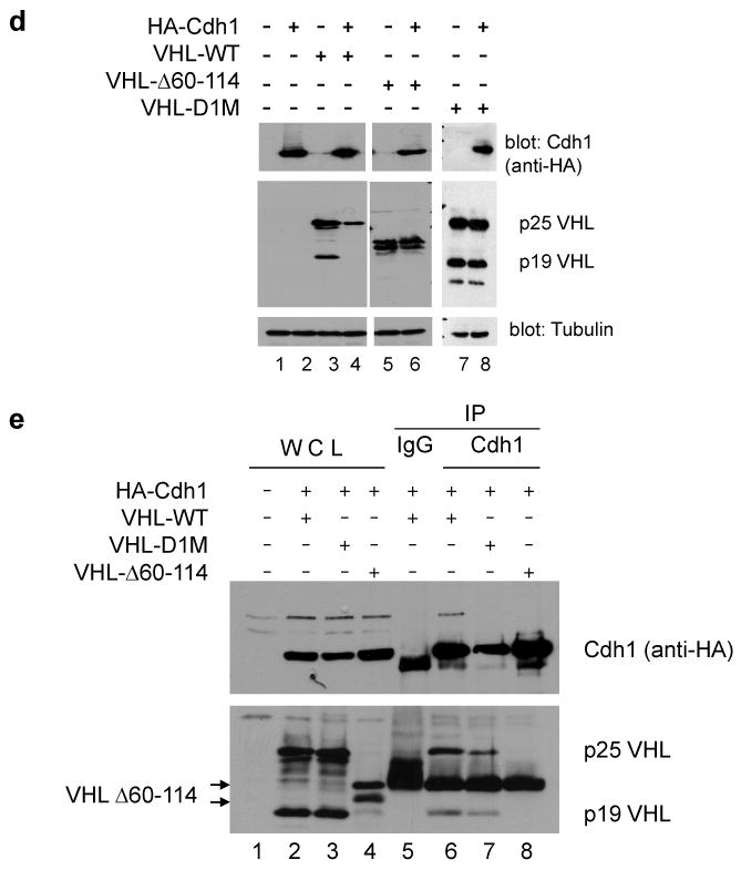
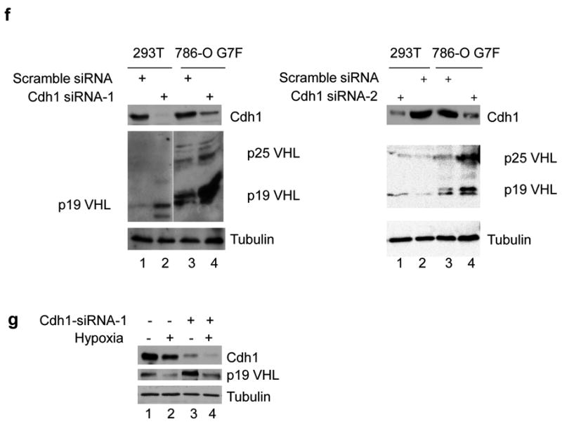
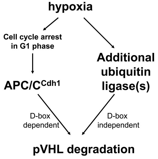
Inactivation of von Hippel-Lindau tumor-suppressor protein (pVHL) is associated with von Hippel-Lindau disease, an inherited cancer syndrome, as well as the majority of patients with sporadic clear cell renal cell carcinoma (RCC). Although the involvement of pVHL in oxygen sensing through targeting hypoxia-inducible factor-α subunits to ubiquitin-dependent proteolysis has been well documented, less is known about pVHL regulation under both normoxic and hypoxic conditions. We found that pVHL levels decreased in hypoxia and that hypoxia-induced cell cycle arrest is associated with pVHL expression in RCC cells. pVHL levels fluctuate during the cell cycle, paralleling cyclin B1 levels, with decreased levels in mitosis and G1. pVHL contains consensus destruction (D) box sequences, and pVHL associates with Cdh1, an activator of the anaphase-promoting complex/cyclosome (APC/C) E3 ubiquitin ligase. We show that pVHL has a decreased half-life in G1, Cdh1 downregulation results in increased pVHL expression, whereas Cdh1 overexpression results in decreased pVHL expression. Taken together, these results suggest that pVHL is a novel substrate of APC/C(Cdh1). D box-independent pVHL degradation was also detected, indicating that other ubiquitin ligases are also activated for pVHL degradation.


Figures

Figure 1
Figure 1. Hypoxia-induced downregulation of pVHL via the ubiquitin-proteasome pathway
(a) HeLa cells, (b) 293T cells, and (c) 786-O G7F RCC cells were cultured in hypoxia in the absence or presence of 10 μM MG132 for the indicated time intervals. The 0 time point represents lysates from normoxic cultures that were prepared at the time of transfer to hypoxia. pVHL expression was detected by Ig32 anti-pVHL western blotting, and blots were probed with tubulin antibody to demonstrate equal loading. (d) Increasing GLUT1 expression levels in 786-O G7F cells that were cultured in hypoxia. (e) 293T cells were treated with 10 μM MG132 and cultured under normoxic or hypoxic conditions for 24 h. Whole cell lysates were prepared with RIPA buffer, and immunoprecipitation and western blotting were performed with anti-VHL (Ig32) and anti-ubiquitin antibody, respectively. HC, immunoglobulin heavy chain.
Figure 2
Figure 2. Post-transcriptional regulation of VHL expression
(a) 786-O G7F RCC cells were pulse labeled with 35S-Methionine for 30 min (P) at which point labeling medium was washed away and replaced with medium containing cold methionine. RIPA lysates were prepared from chase cultures at the indicated time points. A representative control IgG immunoprecipitation from a pulse-labeled lysate is shown. (b) 293T cells were cultured in normoxia (N) or in hypoxia for 6 or 16 h, and VHL and VEGF mRNA expression levels were determined by quantitative real time RT-PCR. Relative quantitation (RQ) was calculated using 18S rRNA amplification as reference. Amplification of cDNA from hypoxic cells is expressed relative to amplification of normoxic cells (N), which was given a value of 1. The data shown here are representative of three independent experiments.
Figure 3
Figure 3. Hypoxia-induced cell cycle arrest and pVHL downregulation
RCC cells were cultured in normoxia or hypoxia for the indicated times and cells were harvested for (a - c) cell cycle analysis by propidium iodide staining and flow cytometry or (d) western blot analysis. Representatives of multiple experiments are shown here. Overall there was less than 10% variance in cell numbers at the G1, S, and G2/M phases of the cell cycle among the various experiments. Western blots were probed with Ig32 pVHL antibody or tubulin antibody to demonstrate equal loading.
Figure 4
Figure 4. pVHL levels fluctuate through the cell cycle
Cell cycle synchronization of HeLa and 786-O G7F RCC cells was performed using double thymidine block or thymidine plus nocodazole block as described in the Materials and Methods. Cells were released from synchronization and harvested at the indicated times. (a) Expression of the indicated proteins in HeLa cells or 786-O G7F RCC cells was examined by western blotting, and β-actin detection was used to demonstrate equal loading. (b) Analyses of HeLa cells were performed to confirm cell cycle arrest and release using propidium iodide staining and flow cytometry. Histograms are shown as well as the corresponding numbers of cells in G1, S, and the G2/M phases. The 0 time point indicates cells arrested with the indicated agents but not released from block. A representative experiment of three performed is shown. Similar results were obtained using 786-O G7F RCC cells (data not shown). (c) HeLa cells were synchronized by thymidine plus nocodazole block. Three hrs after release pulse-chase studies were performed as described in Figure 2 and the Materials and Methods.
Figure 5
Figure 5. pVHL interacts with Cdh1
(a) Endogenous pVHL was co-immunoprecipitated with anti-Cdh1 antibody from 293T cell extracts. Western blots were probed with Ig32 anti-pVHL antibody. (b) 293T cells were transfected with vectors expressing HA-Cdh1 and pVHL-myc/His alone or together and complexes were isolated using metal affinity resin. Western blots were probed with either anti-HA (top panel) or anti-myc (bottom panel). Cdh1 was co-immunoprecipitated with FL181 anti-pVHL antibody from pVHL-expressing (c) 786-O G7F RCC cells or (d) WT7 RC cells but not from pVHL-negative 786-O RCC cells. In panels c and d western blots were probed with either anti-Cdh1 (top panel) or anti-pVHL (Ig32) (bottom panel).
Figure 6
Figure 6. pVHL contains Cdh1 recognition sequences and is a novel substrate of APC/CCdh1
(a) The pVHL protein sequence contains two consensus Destruction boxes (D1 and D2) that are similar to those found in known APC/CCdh1 target proteins. (b) The pVHL D1 and D2 sequences are evolutionarily conserved. Sequence alignment and generation of a consensus sequence was performed using Vector NTI. (c) VHL gene point mutations that have been identified in the D1 (codons 60-67) and D2 (codons 82-90) sequences were obtained from the VHL gene mutation database (http://www.umd.be). X signifies a nonsense mutation. (d) 293T cells were transfected with vectors expressing myc/His-tagged wild type pVHL or Destruction box mutants alone or together with HA-Cdh1. RIPA extracts were prepared and protein expression levels were determined by western blotting. (e) 293T cells were co-transfected with vectors expressing HA-Cdh1 and myc/His-tagged VHL constructs. Lysates were prepared in NP40 lysis buffer and co-immunoprecipitations were performed with anti-Cdh1 antibody. Western blots were probed with anti-HA (top) or anti-myc (bottom) antibodies. (f) 293T and 786-O G7F cells were transfected with two different siRNA's targeting Cdh1 or a scrambled control as described in the Materials and Methods. Cdh1 and pVHL expression were detected by western blot with anti-Cdh1 (top panel) or Ig32 pVHL (middle panel) antibodies. (g) 293T cells were cultured in normoxia or hypoxia in the presence of Cdh1 siRNA or control siRNA, and Cdh1 and pVHL (Ig32) expression levels were detected by western blotting. Western blots were probed with tubulin antibody to demonstrate equal loading.
Figure 7
Figure 7. Destruction box-dependent and –independent degradation of pVHL
HeLa cells were (a, b) synchronized in G1 phase or (c) cultured in hypoxia for 24 h, followed by hypotonic lysis as described in the Materials and methods. APC/C depletion was performed by immunoprecipitation with anti-cdc27 antibody (see Supplementary Figure 2). The indicated wild type or mutant pVHL proteins were radiolabeled through in vitro protein synthesis, incubated in the HeLa cell extracts for the indicated time intervals, and reactions were stopped by removing an aliquot, mixing with Laemmli protein sample buffer, and heating to 95°C. The 0 time points indicate the addition of radiolabeled protein to the HeLa extracts and immediate removal of an aliquot. The band densities of input radiolabeled pVHL were analyzed using Bio-Rad Quantity One imaging software.
Figure 8
Figure 8. Model for pVHL degradation in hypoxia
Destruction box-dependent pVHL degradation by APC/CCdh1 is proposed to occur as a result of hypoxia-induced cell cycle arrest at G1 phase, while Destruction box-independent pVHL degradation may result through the action of additional ubiquitin ligases.

Similar articles

Cited by

References

    1. Baker DJ, Dawlaty MM, Galardy P, van Deursen JM. Mitotic regulation of the anaphase-promoting complex. Cellular & Molecular Life Sciences. 2007;64:589–600. - PMC - PubMed
    1. Beroud C, Collod-Beroud G, Boileau C, Soussi T, Junien C. UMD (Universal mutation database): a generic software to build and analyze locus-specific databases. Hum Mutat. 2000;15:86–94. - PubMed
    1. Chi JT, Wang Z, Nuyten DS, Rodriguez EH, Schaner ME, Salim A, et al. Gene expression programs in response to hypoxia: cell type specificity and prognostic significance in human cancers. PLoS Med. 2006;3:e47. - PMC - PubMed
    1. de Paulsen N, Brychzy A, Fournier MC, Klausner RD, Gnarra JR, Pause A, et al. Role of transforming growth factor-alpha in von Hippel--Lindau (VHL)(-/-) clear cell renal carcinoma cell proliferation: a possible mechanism coupling VHL tumor suppressor inactivation and tumorigenesis. Proc Natl Acad Sci U S A. 2001;98:1387–92. - PMC - PubMed
    1. Deshaies RJ, Joazeiro CA. RING domain E3 ubiquitin ligases. Annu Rev Biochem. 2009;78:399–434. - PubMed

Publication types

MeSH terms

Substances