. 2010 Jul 13;1(4):38.
doi: 10.1038/ncomms1037.

PI(3,5)P(2) controls membrane trafficking by direct activation of mucolipin Ca(2+) release channels in the endolysosome

Affiliations

PI(3,5)P(2) controls membrane trafficking by direct activation of mucolipin Ca(2+) release channels in the endolysosome

Xian-ping Dong et al. Nat Commun. .

Abstract

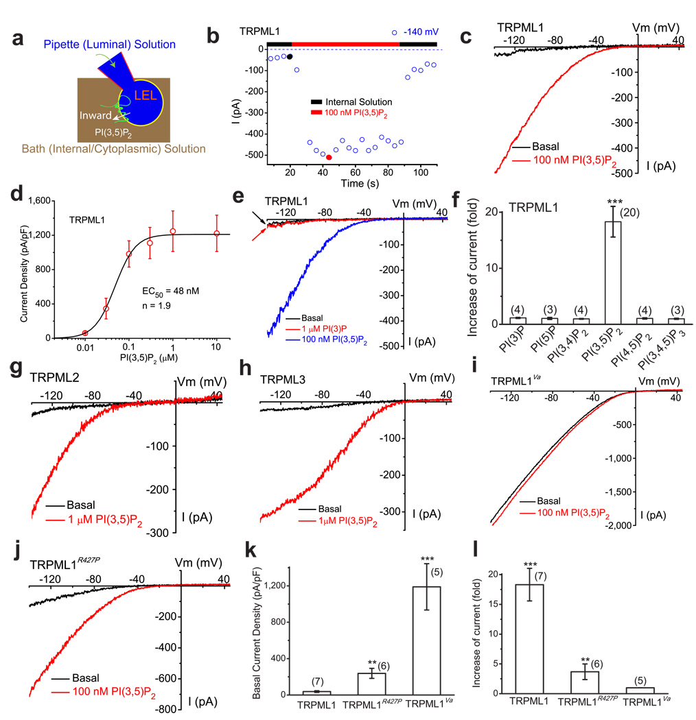
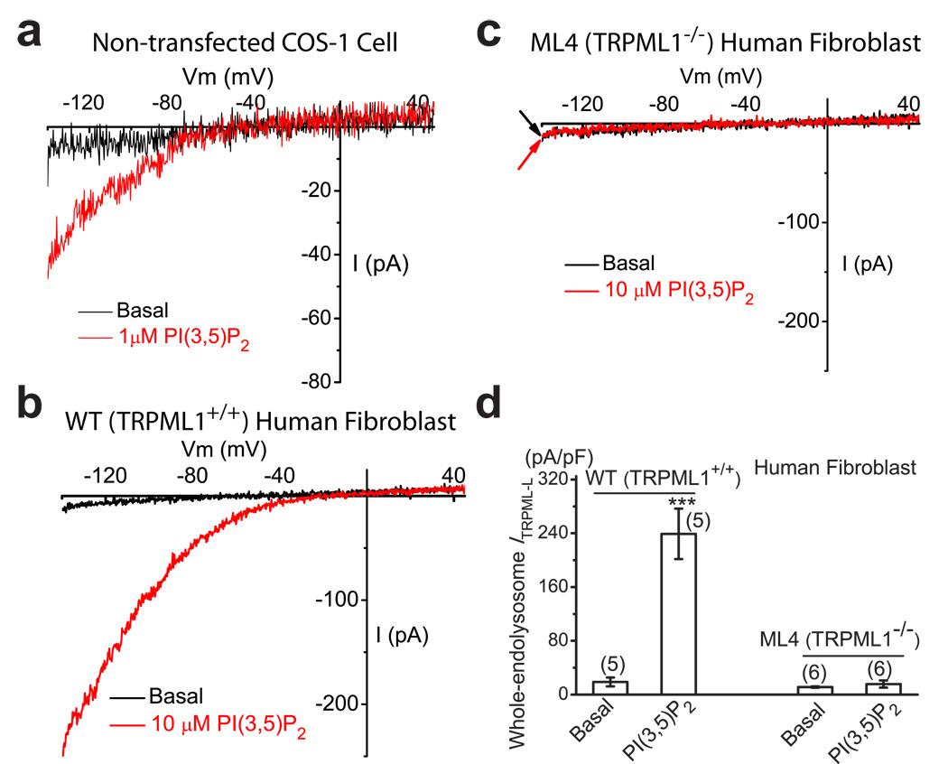
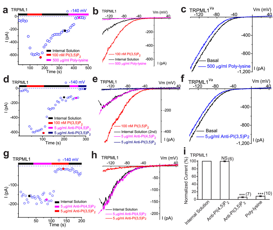
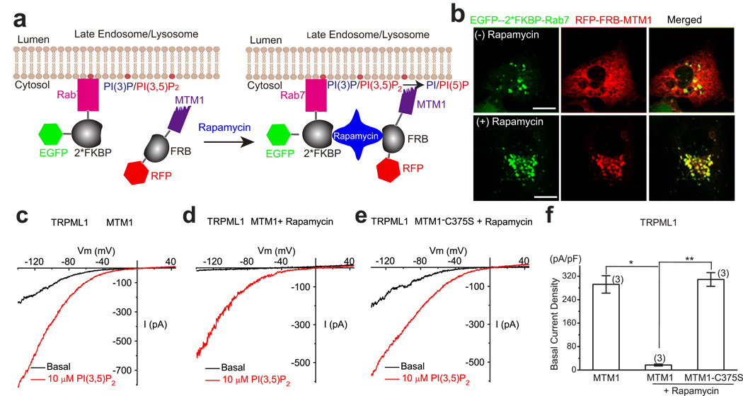
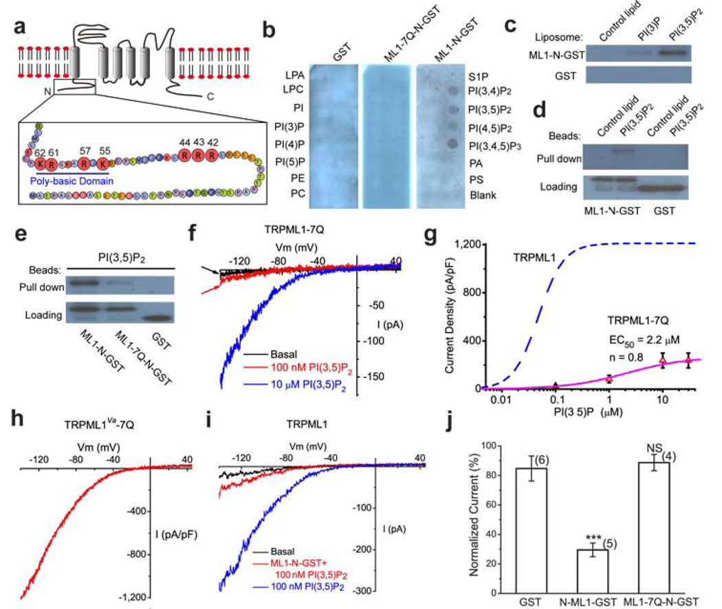
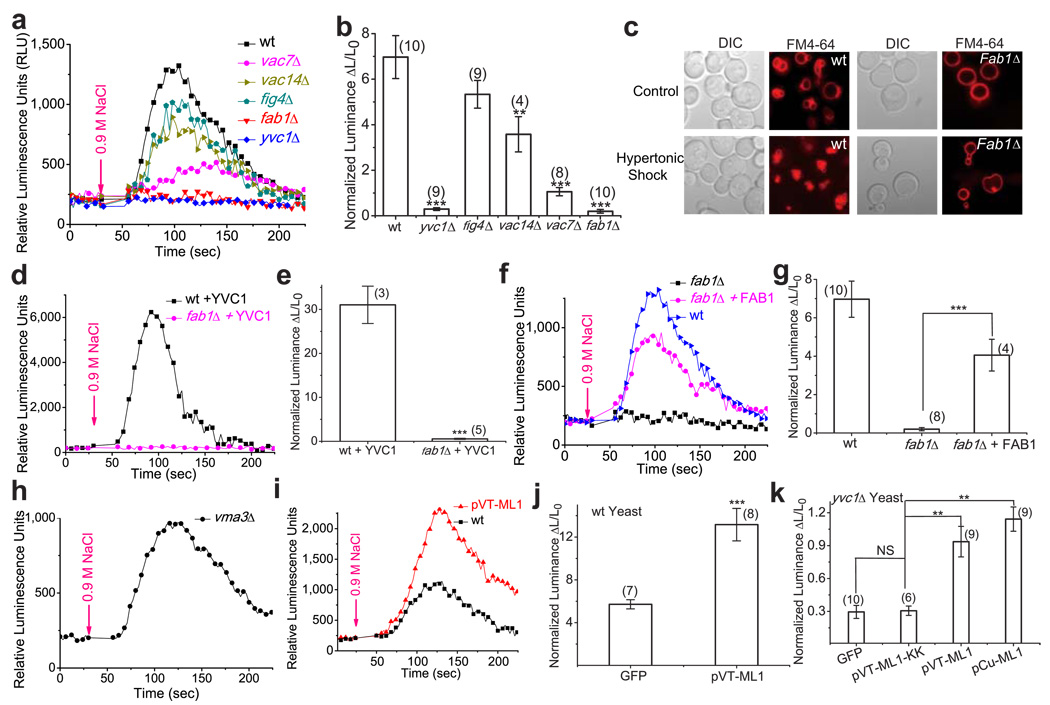
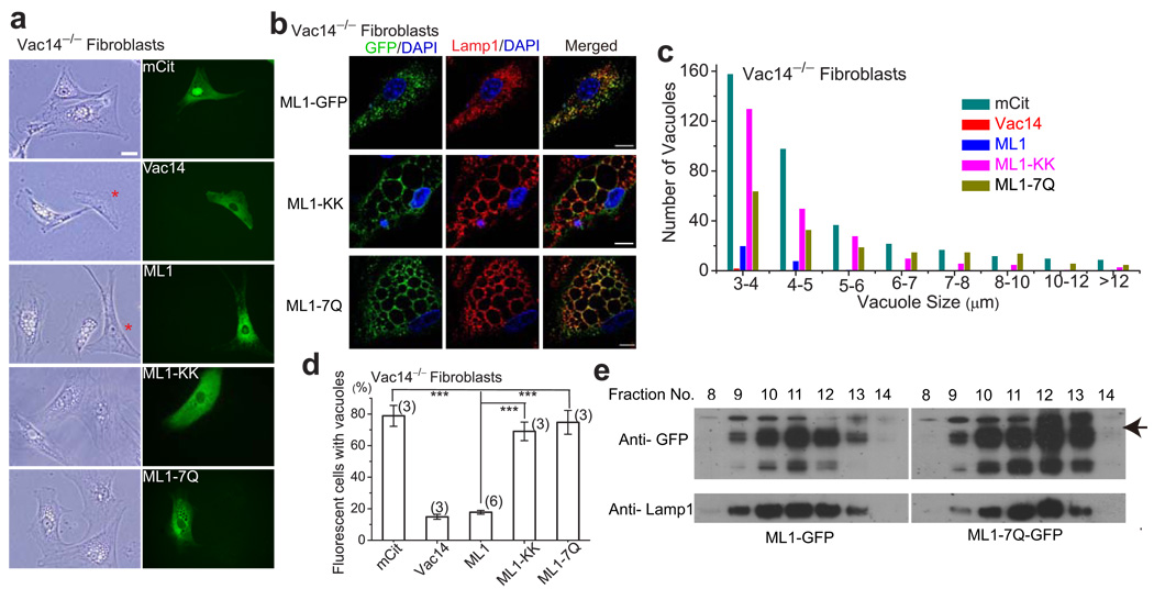
Membrane fusion and fission events in intracellular trafficking are controlled by both intraluminal Ca(2+) release and phosphoinositide (PIP) signalling. However, the molecular identities of the Ca(2+) release channels and the target proteins of PIPs are elusive. In this paper, by direct patch-clamping of the endolysosomal membrane, we report that PI(3,5)P(2), an endolysosome-specific PIP, binds and activates endolysosome-localized mucolipin transient receptor potential (TRPML) channels with specificity and potency. Both PI(3,5)P(2)-deficient cells and cells that lack TRPML1 exhibited enlarged endolysosomes/vacuoles and trafficking defects in the late endocytic pathway. We find that the enlarged vacuole phenotype observed in PI(3,5)P(2)-deficient mouse fibroblasts is suppressed by overexpression of TRPML1. Notably, this PI(3,5)P(2)-dependent regulation of TRPML1 is evolutionarily conserved. In budding yeast, hyperosmotic stress induces Ca(2+) release from the vacuole. In this study, we show that this release requires both PI(3,5)P(2) production and a yeast functional TRPML homologue. We propose that TRPMLs regulate membrane trafficking by transducing information regarding PI(3,5)P(2) levels into changes in juxtaorganellar Ca(2+), thereby triggering membrane fusion/fission events.

Keywords: Ca2+ release channel; Fab1; PI(3,5)P2; PIKfyve; TRP channel; Whole-endolysosome recording; endosome; lysosome; membrane trafficking; phosphoinositide; type IV Mucolipidosis; vacuole.


Figures

Figure 1
Figure 1. PI(3,5)P2 activates recombinant TRPML channels in the endolysosomal membranes
a) Illustration of a whole-endolysosome recording configuration. Pipette (luminal) solution was a standard external (Tyrode’s) solution adjusted to pH 4.6 to mimic the acidic environment of the lysosome lumen. Bath (internal/cytoplasmic) solution was a K+-based solution (140 mM K+-gluconate). Putative membrane topology of TRPML channels is illustrated in the late endosome and lysosome (LEL). Note that the inward current indicates cations flowing out of the endolysosome (see red arrow for the direction). b) Bath application of PI(3,5)P2 (diC8, 100 nM) activated inwardly rectifying whole-endolysosome TRPML1-mediated current (ITRPML1) in an enlarged endolysosome/vacuole from a TRPML1-EGFP-expressing Cos-1 cell that was pre-treated with vacuolin-1. ITRPML1 was elicited by repeated voltage ramps (−140 to +140 mV; 400 ms) with a 4-s interval between ramps. ITRPML1 exhibited a small basal current prior to PI(3,5)P2 application; bath application of PI(3,5)P2 to the cytoplasmic side of the endolysosome resulted in maximal activation of 18-fold of baseline within a minute, measured at −140 mV of ITRPML1. c) Representative traces of ITRPML1 before (black) and after (red) PI(3,5)P2 at two time points, as shown in a (black and red circles). Only a portion of the voltage protocol is shown; holding potential = 0 mV. d) Dose-dependence of PI(3,5)P2-dependent activation (EC50 = 48 nM, n = 1.9). e) PI(3)P (1 µM) failed to activate ITRPML1. f) Specific activation of TRPML1 by PI(3,5)P2 ( in 100 nM), but not other diC8 PIPs (all in 1 µM). g) Activation of whole-endolysosome ITRPML2 by PI(3,5)P2 (1 µM). h) Activation of whole-endolysosome ITRPML3 by PI(3,5)P2 (1 µM). i) Whole-endolysosome ITRPML1-Va exhibited high basal activity but was insensitive to PI(3,5)P2 (100 nM). j) Whole-endolysosome ITRPML1-R427P was weakly activated by PI(3,5)P2. k) Basal current amplitudes of whole-endolysosome ITRPML1, ITRPML1-R427P, and ITRPML1-Va. l) Effects of PI(3,5)P2 on ITRPML1, ITRPML1-R427P, and ITRPML1-Va. For histogram graphs of all figures including panels (f, k, l) of this figure, data are presented as the mean ± standard error of the mean (SEM); the n numbers are in parentheses. Statistical comparisons were made using analysis of variance (ANOVA): P value < 0.05 was considered statistically significant and indicated with asterisks (*, 0.01< P < 0.05; **, P < 0.01; ***, P < 0.001).
Figure 2
Figure 2. PI(3,5)P2 activates endogenous TRPML-like currents in the endolysosomal membranes
a) An endogenous inwardly rectifying TRPML-like current (ITRPML1-L) activated by PI(3,5)P2 (10 µM) in a vacuole isolated from a non-transfected Cos-1 cell. b) Large PI(3,5)P2-activated ITRPML1-L in a vacuole isolated from wild type (WT) human skin fibroblast cell. c) Lack of significant PI(3,5)P2-activated ITRPML1-L in a vacuole isolated from a human ML4 (TRPML1−/−) skin fibroblast cell. d) Endogenous PI(3,5)P2-activated ITRPML-L in WT and TRPML1−/− human fibroblasts. For statistical analysis: ***, P < 0.001; NS (non-significant), P > 0.05).
Figure 3
Figure 3. A decrease in PI(3,5)P2 by chelating agents suppresses TRPML1 channel activity in the endolysosomal membrane
a) Post- PI(3,5)P2 quasi-steady state ITRPML1 was inhibited by bath (internal/cytoplasmic) application of poly-lysine (500 µg/ml) to an enlarged endolysosome from a TRPML1-EGFP-expressing Cos-1 cell. ITRPML1 increased with addition of 100 nM PI(3,5)P2, but gradually reduced to a quasi-steady state level upon washout. b) Representative traces of ITRPML1 at three time points, as shown in a: upon PI(3,5)P2 application (red), washout (black), and poly-lysine application (magenta). c) Lack of poly-lysine (500 µg/ml) effect on whole-endolysosome ITRPML1-Va. d) Post- PI(3,5)P2 quasi-steady state ITRPML1 was inhibited by bath application of neutralizing anti-PI(3,5)P2(5 µg/ml), but not anti-PI(4,5)P2 (5 µg/ml). e) Representative traces of ITRPML1 at three time points, as shown in d. f) Lack of anti- PI(3,5)P2 (5 µg/ml) effect on whole-endolysosome ITRPML1-Va. g, h) Large basal pre- PI(3,5)P2 ITRPML1 was inhibited by bath application of neutralizing anti-PI(3,5)P2 (5 µg/ml), but not anti-PI(4,5)P2. i) ITRPML1 was inhibited by more than 90% by bath (internal/cytoplasmic) application of poly-lysine (500 µg/ml) or PI(3,5)P2 antibody.
Figure 4
Figure 4. A decrease in PI(3,5)P2 level by a translocatable lipid phosphatase suppresses TRPML1 channel activity in the endolysosomal membrane
a) Recruitment of MTM1 to endolysosomal membranes by rapamycin-dependent heterodimerization of RFP-FRB-MTM1 and EGFP-2*FKBP-Rab7. Rab7 is a LEL-specific Rab protein. MTM1 is a PI-3 phosphatase that can convert PI(3,5)P2 and PI(3)P into PI(5)P and PI, respectively. b) Rapamycin-dependent heterodimerization of RFP-FRB-MTM1 and EGFP-2*FKBP-Rab7 alters subcellular localization of MTM1. Cos-1 cells were transfected with both RFP-FRB-MTM1 and EGFP-2*FKBP-Rab7. Rapamycin (500 nM; 20 min) treatment promotes co-localization of MTM1-RFP with Rab7-EGFP. Scale Bar = 10 µm. c-f) The effects of MTM1 on ITRPML1. Cos-1 cells were co-transfected with human TRPML1-myc, RFP-FRB-MTM1 or RFP-FRB-MTM1-C375S, and EGFP-2*FKBP-Rab7. MTM1 was recruited to LEL membranes by rapamycin (500 nM) -dependent heterodimerization of RFP-FRB-MTM1 and EGFP-2*FKBP-Rab7. c) ITRPML1 in MTM1-transfected cells before rapamycin treatment. d) ITRPML1 in MTM1-transfected cells after rapamycin treatment. e) ITRPML1 in MTM1-C357S-transfected cells after rapamycin treatment. f) Differential effects of WT and inactive mutant (C375S) MTM1 on basal whole-endolysosome ITRPML1 For statistical analysis: *, 0.01 < P < 0.05; **, P < 0.01).
Figure 5
Figure 5. Direct binding of PI(3,5)P2 to the TRPML1 N-terminus requires multiple positively-charged amino acid residues
a) The cytoplasmic N-terminus of TRPML1 contains a poly-basic region and clusters of positively charged amino acid residues as potential PI(3,5)P2 binding sites. The positively charged amino acid residues (Arg and Lys) that were mutated into neutral amino acids Gln (Q) in this study are shown with enlarged circles and their amino acid residue numbers. b) Protein-lipid overlays. The strip contained 15 different types of lipids: PA, phosphatidic acid; S1P, sphingosine-1-phosphate. Three purified proteins were used to probe the strip: GST alone (left panel), GST-fused to the N-terminal fragment of TRPML1 (ML1-N-GST; right panel), and Gln-substituted mutant of ML1-N-GST (ML1-7Q-N-GST; middle). Proteins were detected with anti-GST antibodies. c) Liposome pull-down assay. Liposomes were incubated with purified GST-fusion proteins, centrifuged, and associated proteins visualized by Western blot with GST antibodies. d) Binding of GST-ML1-N to agarose beads conjugated to PI(3,5)P2, but not control lipids; GST alone failed to pull down PI(3,5)P2-conjugated beads. e) Compared to GST-ML1-N, GST-ML1-7Q-N exhibited significantly weaker binding to PI(3,5)P2-conjugated agarose beads. f) Whole-endolysosome ITRPML1- 7Q was weakly activated by high concentrations of PI(3,5)P2. g) PI(3,5)P2 dose dependence of ITRPML1- 7Q. Dotted line indicates the dose dependence of ITRPML1 (repotted from Fig. 1d). h) Large basal whole-endolysosome ITRPML1-Va-7Q. Charge-removing Gln substitutions (7Q) were introduced into the gain-of-function Va background. i) GST-ML1-N peptide (5 µg/ml) reduced PI(3,5)P2-dependent activation of whole-endolysosome ITRPML1·. j) Charge-removing Gln-substituted substitutions (7Q) abolished the inhibitory effect of GST-ML1-N peptide.
Figure 6
Figure 6. Ca2+ release from yeast vacuoles after hyperosmostic shock is dependent on PI(3,5)P2 production
a) Representative Ca2+ response measured with aequorin-mediated luminescence in wild-type (wt), yvc1Δ, and PI(3, 5P)2-deficient strains (vac7Δ, vac14Δ, fig4Δ, fab1Δ) upon hyperosmotic shock induced by addition of 0.9 M NaCl. b) Quantitative data to show luminescence responses upon hyperosmotic shock in different yeast strains. Fold-response was normalized to basal luminescence prior to shock. c) Hyperosmotic shock increases the number of vacuoles and decreases the vacuolar volume in wild type, but not fab1 mutant yeast strains. Both WT and fab1Δ cells were labeled with FM4-64 dye to visualize vacuole volume and the number of vacuole lobes. Cells were treated with 0.45M NaCl and viewed by fluorescence microscopy. d, e) Overexpression of YVC1 in fab1Δ cells failed to restore hyperosmolarity-induced Ca2+ response. f, g) Overexpression of FAB1 in fab1Δ cells restored hyperosmolarity-induced Ca2+ response. h, i) Overexpression of TRPML1 in WT yeast cells increased hyperosmolarity-induced Ca2+ response. j) Hyperosmotic shock induces vacuolar Ca2+ release in vma3 mutant yeast strains. k) Overexpression of TRPML1, but not pore mutant TRPML1-KK (in pCu and pVT expression vectors), in yvc1Δ yeast cells resulted in small but significant hyperosmolarity-induced Ca2+ response.
Figure 7
Figure 7. Overexpression of TRPML1 rescues the enlarged endolysosome phenotype of PI(3,5)P2-deficient mouse fibroblasts
a, b) The effects of overexpression of WT TRPML1 and pore (ML1-KK) or PI(3,5)P2-insensitive (ML1-7Q) mutant TRPML1 on the number and size of the vacuoles in Vac14−/− fibroblasts. Cultured Vac14−/− mouse fibroblast cells exhibited variable numbers (1–20) of large (> 3 µm) vacuoles/endolysosomes. Non-vacuolated cells are indicated with asterisk. Scale Bar = 20 µm. b) TRPML1, ML1-KK, and ML1-7Q proteins were co-localized in Lamp1-positive compartments of Vac14−/− fibroblast cells. c) Large vacuoles in 75% of vector (mCit) -transfected Vac14−/− fibroblast cells. Overexpression of Vac14-mCit or EGFP-ML1 reduced the percentage (of enlarged vacuoles) to approximately 15%, while the 75% of EGFP-ML1-KK or EGFP-ML1-7Q-transfected cells contained enlarged vacuoles. d) Histogram analysis of the vacuole size/number in Vac14−/− fibroblasts transfected with indicated constructs. e) Fractionation analysis reveals co-localization of TRPML1 and TRPML1-7Q with Lamp-1. Gradient cellular fractionations were obtained using ultracentrifugation. Both TRPML1 and TRPML1-7Q proteins were concentrated in Lamp1-rich fractionations.

Similar articles

Cited by

References

    1. Hay JC. Calcium: a fundamental regulator of intracellular membrane fusion? EMBO reports. 2007;8:236–240. - PMC - PubMed
    1. Peters C, Mayer A. Ca2+/calmodulin signals the completion of docking and triggers a late step of vacuole fusion. Nature. 1998;396:575–580. - PubMed
    1. Luzio JP, Pryor PR, Bright NA. Lysosomes: fusion and function. Nature reviews. 2007;8:622–632. - PubMed
    1. Dong XP, Wang X, Xu H. TRP channels of intracellular membranes. Journal of neurochemistry. 2010;113:313–328. - PMC - PubMed
    1. Cheng X, Shen D, Samie M, Xu H. Mucolipins: Intracellular TRPML1-3 channels. FEBS letters. 2010 - PMC - PubMed

Publication types

Substances