

First-visit diagnosis of preperimetric glaucoma
- PMID: 20802808
- PMCID: PMC2928913
- DOI: 10.2174/1874364101004010022
First-visit diagnosis of preperimetric glaucoma
Abstract
Purpose: To present a revised interpretation of the work-up data that enabled diagnosis of preperimetric glaucoma (PPG) at the first examination.
Methods: a) Literature analysis on PPG; b) 6-year follow-up of a glaucoma-suspect patient.
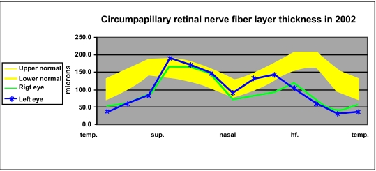
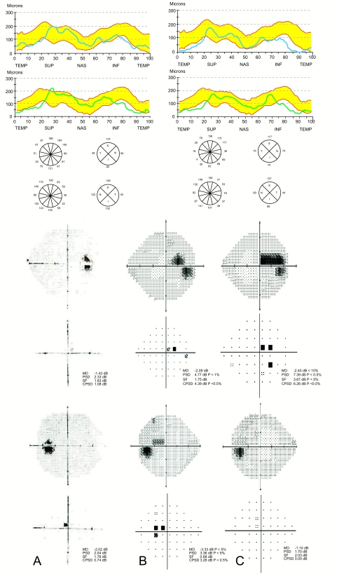
Results: TWO NEW CONCEPTS MAY BE ADAPTED: (a) the objective finding of retinal nerve fiber layer (RNFL) thinning below the normal border in the opposing typical glaucoma locations, the inferior and superior quadrants, and in a non-diffuse pattern, appears asymptomatically and simultaneously only in glaucoma; and (b) the imaging-related RNFL thickness may be considered the reference glaucoma standard, whereas the suspicious early glaucomatous optic neuropathy, having a potential diagnostic inaccuracy, would serve as a complementary revealing finding. That approach enabled, in retrospect, a first-visit diagnosis of low-tension PPG in the patient. Diagnosis was confirmed after 6 years, when cecocentral scotoma and further RNFL thinning emerged despite treatment.
Conclusions: A revised approach enabled PPG diagnosis during the first visit.
Keywords: Preperimetric glaucoma; early glaucoma; low-tension glaucoma; optical coherence tomography; retinal nerve fiber layer..
Figures
Similar articles
-
Development of Glaucomatous Visual Field Defects in Preperimetric Glaucoma Patients Within 3 Years of Diagnosis.J Glaucoma. 2016 Jun;25(6):e591-5. doi: 10.1097/IJG.0000000000000260. J Glaucoma. 2016. PMID: 25943739
-
Evaluation of ganglion cell-inner plexiform layer thickness in the diagnosis of preperimetric glaucoma and comparison to retinal nerve fiber layer.Indian J Ophthalmol. 2021 May;69(5):1113-1119. doi: 10.4103/ijo.IJO_965_20. Indian J Ophthalmol. 2021. PMID: 33913844 Free PMC article.
-
Diagnostic Ability of Wide-field Retinal Nerve Fiber Layer Maps Using Swept-Source Optical Coherence Tomography for Detection of Preperimetric and Early Perimetric Glaucoma.J Glaucoma. 2017 Jun;26(6):577-585. doi: 10.1097/IJG.0000000000000662. J Glaucoma. 2017. PMID: 28368998
-
Comparison of glaucoma-diagnostic ability between wide-field swept-source OCT retinal nerve fiber layer maps and spectral-domain OCT.Eye (Lond). 2018 Sep;32(9):1483-1492. doi: 10.1038/s41433-018-0104-5. Epub 2018 May 23. Eye (Lond). 2018. PMID: 29789659 Free PMC article.
-
Ability of different scanning protocols of spectral domain optical coherence tomography to diagnose preperimetric glaucoma.Invest Ophthalmol Vis Sci. 2013 Nov 1;54(12):7252-7. doi: 10.1167/iovs.13-12731. Invest Ophthalmol Vis Sci. 2013. PMID: 24114539
Cited by
-
Diagnostic performance of optical coherence tomography macular ganglion cell inner plexiform layer and retinal nerve fiber layer thickness in glaucoma suspect and early glaucoma patients at St. Paul's hospital millennium medical college, Addis Ababa, Ethiopia.PLoS One. 2023 Jan 31;18(1):e0263959. doi: 10.1371/journal.pone.0263959. eCollection 2023. PLoS One. 2023. PMID: 36719894 Free PMC article.
-
Differentiating Multiple Sclerosis and Glaucoma With Sectoral Pattern Analysis of Peripapillary Nerve Fiber Layer.Transl Vis Sci Technol. 2024 Nov 4;13(11):11. doi: 10.1167/tvst.13.11.11. Transl Vis Sci Technol. 2024. PMID: 39535747 Free PMC article.
-
Evaluation of RETICs Glaucoma Diagnostic Calculators in Preperimetric Glaucoma.Transl Vis Sci Technol. 2018 Nov 30;7(6):13. doi: 10.1167/tvst.7.6.13. eCollection 2018 Nov. Transl Vis Sci Technol. 2018. PMID: 30519498 Free PMC article.
-
Development of visual field defect after first-detected optic disc hemorrhage in preperimetric open-angle glaucoma.Jpn J Ophthalmol. 2017 Jul;61(4):307-313. doi: 10.1007/s10384-017-0509-x. Epub 2017 Mar 29. Jpn J Ophthalmol. 2017. PMID: 28357611
-
Color visual acuity in preperimetric glaucoma and open-angle glaucoma.PLoS One. 2019 Apr 17;14(4):e0215290. doi: 10.1371/journal.pone.0215290. eCollection 2019. PLoS One. 2019. PMID: 30995280 Free PMC article. Clinical Trial.
References
-
- Sommer A, Katz J, Quigley HA, Miller NR, Robin AL, Richter RC. Clinically detectable nerve fiber atrophy precedes the onset of glaucomatous field loss. Arch Ophthalmol. 1991;109:77–83. - PubMed
-
- Medeiros FA, Zangwill LM, Bowd C, Sample PA, Weinreb RN. Use of progressive glaucomatous optic disk change as the reference standard for evaluation of diagnostic tests in glaucoma. Am J Ophthalmol. 2005;139:1010–8. - PubMed
-
- Airaksinen PJ, Tuulonen A, Werner EB. In: Clinical evaluation of the optic disc and retinal nerve fiber layer. Ritch R, Shields MB, Krupin T, editors. St. Louis: Mosby: The Glaucomas; 1996. pp. 617–57.
-
- Blumenthal EZ, Williams JM, Weinreb RN, Girkin CA, Berry CC, Zangwill LM. Reproducibility of nerve fiber layer thickness measurements by use of optical coherence tomography. Ophthalmology. 2000;107:2278–82. - PubMed
LinkOut - more resources
Full Text Sources