. 2010 Jul 6;1(3):e00102-10.
doi: 10.1128/mBio.00102-10.

Indirect pathogenicity of Haemophilus influenzae and Moraxella catarrhalis in polymicrobial otitis media occurs via interspecies quorum signaling

Affiliations

Indirect pathogenicity of Haemophilus influenzae and Moraxella catarrhalis in polymicrobial otitis media occurs via interspecies quorum signaling

Chelsie E Armbruster et al. mBio. .

Abstract

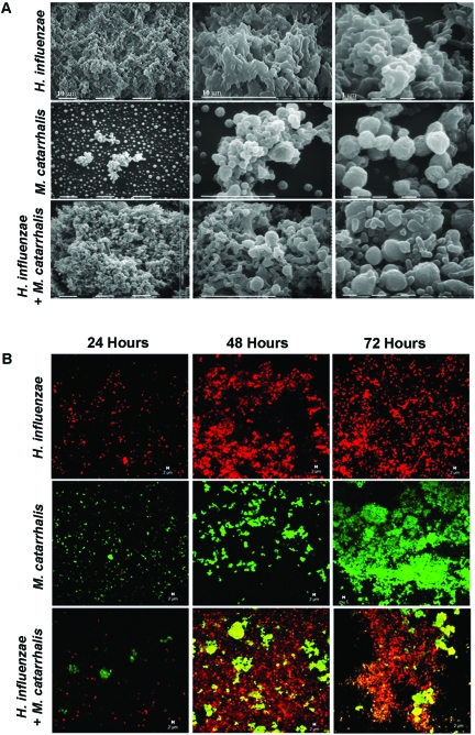
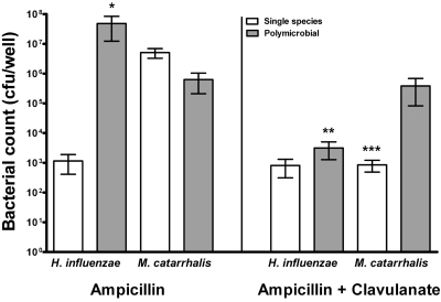
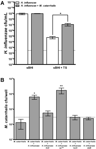
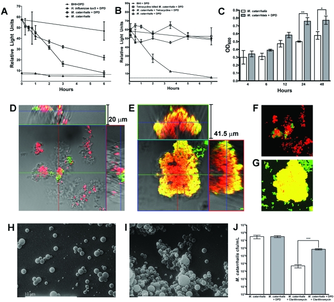
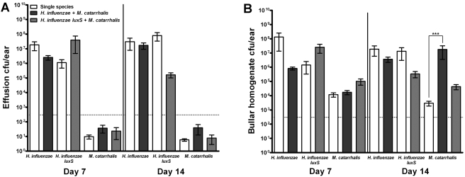
Otitis media (OM) is among the leading diseases of childhood and is caused by opportunists that reside within the nasopharynx, such as Haemophilus influenzae and Moraxella catarrhalis. As with most airway infections, it is now clear that OM infections involve multiple organisms. This study addresses the hypothesis that polymicrobial infection alters the course, severity, and/or treatability of OM disease. The results clearly show that coinfection with H. influenzae and M. catarrhalis promotes the increased resistance of biofilms to antibiotics and host clearance. Using H. influenzae mutants with known biofilm defects, these phenotypes were shown to relate to biofilm maturation and autoinducer-2 (AI-2) quorum signaling. In support of the latter mechanism, chemically synthesized AI-2 (dihydroxypentanedione [DPD]) promoted increased M. catarrhalis biofilm formation and resistance to antibiotics. In the chinchilla infection model of OM, polymicrobial infection promoted M. catarrhalis persistence beyond the levels seen in animals infected with M. catarrhalis alone. Notably, no such enhancement of M. catarrhalis persistence was observed in animals infected with M. catarrhalis and a quorum signaling-deficient H. influenzae luxS mutant strain. We thus conclude that H. influenzae promotes M. catarrhalis persistence within polymicrobial biofilms via interspecies quorum signaling. AI-2 may therefore represent an ideal target for disruption of chronic polymicrobial infections. Moreover, these results strongly imply that successful vaccination against the unencapsulated H. influenzae strains that cause airway infections may also significantly impact chronic M. catarrhalis disease by removing a reservoir of the AI-2 signal that promotes M. catarrhalis persistence within biofilm.


Figures

FIG 1
FIG 1
H. influenzae and M. catarrhalis form polymicrobial biofilms in vitro. Stationary biofilms were established in chamber slides for visualization of bacteria by SEM and confocal laser scanning microscopy (CLSM). (A) Samples of H. influenzae and M. catarrhalis single-species or polymicrobial biofilms were taken at 48 h and prepared for SEM. Images shown are at three different levels of magnification. (B) CLSM was performed on 24-, 48-, and 72-h biofilms following staining of H. influenzae (red) and M. catarrhalis (green).
FIG 2
FIG 2
Beta-lactam protection in a polymicrobial biofilm. Stationary biofilms of H. influenzae Rd and/or M. catarrhalis were established on chamber slides for 24 h and treated with 100 µg/ml ampicillin or ampicillin with 25 µg/ml clavulanate for an additional 24 h. Biofilms were resuspended in sterile PBS, serially diluted, and plated on sBHI medium plus clarithromycin and BHI medium plates for enumeration of viable H. influenzae Rd and M. catarrhalis bacteria, respectively. Data are represented as means ± SEM. *, P < 0.05 compared to H. influenzae plus ampicillin; **, P < 0.05 compared to polymicrobial H. influenzae plus ampicillin; ***, P < 0.05 compared to M. catarrhalis plus ampicillin.
FIG 3
FIG 3
Polymicrobial biofilm formation protects H. influenzae and M. catarrhalis from antibiotic treatment. Single-species or polymicrobial stationary biofilms were established for 4 h and treated with 60 µg/ml trimethoprim-sulfamethoxazole (TS) (A) or 6 µg/ml clarithromycin (B) for 24 h. Biofilms were resuspended in sterile PBS, serially diluted, and plated on sBHI medium plus clarithromycin or BHI medium for enumeration of viable H. influenzae and M. catarrhalis bacteria, respectively. Data are represented as the mean results from three combined experiments, with two replicates per experiment. Error bars represent SEM. *, P < 0.05.
FIG 4
FIG 4
AI-2 promotes M. catarrhalis biofilm development and antibiotic resistance. (A) M. catarrhalis was cultured in BHI medium or BHI medium supplemented with 0.2 µM synthetic AI-2 (DPD) to determine AI-2 production and depletion, as measured by Vibrio harveyi bioluminescence. H. influenzae luxS was cultured in sBHI medium supplemented with DPD to measure depletion. An uninoculated control of BHI medium with DPD shows the minimal degradation of the AI-2 signal during 6 h of incubation at 37°C. (B) Depletion of DPD by M. catarrhalis biofilms were established for 24 h following incubation with 10 µg/ml tetracycline was measured by bioluminescence over a period of 7 h. (C) M. catarrhalis biofilms were established in the presence or absense of DPD and stained with crystal violet for determination of biofilm biomass at 4, 6, 12, 24, and 48 h. Data represent the mean results from three combined experiments, with three replicate wells per experiment. Error bars represent SEM. (D and E) M. catarrhalis biofilms were established for 24 h in the presence (E) or absence (D) of DPD and stained with a viability kit for CLSM visualization of surface coverage and biofilm thickness. (F and G) Z-series images from panels D and E were compressed to show total viable and nonviable staining of biofilms established in the presence (G) or absence (F) of DPD. (H and I) SEM images of 24-h M. catarrhalis biofilms established with (I) or without (H) DPD. (J) M. catarrhalis biofilms were established for 4 h in the presence or absence of DPD and then treated with 6 µg/ml clarithromycin for 24 h and plated for enumeration of viable bacteria. Data represent the means from three replicates ± SEM. *, P < 0.05; **, P < 0.01; ***, P < 0.001.
FIG 5
FIG 5
Polymicrobial infection augments M. catarrhalis persistence in vivo. Chinchillas were infected with 103 CFU of H. influenzae or H. influenzae luxS, 104 CFU of M. catarrhalis, or a mixture of both species. (A) Middle ear effusion fluids were removed for enumeration of viable H. influenzae and M. catarrhalis bacteria by plating on sBHI medium plus clarithromycin or BHI medium, respectively. (B) Bullae were removed at each time point and homogenized for enumeration of viable H. influenzae and M. catarrhalis bacteria, as described above. Data represent the mean results from four experiments ± SEM. ***, P < 0.005 compared to the number of CFU from M. catarrhalis single-species bullar homogenate.

References

    1. Klein J. O. 2000. The burden of otitis media. Vaccine 19:S2–S8 - PubMed
    1. Mandel E. M., Doyle W. J., Winther B., Alper C. M. 2008. The incidence, prevalence and burden of OM in unselected children aged 1–8 years followed by weekly otoscopy through the “common cold” season. Int. J. Pediatr. Otorhinolaryngol. 72:491–499 - PMC - PubMed
    1. Paradise J. L., Rockette H. E., Colborn D. K., Bernard B. S., Smith C. G., Kurs-Lasky M., Janosky J. E. 1997. Otitis media in 2253 Pittsburgh-area infants: prevalence and risk factors during the first two years of life. Pediatrics 99:318–333 - PubMed
    1. Finkelstein J. A., Davis R. L., Dowell S. F., Metlay J. P., Soumerai S. B., Rifas-Shiman S. L., Higham M., Miller Z., Miroshnik I., Pedan A., Platt R. 2001. Reducing antibiotic use in children: a randomized trial in 12 practices. Pediatrics 108:1–7 - PubMed
    1. Pichichero M. E. 2000. Recurrent and persistent otitis media. Pediatr. Infect. Dis. J. 19:911–916 - PubMed

Publication types

MeSH terms

Substances