Single-prolonged stress induces apoptosis by activating cytochrome C/caspase-9 pathway in a rat model of post-traumatic stress disorder
- PMID: 20803313
- PMCID: PMC11498605
- DOI: 10.1007/s10571-010-9550-8
Single-prolonged stress induces apoptosis by activating cytochrome C/caspase-9 pathway in a rat model of post-traumatic stress disorder
Abstract
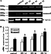
The purpose of this study was to provide a novel insight into the mechanism of how amygdala might participate in PTSD by investigating the changes of cytochrome c oxidase (COX), caspase-9, and caspase-3 in the amygdala of single-prolonged stress (SPS) rats. A total of 80 healthy, male Wistar rats were selected for this study. The models of post-traumatic stress disorder (PTSD) were created by SPS, which is an established animal model for PTSD. The change of COX was detected by light microscope and transmission electron microscopy (TEM). The expression of caspase-9 and caspase-3 in the basolateral amygdala was examined by immunofluorescence and reverse transcription-polymerase chain reaction (RT-PCR). SPS exposure resulted in a significant change of COX in the SPS model groups compared with the normal control group. Evaluation by enzymohistochemistry indicated translocation of COX from mitochondria to cytoplasm. The expression of both caspase-9 and caspase-3 significantly increased 1 day after SPS stimulation, then gradually increased and peaked at SPS 7d. This findings suggest changes of COX, caspase-9, and caspase-3 in the amygdala of SPS rats, which may play important roles in the pathogenesis of PTSD.
Figures


Similar articles
-
Single-prolonged stress induced mitochondrial-dependent apoptosis in hippocampus in the rat model of post-traumatic stress disorder.J Chem Neuroanat. 2010 Nov;40(3):248-55. doi: 10.1016/j.jchemneu.2010.07.001. Epub 2010 Jul 17. J Chem Neuroanat. 2010. PMID: 20624456
-
Single prolonged stress induces dysfunction of endoplasmic reticulum in a rat model of post-traumatic stress disorder.Mol Med Rep. 2015 Aug;12(2):2015-20. doi: 10.3892/mmr.2015.3590. Epub 2015 Apr 2. Mol Med Rep. 2015. PMID: 25846848
-
Single-prolonged stress induces apoptosis in dorsal raphe nucleus in the rat model of posttraumatic stress disorder.BMC Psychiatry. 2012 Nov 27;12:211. doi: 10.1186/1471-244X-12-211. BMC Psychiatry. 2012. PMID: 23181934 Free PMC article.
-
Single Prolonged Stress induces ATF6 alpha-dependent Endoplasmic reticulum stress and the apoptotic process in medial Frontal Cortex neurons.BMC Neurosci. 2014 Oct 21;15:115. doi: 10.1186/s12868-014-0115-5. BMC Neurosci. 2014. PMID: 25331812 Free PMC article.
-
Role of apoptosis in the Post-traumatic stress disorder model-single prolonged stressed rats.Psychoneuroendocrinology. 2018 Sep;95:97-105. doi: 10.1016/j.psyneuen.2018.05.015. Epub 2018 May 12. Psychoneuroendocrinology. 2018. PMID: 29843020 Review.
Cited by
-
Using c-Jun to identify fear extinction learning-specific patterns of neural activity that are affected by single prolonged stress.Behav Brain Res. 2018 Apr 2;341:189-197. doi: 10.1016/j.bbr.2017.12.037. Epub 2017 Dec 29. Behav Brain Res. 2018. PMID: 29292158 Free PMC article.
-
Single-Prolonged Stress: A Review of Two Decades of Progress in a Rodent Model of Post-traumatic Stress Disorder.Front Psychiatry. 2018 May 15;9:196. doi: 10.3389/fpsyt.2018.00196. eCollection 2018. Front Psychiatry. 2018. PMID: 29867615 Free PMC article. Review.
-
Cerebellar and multi-system metabolic reprogramming associated with trauma exposure and post-traumatic stress disorder (PTSD)-like behavior in mice.Neurobiol Stress. 2021 Jan 23;14:100300. doi: 10.1016/j.ynstr.2021.100300. eCollection 2021 May. Neurobiol Stress. 2021. PMID: 33604421 Free PMC article.
-
CpG methylation of brain-derived the neurotrophic factor gene promoter as a potent diagnostic and prognostic biomarker for post-traumatic stress disorder.Int J Clin Exp Pathol. 2018 Oct 1;11(10):5101-5109. eCollection 2018. Int J Clin Exp Pathol. 2018. PMID: 31949588 Free PMC article.
-
Increased neuronal apoptosis in medial prefrontal cortex is accompanied with changes of Bcl-2 and Bax in a rat model of post-traumatic stress disorder.J Mol Neurosci. 2013 Sep;51(1):127-37. doi: 10.1007/s12031-013-9965-z. Epub 2013 Feb 5. J Mol Neurosci. 2013. PMID: 23381833
References
-
- American Psychiatric Association (1994) Diagnostic and statistical manual of mental disorders, 4th edn. DSM-IV. American Psychiatric Press, Washington
-
- Cahill L, McGaugh JL (1998) Mechanisms of emotional arousal and lasting declarative memory. Trends Neurosci 21:294–299 - PubMed
-
- Cryns V, Yuan J (1998) Proteases to die for. Genes Dev 12:1551–1570 - PubMed
-
- Davis M (1994) The role of the amygdala in emotional learning. Int Rev Neurobiol 36:225–266 - PubMed
Publication types
MeSH terms
Substances
LinkOut - more resources
Full Text Sources
Medical
Research Materials

