. 2010 Oct;239(10):2659-73.
doi: 10.1002/dvdy.22405.

Expression, functional, and structural analysis of proteins critical for otoconia development

Affiliations

Expression, functional, and structural analysis of proteins critical for otoconia development

Yinfang Xu et al. Dev Dyn. 2010 Oct.

Erratum in

  • Dev Dyn. 2011 Feb;240(2):457

Abstract

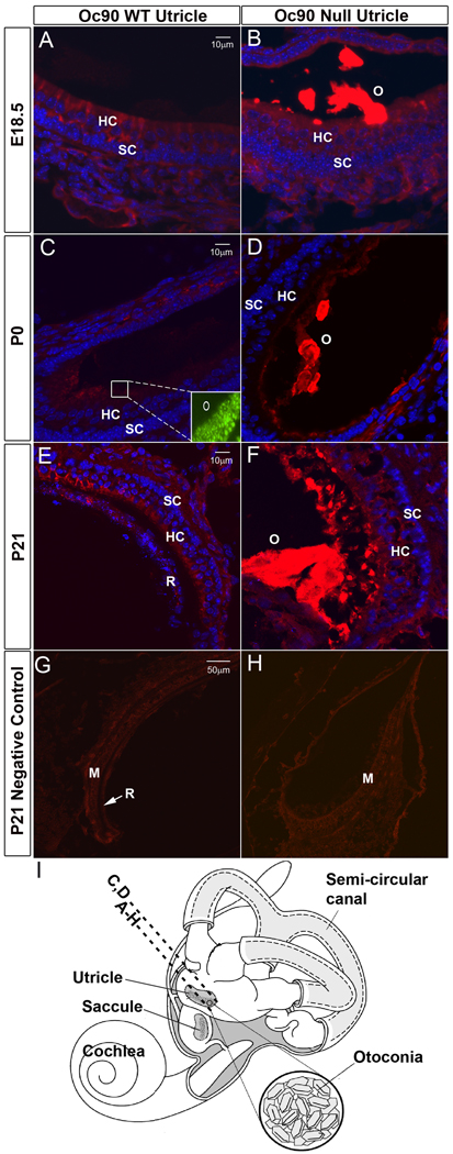
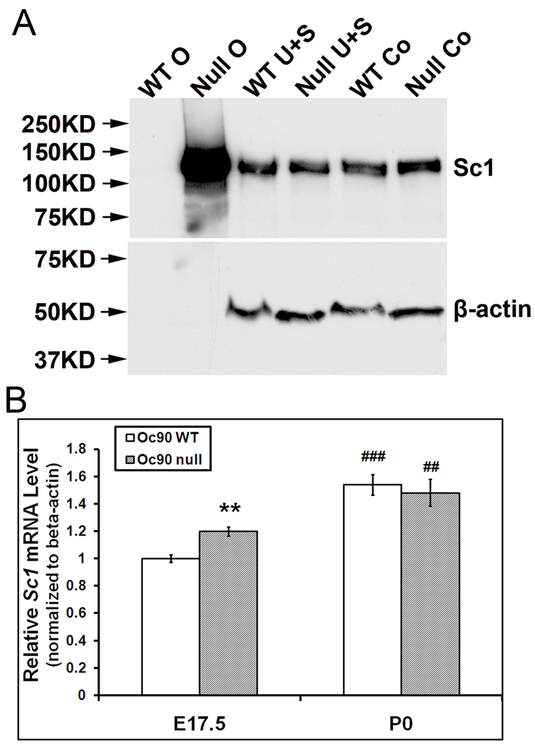
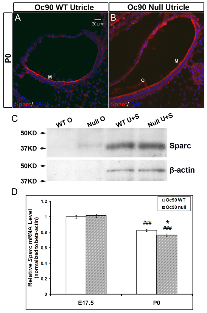
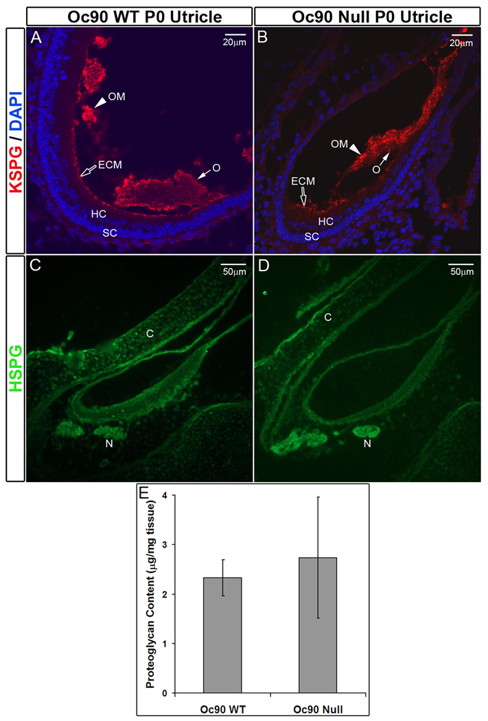
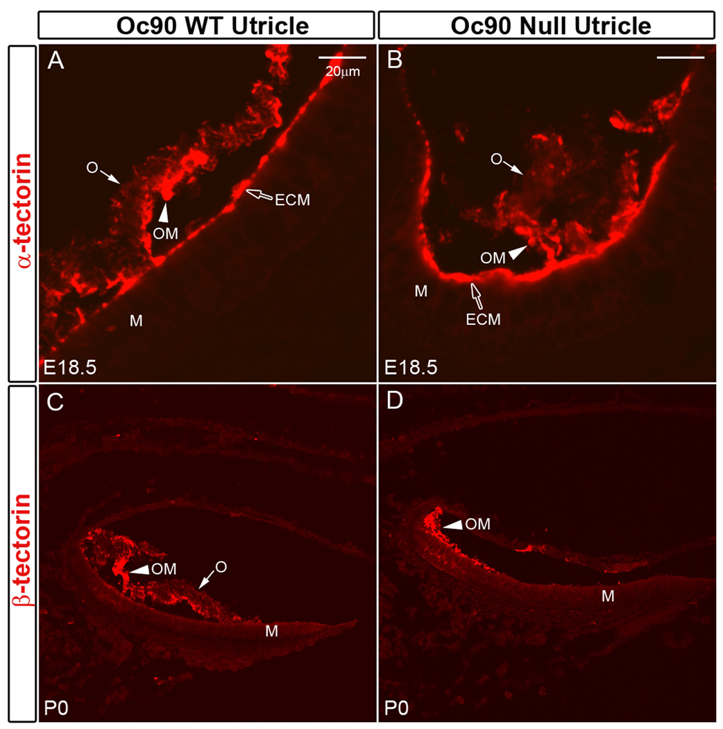
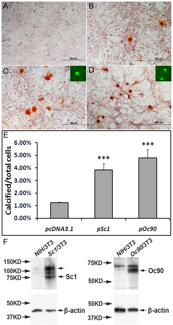
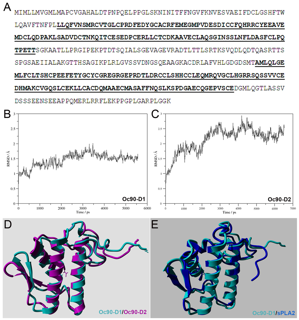
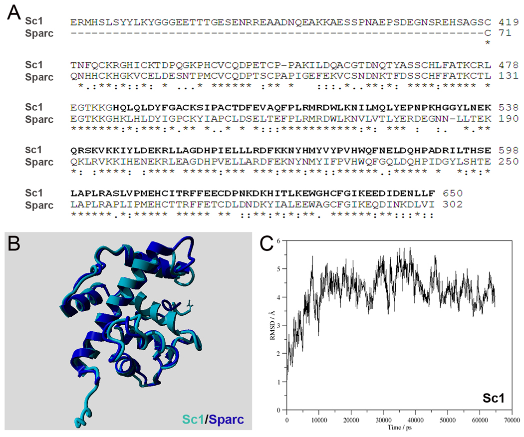
Otoconia, developed during late gestation and perinatal stages, couple mechanic force to the sensory hair cells in the vestibule for motion detection and bodily balance. In the present work, we have investigated whether compensatory deposition of another protein(s) may have taken place to partially alleviate the detrimental effects of Oc90 deletion by analyzing a comprehensive list of plausible candidates, and have found a drastic increase in the deposition of Sparc-like 1 (aka Sc1 or hevin) in Oc90 null versus wt otoconia. We show that such up-regulation is specific to Sc1, and that stable transfection of Oc90 and Sc1 full-length expression constructs in NIH/3T3 cells indeed promotes matrix calcification. Analysis and modeling of Oc90 and Sc1 protein structures show common features that may be critical requirements for the otoconial matrix backbone protein. Such information will serve as the foundation for future regenerative purposes.


Figures

Figure 1
Figure 1
Deposition of Sc1 in Oc90 wt and null otoconia (the utricle is shown). Immunostaining is shown in (A, B) E18.5, (C, D) P0, (E, F) P21 and (G, H, negative controls at P21 using non-immune serum). In (A, C, E), WT otoconia had extremely faint staining that was slightly visible only under the microscope but not in the photographs, whereas the giant crystals (labeled as “O” in B, D, F) in null tissues were intensely stained. Inset in C shows the presence of normal otoconia in the wt tissue section as detected by Oc90 immunostaining. (I) An illustration of the mouse inner ear. The dashed line labeled with A–H shows the approximate location of the sections except C, D, which are located more periphery as marked by the dashed line above. All epithelial cell types in the utricle and saccule participate in otoconia formation. HC, hair cells; SC, supporting cells; M, macula; R, roof.
Figure 2
Figure 2
Otoconial deposition and cellular expression of Sc1 in Oc90 wt and null vestibules. (A) Western blotting of Sc1 in Oc90 wt and null tissues. 6 µg total protein from Oc90 wt otoconia (labeled as “WT O”), 2 µg total protein from null otoconia (“Null O”), and 20 µg total protein from each of the epithelial cellular extracts (U, utricle; S, saccule; Co, cochlea) were loaded. In Oc90 wt otoconia, there was only a faint band after a long exposure; whereas a large amount of Sc1 was detected in Oc90 null otoconia (age P2–3). The same membrane was stripped and re-used for β-actin. β-actin is not present in otoconia, but the equal amount of β-actin detected in each epithelial extract shows that the micro-BCA assay accurately measured the protein content, which was used to control the loading amount. (B) Real-time PCR shows a small but significant increase in Sc1 mRNA in the Oc90 null utricular and saccular epithelia as compared to wt tissues at age E17.5 (**, P<0.01, n=3) but not at P0 (n=3). The relative Sc1 expression levels are higher at P0 than E17.5 in both genotypes (## and ###, P<0.01 and P<0.001, respectively, P0 vs. E17.5 within the same genotype).
Figure 3
Figure 3
Deposition of Sparc in Oc90 wt and null otoconia and its cellular expression (the utricle is shown). Immunostaining is shown in (A, B) P0. Negative controls are similar to Figure 1. WT otoconia show no visible staining, but the giant crystals (“O”) in null tissues are mildly stained. HC, hair cells; SC, supporting cells. (C) Western blotting of Sparc in Oc90 wt and null tissues. Otoconia extracts with equal total protein show a small amount of Sparc in the null otoconia that is absent in wt otoconia (age P15, 7 µg each). The cellular Sparc level is similar between wt and null tissues (20 µg total protein was loaded for each). The same membrane was stripped and re-used for β-actin. β-actin is not present in otoconia, but the equal amount of β-actin detected in each epithelial extract shows that the micro-BCA assay accurately measured the protein content, which was used to control the loading amount. (D) Real-time PCR shows no significant difference in Sparc mRNA levels between wt and null utricular and saccular epithelia at age E17.5 and a small difference at age P0 (* indicates P<0.05, n=4). Expression reduces with age for both genotypes (###, P<0.001, P0 vs. E17.5 within the same genotype).
Figure 4
Figure 4
Presence of KSPG (A, B) and absence of HSPG (C, D) in murine otoconia. (E) DMB proteoglycan assay shows no significant difference in the total epithelial proteoglycan content between Oc90 wt and null utricles and saccules (n=4, P=0.2, age P6). C, cartilage; ECM, extra-cellular matrix; HC, hair cells; M, macula; N, nerve fiber; O, otoconia; OM, otoconial membrane; SC, supporting cells.
Figure 5
Figure 5
Presence/absence of α-tectorin (A, B) and β-tectorin (C, D) in murine otoconia. Expression and deposition of these proteins are not affected in the Oc90 null vestibule. The tectorins are abundant in the extra-cellular matrix (ECM) and otoconial membrane (OM). There is an occasional staining of α-tectorin in the wt crystals. ECM, extracellular matriz; M, Macula; O, otoconia; OM, otoconial membrane.
Figure 6
Figure 6
The effects of Oc90 or Sc1 on ECM calcification. NIH/3T3 cells stably transfected with (C) pSc1 and (D) pOc90 calcified intensely after 5 days of induction with 0.5 mM Ca2+ and 2mM Pi. Inorganic calcium deposits were visualized with ARS staining, proteins with fluorescent immunostaining. Averaged percentages of cells with ECM calcification over total cells were compared for different vectors. Under the same culture conditions, untransfected cells and cells transfected with empty vectors (pcDNA3.1 is shown in B) had similar ratios of calcification, but both had significantly lower ratios than those transfected with Oc90 or Sc1 (Figure 6C & 6D vs. 6B, p<0.001, n=3 experiments × 3~4 survey fields in each). Insets in C & D show that Sc1 and Oc90, respectively, are present in the calcified nodules. Figure 6E shows histograms of the averaged percentages of cells with ECM calcification. No calcification was seen in cells transfected with any of these constructs and cultured under standard media without supplemental Ca2+ and Pi (A). (E) Western blotting of Sc1 and Oc90 in untransfected and transfected cells. 20 µg total protein was loaded in each lane. Each membrane was stripped and re-used for β-actin detection. The upper arrow in the Sc1 lane corresponds to Sc1 dimer in the size of ~130–140 kD.
Figure 7
Figure 7
(A) Amino acid sequence of murine Oc90 (Swiss-Prot id Q9Z0L3). The Pla2l domains are underlined and bolded. (B, C) Backbone RMSD of the first 102 residues as function of simulation time of Oc90-D1 and -D2 (the 1st and 2nd Pla2l domain, respectively). (D) Overlaid time-averaged structures of Oc90-D1 (cyan) and -D2 (purple). (E) Overlaid structures of Oc90-D1 (cyan) and human sPLA2 (blue). The RMSD of the two overlaid structures is 1.517 Å, indicating high structural similarity.
Figure 8
Figure 8
(A) Alignment of the sequences of murine Sc1 (Swiss-Prot id P70663) and murine Sparc (Swiss-Prot id P07214). Residues 489 to 650, for which the homology model was built, are underlined and bolded. (B) Overlaid structures of Sc1 (489–650) (purple) and the known crystal structure of the collagen- and Ca2+-binding domain of human SPARC (blue). The RMSD is 1.023 Å, indicating high structural similarity. (C) Backbone RMSD of Sc1 as function of simulation time.

Similar articles

Cited by

References

    1. Balsamo G, Avallone B, Del GF, Trapani S, Marmo F. Calcification processes in the chick otoconia and calcium binding proteins: patterns of tetracycline incorporation and calbindin-D28K distribution. Hear Res. 2000;148:1–8. - PubMed
    1. Banfi B, Malgrange B, Knisz J, Steger K, Dubois-Dauphin M, Krause KH. NOX3, a superoxide-generating NADPH oxidase of the inner ear. J Biol Chem. 2004;279:46065–46072. - PubMed
    1. Bellahcene A, Castronovo V, Ogbureke KU, Fisher LW, Fedarko NS. Small integrin-binding ligand N-linked glycoproteins (SIBLINGs): multifunctional proteins in cancer. Nat Rev Cancer. 2008;8:212–226. - PMC - PubMed
    1. Boanini E, Torricelli P, Gazzano M, Giardino R, Bigi A. Nanocomposites of hydroxyapatite with aspartic acid and glutamic acid and their interaction with osteoblast-like cells. Biomaterials. 2006;27:4428–4433. - PubMed
    1. Bolander ME, Young MF, Fisher LW, Yamada Y, Termine JD. Osteonectin cDNA sequence reveals potential binding regions for calcium and hydroxyapatite and shows homologies with both a basement membrane protein (SPARC) and a serine proteinase inhibitor (ovomucoid) Proc Natl Acad Sci U S A. 1988;85:2919–2923. - PMC - PubMed

Publication types

MeSH terms

LinkOut - more resources