

Review
doi: 10.1002/cm.20472.
Rho-kinase/ROCK: A key regulator of the cytoskeleton and cell polarity
Affiliations
- PMID: 20803696
- PMCID: PMC3038199
- DOI: 10.1002/cm.20472
Item in Clipboard
Review
Rho-kinase/ROCK: A key regulator of the cytoskeleton and cell polarity
Cytoskeleton (Hoboken).
2010 Sep.
Free PMC article
Abstract
Rho-associated kinase (Rho-kinase/ROCK/ROK) is an effector of the small GTPase Rho and belongs to the AGC family of kinases. Rho-kinase has pleiotropic functions including the regulation of cellular contraction, motility, morphology, polarity, cell division, and gene expression. Pharmacological analyses have revealed that Rho-kinase is involved in a wide range of diseases such as vasospasm, pulmonary hypertension, nerve injury, and glaucoma, and is therefore considered to be a potential therapeutic target. This review focuses on the structure, function, and modes of activation and action of Rho-kinase.
2010 Wiley-Liss, Inc.
Figures
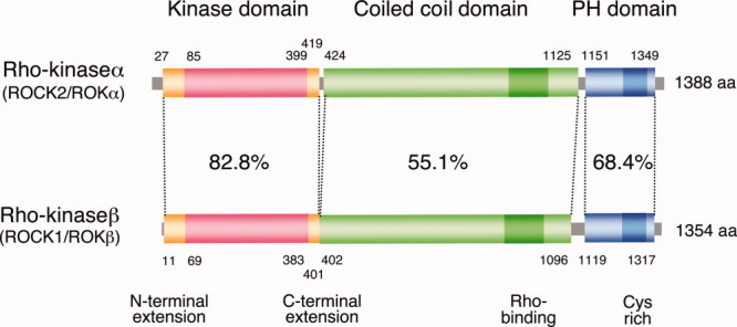
Schematic diagrams of the domain structure of Rho-kinases. Amino acid sequence identities for each domain are indicated.
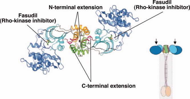
Rho-kinase forms a head-to head homodimer through its N- and C-terminal extensions. Fasudil binds to the ATP-binding cleft. Predicted whole structure of Rho-kinase is also shown, in which Rho-kinase forms parallel dimmer through both the extensions outside of catalytic domain and central coiled-coil regions. Arrows indicate the active centers of Rho-kinase. Reprinted from Structure, Vol 14(3), 2006, Yamaguchi et al., DOI: 10.1016/j.str.2005.11.024 ; ©2005, with permission from Elsevier.
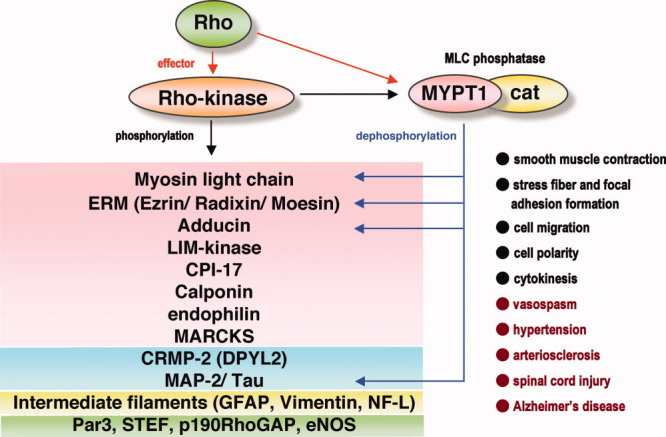
Rho-kinase inhibits the MLC phosphatase activity through both phosphorylation of MYPT1 of MLC phosphatase and phosphorylation of CPI17, an inhibitory protein of myosin phosphatase. Rho-kinase and MLC phosphatase share their substrates, such as MLC, ERM proteins, adducin and MAPs, and thought to regulate the level of phosphorylation. The substrates reported to be phosphorylated by Rho-kinase are illustrated; actin-bindig/regulating proeins (red), MT-binding/regulating proteins (blue), intermediate filaments (yellow), and proteins in other signaling pathways (green). Physiological (black) and pathological (red) processes in which Rho-kinase is involved are listed at the bottom right.
References
-
- Ai S, Kuzuya M, Koike T, Asai T, Kanda S, Maeda K, Shibata T, Iguchi A. Rho-Rho kinase is involved in smooth muscle cell migration through myosin light chain phosphorylation-dependent and independent pathways. Atherosclerosis. 2001;155:321–327. - PubMed
-
- Amano M, Ito M, Kimura K, Fukata Y, Chihara K, Nakano T, Matsuura Y, Kaibuchi K. Phosphorylation and activation of myosin by Rho-associated kinase (Rho-kinase) J Biol Chem. 1996;271:20246–20249. - PubMed
-
- Amano M, Chihara K, Kimura K, Fukata Y, Nakamura N, Matsuura Y, Kaibuchi K. Formation of actin stress fibers and focal adhesions enhanced by Rho-kinase. Science. 1997;275:1308–1311. - PubMed
-
- Amano M, Chihara K, Nakamura N, Fukata Y, Yano T, Shibata M, Ikebe M, Kaibuchi K. Myosin II activation promotes neurite retraction during the action of Rho and Rho-kinase. Genes Cells. 1998;3:177–188. - PubMed
Publication types
MeSH terms
Substances
LinkOut - more resources
Full Text Sources
Other Literature Sources
Molecular Biology Databases
