. 2010 Oct;78(2):320-30.
doi: 10.1111/j.1365-2958.2010.07330.x. Epub 2010 Aug 20.

Substrate path in the AcrB multidrug efflux pump of Escherichia coli

Affiliations

Substrate path in the AcrB multidrug efflux pump of Escherichia coli

Fasahath Husain et al. Mol Microbiol. 2010 Oct.

Abstract

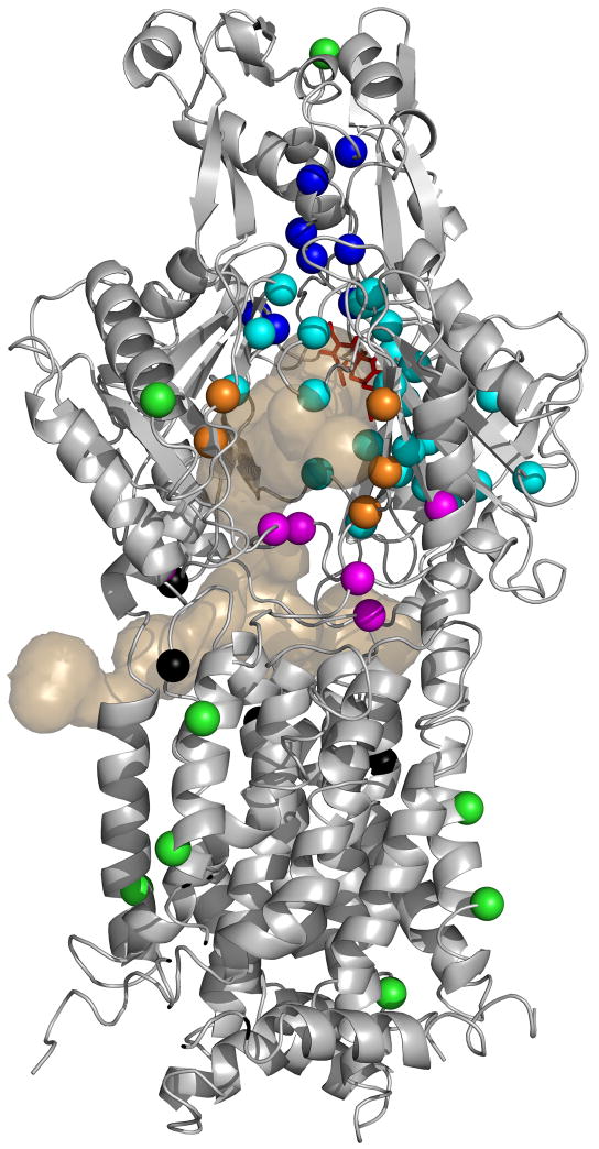
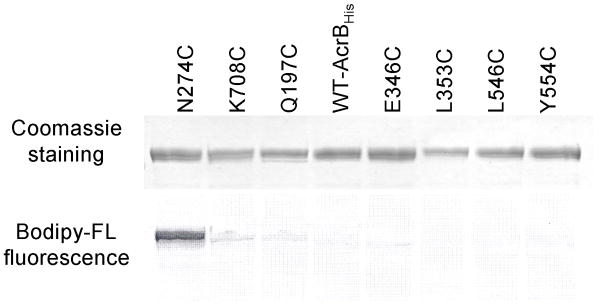
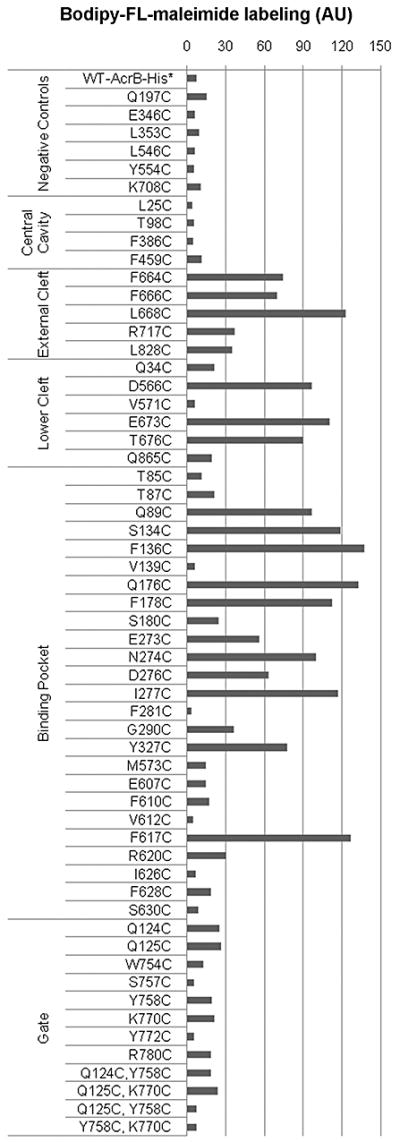
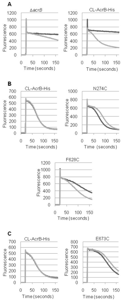
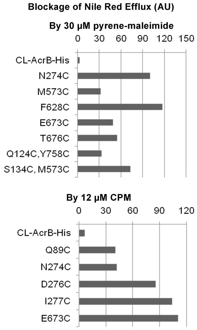
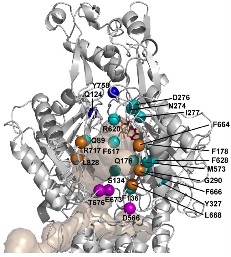
A major tripartite multidrug efflux pump of Escherichia coli, AcrAB-TolC, confers resistance to a wide variety of compounds. The drug molecule is captured by AcrB probably from the periplasm or the periplasm/inner membrane interface, and is passed through AcrB and then TolC to the medium. Currently, there exist numerous crystallographic and mutation data concerning the regions of AcrB and its homologues that may interact with substrates. Starting with these data, we devised fluorescence assays in whole cells to determine the entire substrate path through AcrB. We tested 48 residues in AcrB along the predicted substrate path and 25 gave positive results, based on the covalent labelling of cysteine residues by a lipophilic dye-maleimide and the blocking of Nile red efflux by covalent labelling with bulky maleimide reagents. These residues are all located in the periplasmic domain, in regions we designate as the lower part of the large external cleft, the cleft itself, the crystallographically defined binding pocket, and the gate between the pocket and the funnel. Our observations suggest that the substrate is captured in the lower cleft region of AcrB, then transported through the binding pocket, the gate and finally to the AcrB funnel that connects AcrB to TolC.


Figures

Figure 1
Figure 1. AcrB residues examined in this study
Structure of the binding protomer of AcrB (from PDB file 2DRD) is shown in a grey diagram. The top half of the molecule is the periplasmic domain (with the TolC-binding subdomain [Tamura et al., 2005] on top), and the bottom half represents the transmembrane domain embedded in the inner membrane. The view is from outside of the trimer. The bound minocycline is shown as a red stick model. The light tan-colored surface diagram shows the connection of this binding pocket to the outside, through three wide channels visualized by the program CAVER (Petrek et al., 2006). One channel exits closest to the reader in the large external cleft; the second one exits to the left into the central cavity, and the third one exits to the right into a vestibule. (These features are similar to those described already [Murakami et al., 2006; Sennhauser et al., 2007]). Residues chosen for conversion to Cys are shown in color-coded spheres. They are from the gate (deep blue), binding pocket (light blue), cleft (orange), bottom of the cleft (mauve), and central cavity (black). Green spheres indicate the residues located outside the probable drug pathway and used as controls. For details, see text.
Figure 2
Figure 2. Examples of the staining of CL-AcrBHis mutants by Bodipy-FL-maleimide
The overnight-grown whole cells at OD660 3.5 were stained with 6 μM Bodipy-FL-maleimide for 1 h. The CL-AcrBHis was isolated from 5 ml of cells and was eluted with 100 μl of elution buffer. A portion (7.5 μl) of the eluate was resolved on SDS-PAGE. The resolved AcrB bands were observed for staining by Bodipy-FL-maleimide using Typhoon phosphorimager, and the same gel was then stained using the Coomassie stain. The WT-AcrBHis contains two native cysteines, Cys493 and Cys887. The other alterations are made in CL-AcrBHis, which does not have native cysteines.
Figure 3
Figure 3. Covalent labeling of AcrB mutants in whole cells with 6 μM Bodipy-FL-maleimide
CL-AcrBHis (with no native cysteines) was used to alter the selected residues to cysteines. The extent of labeling was calculated by correcting for the variations in the protein expression level by dividing the fluorescence with the amount of protein, revealed by the Coomassie staining (see Experimental Procedures). The extent of labeling was normalized by using that of N274C as 100. We used 30 as the arbitrary cut-off point to distinguish labeled and unlabeled mutant proteins. WT-AcrBHis has two native cysteines Cys493 and Cys887 outside the presumed substrate path, and thus served as one negative control. AU: arbitrary unit.
Figure 4
Figure 4. AcrB-mediated Nile Red efflux
Nile red efflux was followed as described in Experimental Procedures. Intensity of fluorescence emission is shown in arbitrary units. A. Efflux in strains expressing no AcrB (ΔacrB) or expressing CL-AcrBHis, with (dark line) or without (light line) 0.4 % glucose. B. Effect of pyrene-maleimide on Nile Red efflux. Efflux time course in the presence of 0.4% glucose is shown, as examples, in strains expressing CL-AcrBHis and its mutants (N274C and F628C). Experiments were without pyrene-maleimide (dark line) or with 30 μM pyrene-maleimide (light line). C. Effect of CPM on Nile Red efflux. Time course of Nile Red efflux is shown in CL-AcrBHis and the E673C mutant, as typical examples. All samples contained 0.4% glucose. Experiments were without CPM (dark line) or with 12 μM CPM (light line).
Figure 5
Figure 5. Extent of Blockage of Nile Red efflux in various AcrB mutants by 30 μM pyrene-maleimide or 12 μM CPM
The extent of blockage, calculated as described in Experimental Procedures, is shown as a percentage of blockage seen in reference mutants (N274C for pyrene-maleimide and E673C for CPM). We set the arbitrary cut-off point at 30%. In order to save space, we show only those mutants that showed significant blockage, although we tested all mutants described earlier. The efflux activity of mutant F628C, which was minimal, was significantly stimulated by pyrene-maleimide (Fig. 4B); although this is an inverse of blockage, corresponding to a negative number, it is shown as a positive value here to save space.
Figure 6
Figure 6. Positions of residues involved in substrate transport through AcrB
Those residues that gave positive response in Bodipy-FL-maleimide assay or in CPM/pyrene maleimide assay or both are identified and shown as spheres, colored magenta for those in the bottom of cleft, orange for those in cleft, light blue for those in the binding pocket, and deep blue for those in the gate, as in Fig. 1. E273 is hidden behind the cluster of D276, N274, and I277. The Binding Protomer of AcrB (based on the PDB file 2DRD) is shown as a gray ribbon model, oriented as in Fig. 1, so that its TolC-binding region is on top and we look into the protein from the outside of the trimer structure. Tunnels leading to the binding pocket, as well as the bound minocycline found in the pocket, are also shown as in Fig. 1.

References

    1. Bohnert JA, Karamian B, Nikaido H. Optimized Nile Red efflux assay of AcrAB-TolC multidrug efflux system shows competition between substrates. Antimicrob Agents Chemother. 2010 in press. - PMC - PubMed
    1. Bohnert JA, Kern WV. Selected arylpiperazines are capable of reversing multidrug resistance in Escherichia coli overexpressing RND efflux pumps. Antimicrob Agents Chemother. 2005;49:849–852. - PMC - PubMed
    1. Bohnert JA, Schuster S, Fähnrich E, Trittler R, Kern WV. Altered spectrum of multidrug resistance associated with a single point mutation in the Escherichia coli RND-type MDR efflux pump YhiV (MdtF) J Antimicrob Chemother. 2007;59:1216–1222. - PubMed
    1. Bohnert JA, Schuster S, Seeger MA, Fähnrich E, Pos KM, Kern WV. Site-directed mutagenesis reveals putative substrate binding residues in the Escherichia coli RND efflux pump AcrB. J Bacteriol. 2008;190:8225–8229. - PMC - PubMed
    1. Dastidar V, Mao W, Lomovskaya O, Zgurskaya HI. Drug-induced conformational changes in multidrug efflux transporter AcrB from Haemophilus influenzae. J Bacteriol. 2007;189:5550–5558. - PMC - PubMed

Publication types

MeSH terms

LinkOut - more resources