Immunotherapy targeting fibroblast activation protein inhibits tumor growth and increases survival in a murine colon cancer model
- PMID: 20804499
- PMCID: PMC11158467
- DOI: 10.1111/j.1349-7006.2010.01695.x
Immunotherapy targeting fibroblast activation protein inhibits tumor growth and increases survival in a murine colon cancer model
Abstract
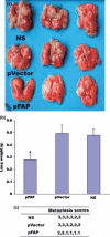
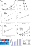
Murine studies have shown that immunological targeting of fibroblast activation protein (FAP) can elicit protective immunity in the absence of significant pathology. Fibroblast activation protein is a product overexpressed by tumor-associated fibroblasts (TAF) and is the predominant component of the stoma in most types of cancer. Tumor-associated fibroblasts differ from normal adult tissue fibroblasts, and instead resemble transient fetal and wound healing-associated fibroblasts. Tumor-associated fibroblasts are critical regulators of tumorigenesis, but differ from tumor cells by being more genetically stable. Therefore, in comparison to tumor cells, TAF may represent more viable therapeutic targets for cancer immunotherapy. To specifically target TAF, we constructed a DNA vaccine directed against FAP. This vaccine significantly suppressed primary tumor and pulmonary metastases primarily through CD8(+) T-cell-mediated killing in tumor-bearing mice. Most importantly, tumor-bearing mice vaccinated against FAP exhibited a 1.5-fold increase in lifespan and no significant pathology. These results suggest that FAP, a product preferentially expressed by TAF, could function as an effective tumor rejection antigen.
© 2010 Japanese Cancer Association.
Figures


Similar articles
-
Anti-tumor effects of DNA vaccine targeting human fibroblast activation protein α by producing specific immune responses and altering tumor microenvironment in the 4T1 murine breast cancer model.Cancer Immunol Immunother. 2016 May;65(5):613-24. doi: 10.1007/s00262-016-1827-4. Epub 2016 Mar 28. Cancer Immunol Immunother. 2016. PMID: 27020681 Free PMC article.
-
Improvement of anti-tumor immunity of fibroblast activation protein α based vaccines by combination with cyclophosphamide in a murine model of breast cancer.Cell Immunol. 2016 Dec;310:89-98. doi: 10.1016/j.cellimm.2016.08.006. Epub 2016 Aug 16. Cell Immunol. 2016. PMID: 27545090
-
A whole-cell tumor vaccine modified to express fibroblast activation protein induces antitumor immunity against both tumor cells and cancer-associated fibroblasts.Sci Rep. 2015 Sep 23;5:14421. doi: 10.1038/srep14421. Sci Rep. 2015. PMID: 26394925 Free PMC article.
-
The application of the fibroblast activation protein α-targeted immunotherapy strategy.Oncotarget. 2016 May 31;7(22):33472-82. doi: 10.18632/oncotarget.8098. Oncotarget. 2016. PMID: 26985769 Free PMC article. Review.
-
[FIBROBLAST ACTIVATION PROTEIN (FAP) AS A POSSIBLE TARGET OF THE ANTITUMOR STRATEGY.].Mol Gen Mikrobiol Virusol. 2016;34(3):90-97. Mol Gen Mikrobiol Virusol. 2016. PMID: 30383930 Review. Russian.
Cited by
-
Fibroblast Activation Protein Expression in Sarcomas.Sarcoma. 2023 Jun 9;2023:2480493. doi: 10.1155/2023/2480493. eCollection 2023. Sarcoma. 2023. PMID: 37333052 Free PMC article.
-
The role of cancer-associated myofibroblasts in intrahepatic cholangiocarcinoma.Nat Rev Gastroenterol Hepatol. 2011 Nov 29;9(1):44-54. doi: 10.1038/nrgastro.2011.222. Nat Rev Gastroenterol Hepatol. 2011. PMID: 22143274 Review.
-
How Signaling Molecules Regulate Tumor Microenvironment: Parallels to Wound Repair.Molecules. 2017 Oct 26;22(11):1818. doi: 10.3390/molecules22111818. Molecules. 2017. PMID: 29072623 Free PMC article. Review.
-
State of the Art in CAR-T Cell Therapy for Solid Tumors: Is There a Sweeter Future?Cells. 2024 Apr 23;13(9):725. doi: 10.3390/cells13090725. Cells. 2024. PMID: 38727261 Free PMC article. Review.
-
Targeting stroma to treat cancers.Semin Cancer Biol. 2012 Feb;22(1):41-9. doi: 10.1016/j.semcancer.2011.12.008. Epub 2011 Dec 24. Semin Cancer Biol. 2012. PMID: 22212863 Free PMC article. Review.
References
-
- Marincola FM, Jaffee EM, Hicklin DJ et al. Escape of human solid tumors from T‐cell recognition: molecular mechanisms and functional significance. Adv Immunol 2000; 74: 181–273. - PubMed
-
- Cohen S, Regev A, Lavi S. Small polydispersed circular DNA (spcDNA) in human cells: association with genomic instability. Oncogene 1997; 14: 977–85. - PubMed
-
- Vitale M, Rezzani R, Rodella L et al. HLA class I antigen and transporter associated with antigen processing (TAPI and TAP2) down‐regulation in high‐grade primary breast carcinoma lesions. Cancer Res 1998; 58: 737–42. - PubMed
-
- Shaheen RM, Davis DW, Liu W et al. Antiangiogenic therapy targeting the tyrosine kinase receptor for vascular endothelial growth factor receptor inhibits the growth of colon cancer liver metastasis and induces tumor and endothelial cell apoptosis. Cancer Res 1999; 59: 5412–6. - PubMed
-
- Reed JC. Apoptosis‐targeted therapies for cancer. Cancer Cell 2003; 3: 17–22. - PubMed
Publication types
MeSH terms
Substances
LinkOut - more resources
Full Text Sources
Other Literature Sources
Research Materials
Miscellaneous

