

Lysophosphatidic acid stimulates gastric cancer cell proliferation via ERK1-dependent upregulation of sphingosine kinase 1 transcription
- PMID: 20804754
- PMCID: PMC2947956
- DOI: 10.1016/j.febslet.2010.08.035
Lysophosphatidic acid stimulates gastric cancer cell proliferation via ERK1-dependent upregulation of sphingosine kinase 1 transcription
Abstract
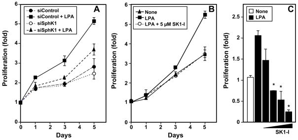
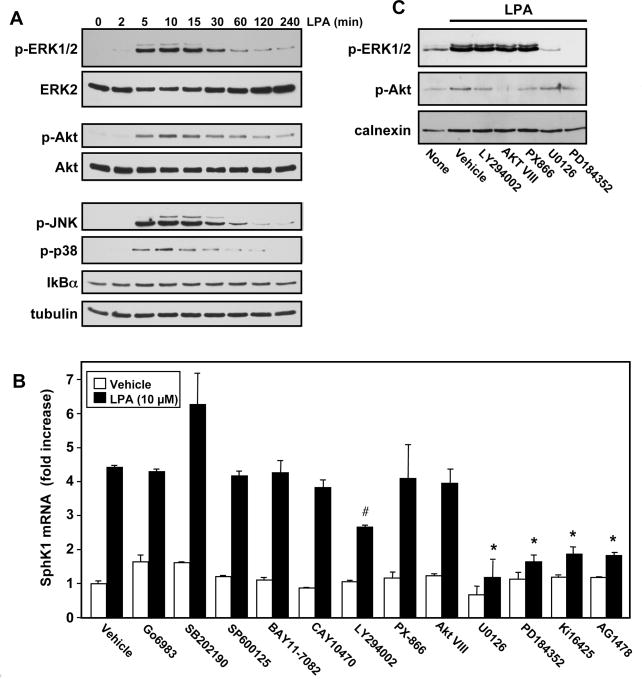
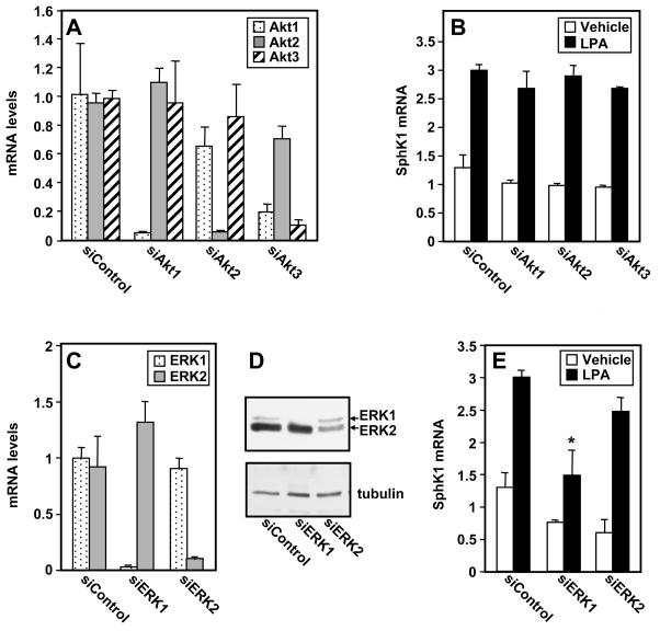
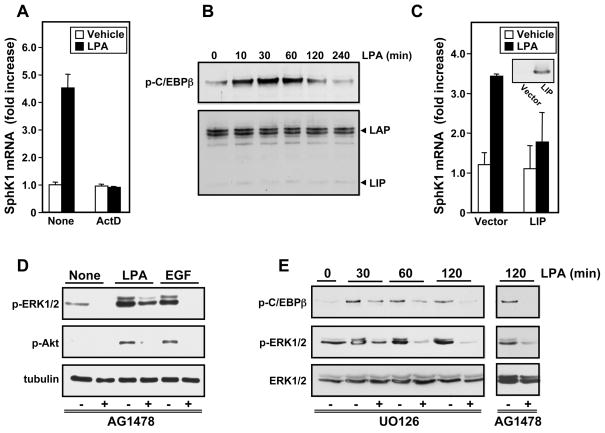
In MKN1 gastric cancer cells, lysophosphatidic acid (LPA) upregulates expression of sphingosine kinase 1 (SphK1) and its downregulation or inhibition suppresses LPA mediated proliferation. Although LPA activates numerous signaling pathways downstream of its receptors, including extracellular-signal-regulated kinase 1/2, p38, JNK, and Akt, and the transactivation of the epidermal growth factor receptor, pharmacological and molecular approaches demonstrated that only activation of ERK1, in addition to the CCAAT/enhancer-binding protein β transcription factor, is involved in transcriptional upregulation of SphK1 by LPA. Our data implicate ERK1 as an important mediator of LPA signaling leading to upregulation of SphK1 and point to SphK1 and sphingosine-1-phosphate production as potential therapeutic targets in gastric cancer.
Copyright © 2010 Federation of European Biochemical Societies. Published by Elsevier B.V. All rights reserved.
Figures
Similar articles
-
Cross-talk between LPA1 and epidermal growth factor receptors mediates up-regulation of sphingosine kinase 1 to promote gastric cancer cell motility and invasion.Cancer Res. 2008 Aug 15;68(16):6569-77. doi: 10.1158/0008-5472.CAN-08-0411. Cancer Res. 2008. PMID: 18701480 Free PMC article.
-
Sphingosine kinase 1 enhances colon cancer cell proliferation and invasion by upregulating the production of MMP-2/9 and uPA via MAPK pathways.Int J Colorectal Dis. 2012 Dec;27(12):1569-78. doi: 10.1007/s00384-012-1510-y. Epub 2012 Jun 9. Int J Colorectal Dis. 2012. PMID: 22684547
-
SphK1 confers resistance to apoptosis in gastric cancer cells by downregulating Bim via stimulating Akt/FoxO3a signaling.Oncol Rep. 2014 Oct;32(4):1369-73. doi: 10.3892/or.2014.3391. Epub 2014 Aug 7. Oncol Rep. 2014. PMID: 25109605 Free PMC article.
-
The Tumorigenic Effect of Sphingosine Kinase 1 and Its Potential Therapeutic Target.Cancer Control. 2020 Jan-Dec;27(1):1073274820976664. doi: 10.1177/1073274820976664. Cancer Control. 2020. PMID: 33317322 Free PMC article. Review.
-
Sphingosine Kinase 1 and Sphingosine-1-Phosphate Signaling in Colorectal Cancer.Int J Mol Sci. 2017 Oct 8;18(10):2109. doi: 10.3390/ijms18102109. Int J Mol Sci. 2017. PMID: 28991193 Free PMC article. Review.
Cited by
-
KAI1/CD82 gene and autotaxin-lysophosphatidic acid axis in gastrointestinal cancers.World J Gastrointest Oncol. 2022 Aug 15;14(8):1388-1405. doi: 10.4251/wjgo.v14.i8.1388. World J Gastrointest Oncol. 2022. PMID: 36160748 Free PMC article. Review.
-
Migration of gastric cancer cells in response to lysophosphatidic acid is mediated by LPA receptor 2.Oncol Lett. 2013 Mar;5(3):1048-1052. doi: 10.3892/ol.2013.1107. Epub 2013 Jan 7. Oncol Lett. 2013. PMID: 23426604 Free PMC article.
-
The expression of TMPRSS4 and Erk1 correlates with metastasis and poor prognosis in Chinese patients with gastric cancer.PLoS One. 2013 Jul 29;8(7):e70311. doi: 10.1371/journal.pone.0070311. Print 2013. PLoS One. 2013. PMID: 23922976 Free PMC article.
-
Twofer anti-vascular therapy targeting sphingosine-1-phosphate for breast cancer.Gland Surg. 2012 Aug 1;1(2):80-83. doi: 10.3978/j.issn.2227-684X.2012.07.01. Gland Surg. 2012. PMID: 24855599 Free PMC article. No abstract available.
-
The role of sphingosine-1-phosphate in inflammation and cancer progression.Cancer Sci. 2018 Dec;109(12):3671-3678. doi: 10.1111/cas.13802. Epub 2018 Oct 15. Cancer Sci. 2018. PMID: 30238699 Free PMC article. Review.
References
-
- Shida D, Kitayama J, Yamaguchi H, Okaji Y, Tsuno NH, Watanabe T, Takuwa Y, Nagawa H. Lysophosphatidic acid (LPA) enhances the metastatic potential of human colon carcinoma DLD1 cells through LPA1. Cancer Res. 2003;63:1706–1711. - PubMed
-
- Li W, Yu CP, Xia JT, Zhang L, Weng GX, Zheng HQ, Kong QL, Hu LJ, Zeng MS, Zeng YX, Li M, Li J, Song LB. Sphingosine kinase 1 is associated with gastric cancer progression and poor survival of patients. Clin Cancer Res. 2009;15:1393–1399. - PubMed
-
- Shida D, Kitayama J, Yamaguchi H, Yamashita H, Mori K, Watanabe T, Yatomi Y, Nagawa H. Sphingosine 1-phosphate transactivates c-Met as well as epidermal growth factor receptor (EGFR) in human gastric cancer cells. FEBS Lett. 2004;577:333–338. - PubMed
-
- Galizia G, Lieto E, Orditura M, Castellano P, Mura AL, Imperatore V, Pinto M, Zamboli A, De Vita F, Ferraraccio F. Epidermal growth factor receptor (EGFR) expression is associated with a worse prognosis in gastric cancer patients undergoing curative surgery. World J Surg. 2007;31:1458–1468. - PubMed
Publication types
MeSH terms
Substances
Grants and funding
LinkOut - more resources
Full Text Sources
Medical
Research Materials
Miscellaneous
