. 2010 Sep 3;7(3):319-28.
doi: 10.1016/j.stem.2010.06.022.

Stat3 activation is limiting for reprogramming to ground state pluripotency

Affiliations

Stat3 activation is limiting for reprogramming to ground state pluripotency

Jian Yang et al. Cell Stem Cell. .

Abstract

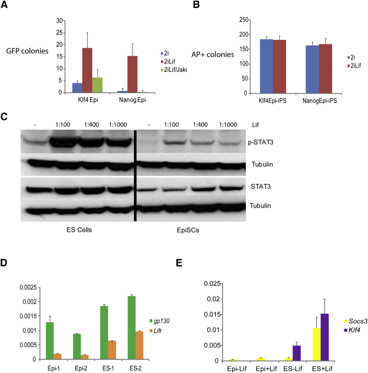
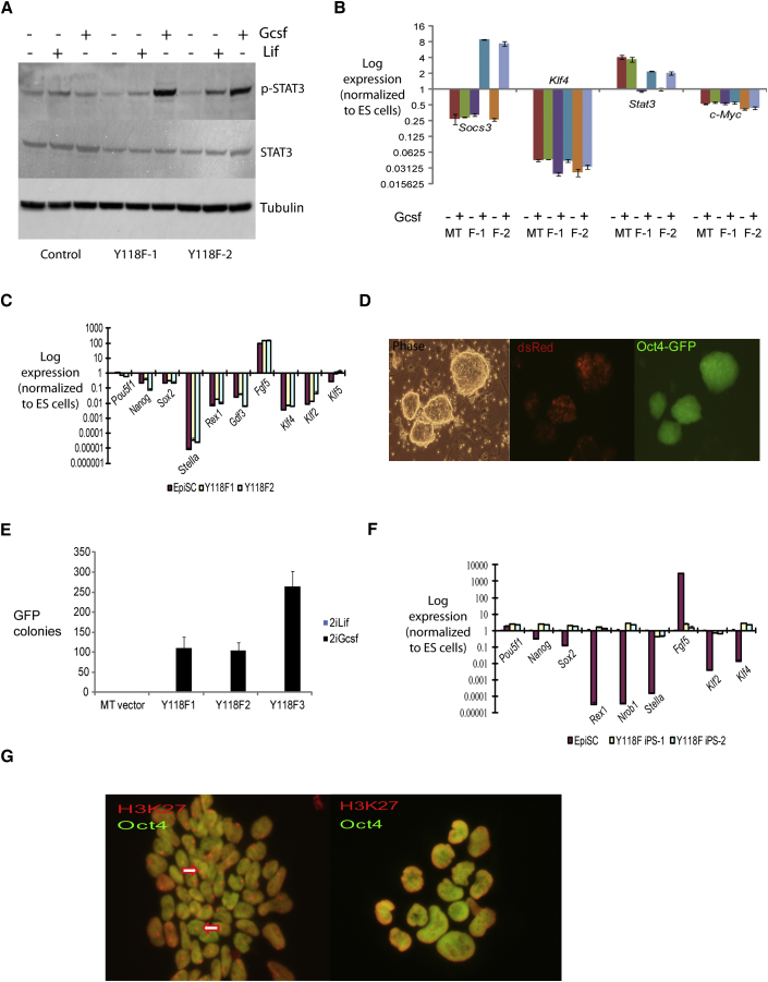
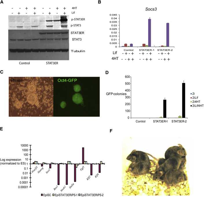
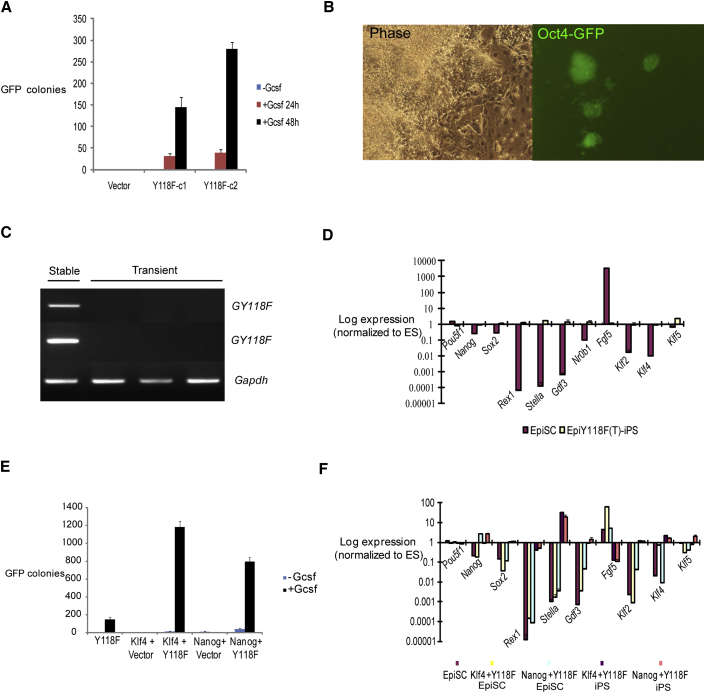
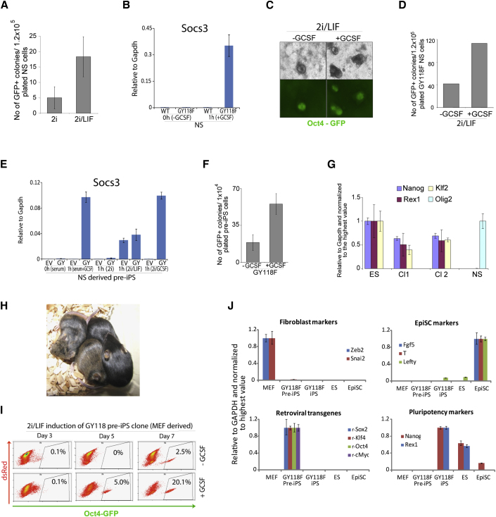
The cytokine leukemia inhibitory factor (Lif) sustains self-renewal of mouse embryonic and induced pluripotent stem cells by activating Jak kinase and the transcription factor Stat3. Here we investigate whether Jak/Stat3 may also contribute to induction of pluripotency. EpiSCs derived from postimplantation embryos express low levels of Lif receptor and Stat3. We introduced into EpiSCs a Jak/Stat3 activating receptor (GY118F) responsive to granulocyte colony stimulating factor (Gcsf). On transfer to ground state culture, in which MAPK signaling and glycogen synthase kinase are inhibited, Gcsf induced transcriptional resetting and functional reprogramming. Activation of a tamoxifen-regulatable fusion, Stat3ER(T2), also converted EpiSCs into chimera-competent iPSCs. We exploited GY118F to increase Jak/Stat3 activity during somatic cell reprogramming. Incompletely reprogrammed cells derived from neural stem cells or fibroblasts responded to Gcsf with elevated frequencies of progression to ground state pluripotency. These findings indicate that Jak/Stat3 participate directly in molecular reprogramming and that activation of this pathway is a limiting component.


Figures

Figure 1
Figure 1
Lif Enhances EpiSC Reprogramming (A) Yield of Oct4-GFP-positive Epi-iPSC colonies from Klf4- or Nanog-transfected EpiSCs transferred to 2i, 2i plus Lif, or 2i plus Lif with JAK inhibitor. (B) Colony formation by Epi-iPSCs in 2i or 2i/Lif. (C) Immunoblot analysis of Stat3 and phosphoStat3 in ESCs and EpiSCs. Cells were cultured in unsupplemented medium for 4 hr, then stimulated with indicated concentrations of Lif for 20 min. (D) qRT-PCR analysis of gp130 and Lifr expression in EpiSCs or ESCs. (E) qRT-PCR analysis of Socs3 and Klf4 expression in EpiSCs or ESCs with or without Lif stimulation for 1 hr. Data on Tbx3 expression are presented in Figure S1. Error bars are standard deviations from the mean of triplicate determinations.
Figure 2
Figure 2
Reprogramming of EpiSCs Transfected with GY118F (A) Western blot analysis of p-Stat3 induction in parental or GY118F EpiSC clones after Lif or Gcsf stimulation for 20 min. (B) qRT-PCR analysis of empty vector (MT) and two clones of GY118F EpiSCs stimulated with Gcsf for 1 hr. (C) Marker gene profile of parental and GY118F EpiSCs cultured in activin/Fgf2. Expression levels are relative to Gapdh and normalized to undifferentiated ESCs. (D) Oct4-GFP-positive colonies obtained from GY118F EpiSCs transferred to 2i/Gcsf. dsRed fluorescence indicates transgene expression. Images were taken after 8 days in 2i/Gcsf. (E) Number of Oct4-GFP-positive colonies after culturing three different clones of GY118F EpiSCs in 2i/Gcsf for 8 days. Data on colony yield in Gcsf alone are presented in Figure S2. (F) qRT-PCR analysis of marker gene expression in GY118F Epi-iPSC clones relative to Gapdh and normalized to ESCs. (G) H3K27me3 staining of female O4G EpiSCs (left) showing nuclear foci (arrowed) and derivative GY118F Epi-iPSCs reprogrammed in 2i/Gcsf (right) with no evident foci. Characterization of Epi-iPSCs after tatCre-mediated excision of the GY118F transgene is presented in Figure S3. Error bars are standard deviations from the mean of triplicate determinations.
Figure 3
Figure 3
Reprogramming EpiSCs by Tamoxifen Activation of Stat3ERT2 (A) Immunoblot analysis of Stat3 and Stat3ERT2 tyrosine phosphorylation in EpiSC transfectants exposed to 4-hydroxytamoxifen (4HT) or Lif for 1 hr. (B) qRT-PCR analysis of Socs3 induction by 4HT and Lif for 1 hr. (C) Oct4-GFP colonies obtained from Stat3ERT2 EpiSCs after 8 days in 2i/Lif plus 4HT. (D) Mean numbers of Oct4-GFP colonies from triplicate wells of Stat3ERT2 EpiSCs after 8 days in 2i with or without Lif and/or 4HT. (E) qRT-PCR analysis of marker gene expression in Stat3ERT2 Epi-iPSC clones relative to Gapdh and normalized to ESCs. (F) Chimeric mice produced from Stat3ERT2 Epi-iPSCs. Error bars are standard deviations from the mean of triplicate determinations.
Figure 4
Figure 4
Jak/Stat3 Acts Early in Reprogramming and Synergizes with Klf4 and Nanog (A) Number of Oct4-GFP colonies from 2 × 10⁴ GY118F EpiSCs cultured in triplicate in 2i/Gcsf 24 hr or 48 hr then switched to 2i/Lif and scored at day 8. (B) Oct4-GFP colonies obtained by transient transfection with GY118F and culture in 2i/Gcsf for 8 days. (C) Genomic PCR (top) and RT-PCR (middle and bottom) analyses of Epi-iPSCs generated by stable or transient transfection. (D) Marker profile of Epi-iPSCs derived from EpiSCs by transient expression of GY118F. (E) Number of Oct4-GFP colonies generated by EpiSCs stably transfected with GY118F and either Klf4 or Nanog then transferred to 2i/Gcsf for 8 days. (F) qRT-PCR analysis of marker gene expression relative to Gapdh and normalized to ESCs for GY118F+Klf4 and GY118F+Nanog double transfected EpiSCs and derivative iPSC clones. Error bars are standard deviations from the mean of triplicate determinations.
Figure 5
Figure 5
Activation of Jak/Stat3 Enhances Somatic Cell Reprogramming (A) Number of Oct4-GFP-positive iPSC colonies generated from NSCs transduced with Oct4, Klf4, and cMyc and cultured in 2i or 2i/Lif. (B) Induction of Socs3 in GY118F-transfected NSCs stimulated with Gcsf for 1 hr. (C) Representative images of iPSC colonies generated from GY118F NSCs transferred 5 days after retroviral transduction into 2i/Lif with or without Gcsf. (D) Number of Oct4-GFP-positive colonies obtained in (C). (E) Induction of Socs3 in GY118F (GY) NSCs derived from incompletely reprogrammed cells (pre-iPSCs) stimulated with Lif or GCSF for 1 hr. Empty vector (EV) transfectants were treated in parallel. (F) Number of Oct4-GFP-positive iPSC colonies from GY118F NSCs passaged at day 5 after reprogramming factor transduction and 2 days later transferred to 2i/Lif with or without GCSF. Empty vector NSC transfectants showed no response to Gcsf. (G) qRT-PCR analysis of the pluripotency markers Nanog, Rex1, and Klf2 and of the NSC marker Olig2 in two iPSC clones (cl) generated in the presence of Gcsf as in (F). (H) Two chimeras and nonchimeric littermates generated after injection of cl1 GY118F NS-iPSCs into C57BL/6 host blastocysts. (I) Frequency of Oct4-GFP-positive cells produced from fibroblast-derived pre-iPSCs transfected with GY118F and cultured in 2i/Lif with or without Gcsf. (J) Marker profile of iPSCs generated in Gcsf from GY118F-transfected fibroblast pre-iPSCs. Error bars in (B), (E), and (F) are standard deviations from the mean of triplicate determinations. In (G) and (J), error bars indicate the range of fold change relative to the sample with highest expression.

Comment in

References

    1. Bao S., Tang F., Li X., Hayashi K., Gillich A., Lao K., Surani M.A. Epigenetic reversion of post-implantation epiblast to pluripotent embryonic stem cells. Nature. 2009;461:1292–1295. - PMC - PubMed
    1. Batlle-Morera L., Smith A., Nichols J. Parameters influencing derivation of embryonic stem cells from murine embryos. Genesis. 2008;46:758–767. - PubMed
    1. Bourillot P.Y., Aksoy I., Schreiber V., Wianny F., Schulz H., Hummel O., Hubner N., Savatier P. Novel STAT3 target genes exert distinct roles in the inhibition of mesoderm and endoderm differentiation in cooperation with Nanog. Stem Cells. 2009;27:1760–1771. - PubMed
    1. Bradley A., Evans M.J., Kaufman M.H., Robertson E. Formation of germ-line chimaeras from embryo-derived teratocarcinoma cell lines. Nature. 1984;309:255–256. - PubMed
    1. Brons I.G., Smithers L.E., Trotter M.W., Rugg-Gunn P., Sun B., Chuva de Sousa Lopes S.M., Howlett S.K., Clarkson A., Ahrlund-Richter L., Pedersen R.A. Derivation of pluripotent epiblast stem cells from mammalian embryos. Nature. 2007;448:191–195. - PubMed

Publication types

MeSH terms

Substances