. 2010 Sep 3;7(3):355-66.
doi: 10.1016/j.stem.2010.07.013.

Functional diversity of ESC-derived motor neuron subtypes revealed through intraspinal transplantation

Affiliations

Functional diversity of ESC-derived motor neuron subtypes revealed through intraspinal transplantation

Mirza Peljto et al. Cell Stem Cell. .

Abstract

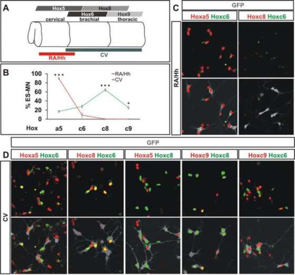
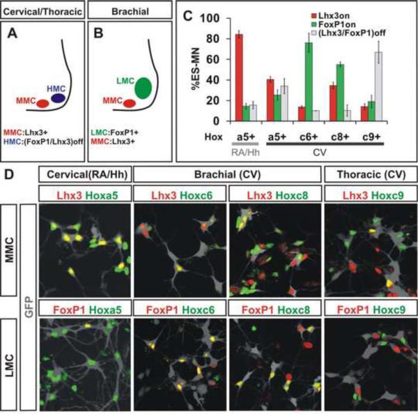
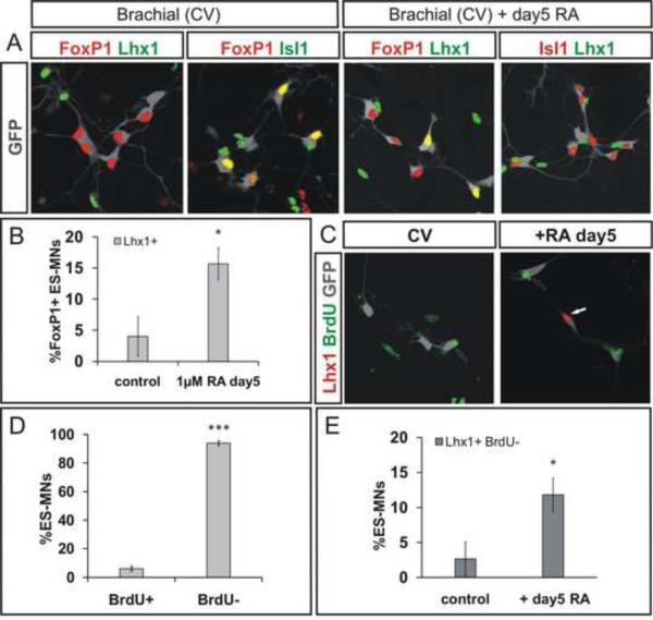
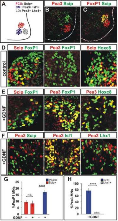
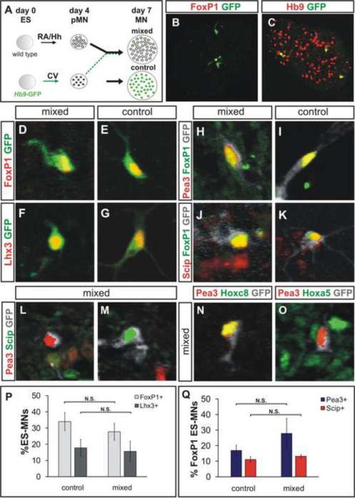
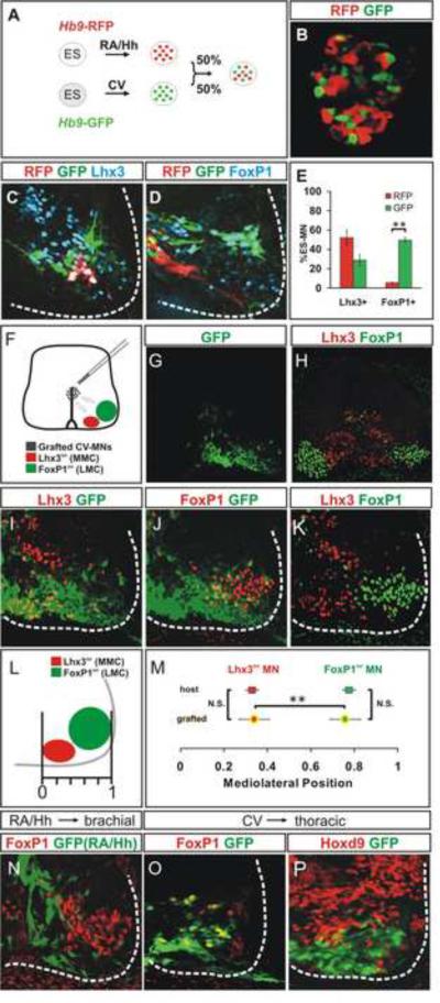
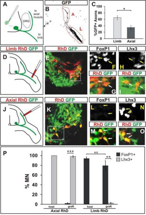
Cultured ESCs can form different classes of neurons, but whether these neurons can acquire specialized subtype features typical of neurons in vivo remains unclear. We show here that mouse ESCs can be directed to form highly specific motor neuron subtypes in the absence of added factors, through a differentiation program that relies on endogenous Wnts, FGFs, and Hh-mimicking the normal program of motor neuron subtype differentiation. Molecular markers that characterize motor neuron subtypes anticipate the functional properties of these neurons in vivo: ESC-derived motor neurons grafted isochronically into chick spinal cord settle in appropriate columnar domains and select axonal trajectories with a fidelity that matches that of their in vivo generated counterparts. ESC-derived motor neurons can therefore be programmed in a predictive manner to acquire molecular and functional properties that characterize one of the many dozens of specialized motor neuron subtypes that exist in vivo.


Figures

Figure 1
Figure 1. Characterization of Hox Gene Expression in ES Motor Neurons
A) Pattern of Hox gene expression in the developing spinal cord. Red and grey lines represent positional identity of ES motor neurons derived under RA/Hh and CV exposure, respectively. B) Quantification of Hox expression in ES motor. ES motor neurons derived under RA/Hh and CV conditions exhibited differences in Hoxa5 (p<0.001), Hoxc8 (p<0.001) and Hoxc9 (p=0.01) expression. Data from three independent experiments (mean ± standard error of the mean, SEM). C) RA/Hh generated Hb9-GFP+ ES motor neurons (grey) expressed Hoxa5, low levels of Hoxc6, but not Hoxc8. D) Most CV differentiated ES motor neurons (grey) expressed Hoxc8, while smaller subsets express Hoxa5, Hoxc6 and Hoxc9. Note mutually exclusive expression of Hoxa5/Hoxc8 and Hoxc6/Hoxc9.
Figure 2
Figure 2. Rostro-Caudal and Columnar Identities of ES Motor Neurons
A) At cervical and thoracic levels, motor neurons are organized into median (MMC) and hypaxial (HMC) motor columns. MMC neurons express Lhx3. HMC neurons lack FoxP1 and Lhx3 expression. B) At brachial level, HMC is replaced by FoxP1+ lateral motor column (LMC). C) Quantification of Hox gene expression in the context of FoxP1+ and Lhx3+ ES motor neurons derived under RA/Hh or CV condition. ANOVA analysis of data presented in Supplemental Table 2. Data from three independent experiments (mean ± SEM). D) FoxP1 and Lhx3 expression in Hoxa5, Hoxc6, Hoxc8, or Hoxc9 expressing Hb9-GFP+ ES motor neurons (grey) derived under RA/Hh or CV conditions.
Figure 3
Figure 3. Retinoid Signaling Specifies Lateral LMC Divisional Identity in Post-mitotic ES Motor Neurons
A) Expression of Lhx1 and Isl1 in CV generated GFP+ ES motor neurons (grey) on day 7 of differentiation under control conditions and following 1μM RA treatment on day 5. Note mutually exclusive expression of Isl1 and Lhx1. B) Increase in Lhx1+ FoxP1+ ES motor neurons after retinoid treatment (p=0.044). Results from three independent experiments (mean ± SEM). C) Labeling of motor neurons born after day 5 in cultures by BrdU treatment on days 5–7. D) A majority of ES motor neurons (94 ± 2%) do not incorporate BrdU supplemented between day 5 and 7 of differentiation. Results from three independent experiments (mean ± SEM). E) RA treatment on day 5 significantly increases the fraction of post-mitotic, BrdU ES motor neurons that express Lhx1 (p=0.028). Results from three independent experiments (mean ± SEM).
Figure 4
Figure 4. ES Motor Neurons Acquire Defined Caudal Brachial Motor Pool Identities
A) Molecularly defined LMC motor pools in caudal brachial spinal cord: Scip+ pool innervates flexor carpi ulnaris (FCU), Pea3+/Isl1+ pool innervates cutaneous maximus (CM), and Pea3+/Lhx1+ motor pool innervates latissimus dorsi (LD) muscles. B) Scip and Pea3 expression in E13.5 mouse spinal cord. C) A subset of FoxP1+ LMC neurons expresses Scip at caudal brachial E13.5 mouse spinal cord. D) Cells in day 6 embryoid bodies express FoxP1, Scip, Pea3 and Hoxc8. A subset of FoxP1+ LMC neurons expresses Scip but not Pea3. Scip+ motor neurons co-express Hoxc8. E) FoxP1+ ES motor neurons acquire Pea3 expression in response to GDNF treatment on day 5–6. Pea3+ motor neurons express Hoxc8. F) Mutually exclusive expression of Pea3 and Scip in ES motor neurons. A majority of Pea3+ ES motor neurons express Isl1 but not Lhx1. G) Quantification of Pea3 and Scip expressing FoxP1+ ES motor neurons in the presence and absence of GDNF. The fraction of Pea3+ but not Scip+ LMC neurons was increased upon GDNF treatment (p<0.001). Results from three independent experiments (mean ± SEM). H) Quantification of Lhx1 and Isl1 expression in Pea3+ motor neurons. Significantly greater fraction of Pea3+ ES motor neurons expressed Isl1 compared to Lhx1 (p<0.001). Results from three independent experiments (mean ± SEM).
Figure 5
Figure 5. Microenvironment-Independent Acquisition of Motor Pool Identities
A) CV differentiated Hb9-GFP ES motor neuron progenitors were dissociated on day 4 and mixed with a five-fold excess of dissociated RA/Hh differentiated wild-type (wt) ES motor neuron progenitors, reaggregated, and cultured until day 7. B) One of three GFP+ ES motor neurons expresses FoxP1. C) ES motor neurons derived from wt ES cells (Hb9+/GFPoff) under RA/Hh condition surround scattered brachial GFP+ ES motor neurons. D–G) ES motor neurons in control and mixed conditions express FoxP1 (D, E) and Lhx3 (F, G). H–K) Day 5 treatment of mixed aggregates with GDNF induced Pea3 (H, I) and Scip (J, K) expression in FoxP1+ LMC ES motor neurons (grey). L, M) Expression of Pea3 and Scip in ES motor neurons (grey) does not overlap in mixed aggregates. N, O) Hoxc8 expressing Pea3+ ES motor neurons (N) are surrounded by Hoxa5+ cervical cells (O). P) Similar fractions of FoxP1+ and Lhx3+ ES motor neurons in control and mixed cultures (p=0.29 and p=0.81, respectively). Results from three independent experiments (mean ± SEM). Q) The fractions of FoxP1+ ES motor neurons expressing Pea3 and Scip were not significantly different in control or mixed cultures (p= 0.40 and p=0.44, respectively). Results from three independent experiments (mean ± SEM).
Figure 6
Figure 6. Column-Specific Settling of Transplanted ES Motor Neurons
A) Hb9-RFP and Hb9-GFP ES cell lines differentiated under RA/Hh and CV conditions, respectively. RFP+ and GFP+ motor neuron aggregates were transplanted into HH stage 15–17 brachial spinal cord and analyzed 3 days later. B) Intermixing of RFP+ and GFP+ ES motor neurons in aggregates. C, D) ES derived neurons in host ventral spinal cord (white outline) express Lhx3 (C) and FoxP1 (D). E) FoxP1 and Lhx3 expression in RFP+ and GFP+ motor neurons 3 days after transplantation. More transplanted GFP+ neurons express FoxP1 (p=0.004). Results from three transplanted embryos (mean ± SEM). F) CV differentiated ES motor neurons transplanted into HH stage 15–17 brachial spinal cord. G) Ventrally localized GFP+ motor neurons three days after transplantation. H–K) Spinal cord section (from G) triple labeled for Lhx3, FoxP1 and GFP. White dashed line delineates spinal cord margin. L) Measuring the settling position of transplanted cells. M) Settling preferences of transplanted and endogenous motor neurons expressing Lhx3 or FoxP1. The settling positions of host and grafted FoxP1+ or Lhx3+ motor neurons are not (N.S.) different (p=0.85, resp. p=0.92). Settling position of transplanted Lhx3+ ES motor neurons is different (p<0.01) from that of FoxP1+ ES motor neurons. Results from four transplanted embryos (mean ± SEM). N) Lack of FoxP1 expression in RA/Hh differentiated ES motor neurons grafted into brachial spinal cord. O, P) FoxP1 expression by CV ES motor neurons transplanted into Hoxd9+ thoracic spinal cord (P).
Figure 7
Figure 7. Axons of LMC and MMC ES Motor Neurons Project to Limb and Axial Muscles
A) Strategy to examine axon pathfinding preference of GFP+ ES motor neurons derived under CV condition three days after grafting into brachial spinal cord (HH stage 15–17). B, C) A greater fraction of transplanted GFP+ (black) axons is detected in the limb (L) nerve compared to axial (A) nerve branch three days after grafting (p=0.011). Results from six transplanted embryos (mean ± SEM). See also Figure S6D, E. D) CV differentiated transplanted motor neurons were backfilled with tetramethylrhodamine-dextran (RhD) from the limb nerve branch. E) Section of a ventral spinal cord containing GFP+ transplanted and RhD+ limb innervating motor neurons (boxed area corresponds to panels F and G). F–I) Transplanted motor neurons retrogradely labeled from the limb (RhD+/GFP+) express FoxP1 (arrows, F, G) but not Lhx3 (arrows, H, I). J, K) Transplanted ES motor neurons backfilled with RhD from the axial nerve branch (boxed area in K corresponds to panels L and M). L–O) ES motor neurons retrogradely labeled from the axial musculature (RhD+/GFP+) expressed Lhx3 (arrows, N, O) but not FoxP1 (arrows, L, M). P) More axially projecting grafted ES motor neurons expressed Lhx3 than FoxP1 (p<0.001). More limb projecting ES motor neurons expressed FoxP1 than Lhx3 (p=0.002). The fractions of endogenous and grafted FoxP1+ motor neurons labeled from the limb, or Lhx3+ motor neurons labeled from the axial muscles are not different (p=0.26, resp. p=0.37). Results from three embryos each for limb and axial backfills (mean ± SEM).

Similar articles

Cited by

References

    1. Agalliu D, Takada S, Agalliu I, McMahon AP, Jessell TM. Motor neurons with axial muscle projections specified by Wnt4/5 signaling. Neuron. 2009;61:708–720. - PMC - PubMed
    1. Andersson E, Tryggvason U, Deng Q, Friling S, Alekseenko Z, Robert B, Perlmann T, Ericson J. Identification of intrinsic determinants of midbrain dopamine neurons. Cell. 2006;124:393–405. - PubMed
    1. Arber S, Ladle DR, Lin JH, Frank E, Jessell TM. ETS gene Er81 controls the formation of functional connections between group Ia sensory afferents and motor neurons. Cell. 2000;101:485–498. - PubMed
    1. Bain G, Kitchens D, Yao M, Huettner JE, Gottlieb DI. Embryonic stem cells express neuronal properties in vitro. Dev Biol. 1995;168:342–357. - PubMed
    1. Buck L, Axel R. A novel multigene family may encode odorant receptors: A molecular basis for odor recognition. Cell. 1991;65:175–187. - PubMed

Publication types

LinkOut - more resources