. 2010 Sep;65(9):775-81.
doi: 10.1136/thx.2010.136374.

Quantitative analysis of high-resolution computed tomography scans in severe asthma subphenotypes

Affiliations

Quantitative analysis of high-resolution computed tomography scans in severe asthma subphenotypes

Sumit Gupta et al. Thorax. 2010 Sep.

Abstract

Background: Severe asthma is a heterogeneous condition. Airway remodelling is a feature of severe asthma and can be determined by the assessment of high-resolution computed tomography (HRCT) scans. The aim of this study was to assess whether airway remodelling is restricted to specific subphenotypes of severe asthma.

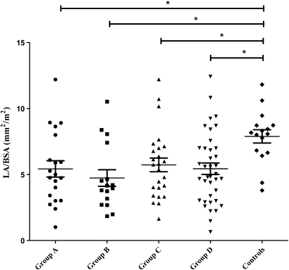
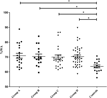
Methods: A retrospective analysis was performed of HRCT scans from subjects who had attended a single-centre severe asthma clinic between 2003 and 2008. The right upper lobe apical segmental bronchus (RB1) dimensions were measured and the clinical and sputum inflammatory characteristics associated with RB1 geometry were assessed by univariate and multivariate regression analyses. Longitudinal sputum data were available and were described as area under the time curve (AUC). Comparisons were made in RB1 geometry across subjects in four subphenotypes determined by cluster analysis, smokers and non-smokers, and subjects with and without persistent airflow obstruction.

Results: Ninety-nine subjects with severe asthma and 16 healthy controls were recruited. In the subjects with severe asthma the RB1 percentage wall area (%WA) was increased (p=0.009) and lumen area (LA)/body surface area (BSA) was decreased (p=0.008) compared with controls but was not different across the four subphenotypes. Airway geometry was not different between smokers and non-smokers and RB1 %WA was increased in those with persistent airflow obstruction. RB1 %WA in severe asthma was best associated with airflow limitation and persistent neutrophilic airway inflammation (model R(2)=0.27, p=0.001).

Conclusions: Airway remodelling of proximal airways occurs in severe asthma and is associated with impaired lung function and neutrophilic airway inflammation.


Conflict of interest statement

Competing interests: CEB has received research grants from MedImmune, AstraZeneca and GlaxoSmithKline and fees for consulting and/or speaking from MedImmune, AstraZeneca, GlaxoSmithKline, Novartis, Aerovant and Genentech. AJW has received research grants from GlaxoSmithKline and Pfizer and is on GlaxoSmithKline advisory board on asthma and related diseases. PH has received an unrestricted educational grant from GlaxoSmithKline. All other authors declare that they have no competing interests of relevance to this manuscript. No pharmaceutical company had any involvement in the planning, design, analysis or interpretation of the current study.

Figures

Figure 1
Figure 1
Comparison of right upper lobe apical segmental bronchus (RB1) lumen area/body surface area (LA/BSA, mm2/m2) of four severe asthma phenotypes and control subjects (p=0.008, ANOVA; *p<0.05, Tukey test to compare all pairs of columns). Group A, patients with severe asthma with a concordant asthma control score and eosinophilic inflammation with a significantly greater bronchodilator response; group B, patients with severe asthma who were predominantly women with high body mass index and evidence of a high asthma control score but very little eosinophilic airway inflammation; group C, patients with severe asthma with a predominantly high asthma control score and very little eosinophilic airway inflammation; group D, patients with severe asthma with predominantly eosinophilic airway inflammation and significantly low asthma control score.
Figure 2
Figure 2
Comparison of right upper lobe apical segmental bronchus (RB1) percentage wall area (%WA) of four severe asthma phenotypes and control subjects (p=0.009, ANOVA; *p<0.05, Tukey test to compare all pairs of columns). Group A, patients with severe asthma with a concordant asthma control score and eosinophilic inflammation with a significantly greater bronchodilator response; group B, patients with severe asthma who were predominantly women with high body mass index and evidence of a high asthma control score but very little eosinophilic airway inflammation; group C, patients with severe asthma with a predominantly high asthma control score and very little eosinophilic airway inflammation; group D, patients with severe asthma with predominantly eosinophilic airway inflammation and significantly low asthma control score.
Figure 3
Figure 3
Correlation between right upper lobe apical segmental bronchus (RB1) percentage wall area (%WA) and postbronchodilator (postBD) forced expiratory volume in 1 s (FEV1)% predicted in patients with severe asthma (r=0.41, p<0.0001).
Figure 4
Figure 4
Correlation between right upper lobe apical segmental bronchus (RB1) percentage wall area (%WA) and sputum neutrophil area under the curve (AUC, %) in patients with severe asthma (r=0.36, p<0.005).

Similar articles

Cited by

References

    1. Wenzel S. Severe asthma in adults. Am J Respir Crit Care Med 2005;172:149–60 - PubMed
    1. Antonicelli L, Bucca C, Neri M, et al. Asthma severity and medical resource utilisation. Eur Respir J 2004;23:723–9 - PubMed
    1. Holgate ST. Epithelium dysfunction in asthma. J Allergy Clin Immunol 2007;120:1233–46 - PubMed
    1. Gupta S, Siddiqui S, Haldar P, et al. Qualitative analysis of high-resolution CT scans in severe asthma. Chest 2009;136:1521–8 - PMC - PubMed
    1. Niimi A, Matsumoto H, Amitani R, et al. Airway wall thickness in asthma assessed by computed tomography: relation to clinical indices. Am J Respir Crit Care Med 2000;162:1518–23 - PubMed

Publication types