Review
. 2011 Jan;39(1):1-18.
doi: 10.1093/nar/gkq742. Epub 2010 Aug 30.

Natural and engineered nicking endonucleases--from cleavage mechanism to engineering of strand-specificity

Affiliations
Review

Natural and engineered nicking endonucleases--from cleavage mechanism to engineering of strand-specificity

Siu-Hong Chan et al. Nucleic Acids Res. 2011 Jan.

Erratum in

  • Nucleic Acids Res. 2011 Mar 1;39(5):1966

Abstract

Restriction endonucleases (REases) are highly specific DNA scissors that have facilitated the development of modern molecular biology. Intensive studies of double strand (ds) cleavage activity of Type IIP REases, which recognize 4-8 bp palindromic sequences, have revealed a variety of mechanisms of molecular recognition and catalysis. Less well-studied are REases which cleave only one of the strands of dsDNA, creating a nick instead of a ds break. Naturally occurring nicking endonucleases (NEases) range from frequent cutters such as Nt.CviPII (^CCD; ^ denotes the cleavage site) to rare-cutting homing endonucleases (HEases) such as I-HmuI. In addition to these bona fida NEases, individual subunits of some heterodimeric Type IIS REases have recently been shown to be natural NEases. The discovery and characterization of more REases that recognize asymmetric sequences, particularly Types IIS and IIA REases, has revealed recognition and cleavage mechanisms drastically different from the canonical Type IIP mechanisms, and has allowed researchers to engineer highly strand-specific NEases. Monomeric LAGLIDADG HEases use two separate catalytic sites for cleavage. Exploitation of this characteristic has also resulted in useful nicking HEases. This review aims at providing an overview of the cleavage mechanisms of Types IIS and IIA REases and LAGLIDADG HEases, the engineering of their nicking variants, and the applications of NEases and nicking HEases.


Figures

Figure 1.
Figure 1.
Cumulative number of putative and non-putative R and fused RM genes from 1969 to 2009. The red shaded region indicates the cumulative number of non-putative R and fused RM genes; the blue shaded region indicates the cumulative number of putative genes.
Figure 2.
Figure 2.
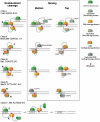
Five classes of restriction endonucleases that recognize asymmetric sequences and their nicking variants. Class I: homodimeric with one catalytic site per protein chain. Class II: monomeric with two catalytic sites per protein chain. Class III: heterodimeric with one catalytic site per protein chain; the large subunit is active in the absence of the small subunit. Class IV: heterodimeric with one catalytic site per protein chain; the two subunits are of comparable size and none of the monomers is active in the absence of the partner. Class V: homodimeric with a half catalytic site (phospholipase D-type) per protein chain. In Classes II and IV, information on the segregation of the DNA-binding domain and the DNA-cleavage domain is not available. Therefore, single semicircles are used to represent a combined DNA binding/cleavage domain. In all cases, mutations that inactivate the cleavage activity are indicated.
Figure 3.
Figure 3.
Comparison of two endonuclease motifs that are heavily represented by monomeric endonuclease domains with significant nicking activity. (A) On the left is the DNA-bound catalytic site of the GIY-YIG endonuclease R.Eco29kI; on the right is the DNA-bound catalytic site of the HNH endonuclease R.Hpy99I. The proposed mechanism and overall catalytic architecture is largely the same (with the exception of the substitution of a tyrosine for a histidine for general base), but are grafted onto two completely separate protein fold topologies. The large purple spheres in the structures are bound magnesium ions; the small light blue spheres are the nucleophilic water molecules. (B) Postulated mechanisms of phosphodiester bond hydrolysis by the GIY-YIG (left) and HNH (right) nuclease active sites.
Figure 4.
Figure 4.
LAGLIDADG endonucleases and engineered nicking I-AniI variant. (A) Members of the LAGLIDADG endonucleases exist both as homodimers that recognize, bind and cleave palindromic and near-palindromic DNA-target sequences (such as I-CreI, left side) and monomeric, pseudo-symmetric monomers that recognize and cleave asymmetric DNA-target sequences (such as I-AniI, right side). These latter monomeric endonucleases often display considerable asymmetry in the relative rates of top versus bottom strand cleavage. (B) A canonical LAGLIDADG catalytic site positions bound metal ions (green spheres labeled 1′ and 2) within direct contact distance to the oxygen atoms of the scissile phosphate, and then surrounds the nucleophilic water (red sphere) with a shell of ordered water molecules that are positioned, in part, through contacts with side chains of peripheral residues. While the identity of those side chains are only moderately conserved, a glutamine residue and/or a lysine residue are usually found to be involved in these contacts. (C) Mutation of these residues in I-AniI generate a variety of behaviors ranging from partial nicking activity (Q171K), to complete conversion to strong nicking activity (K227M; boxed lanes) to a complete loss of endonuclease activity (Q171M/K227M double mutation). Shown on the gel for comparison is activity on the same plasmid substrate by wild-type (WT) I-AniI (which generates a ds break).
Figure 5.
Figure 5.
Rate constants of sequential cleavage of I-SceI WT, K122I and K223I mutant. The enzyme-substrate complex (ES) is converted to the enzyme-bottom-nicked intermediate (ENb) at a rate constant of k1 and then to the enzyme-linear product (EL) at a rate constant of k3. The enzyme-substrate can also be converted to the enzyme-top-nicked intermediate (ENt) at a rate constant of k2 and then to the enzyme-linear product (EL) at a rate constant of k4. The rate constants for mutants K122I and K223I are represented as folds compared to those of the WT enzyme.
Figure 6.
Figure 6.
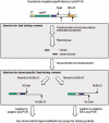
Selection for strand-specific SapI nicking variants. The T7 expression vector for sapIR (pSAPV6) was modified such that nicking by Nt.SapI/Nb.BbvCI or Nb.SapI/Nt.BbvCI resulted in overhangs that can be ligated to adaptors for PCR amplification. A randomly mutated pSAPV6 sapIR library was isolated from an E. coli host without the protection from the cognate MTases so that the plasmid vectors that expressed nicking variants of SapI were nicked in vivo. The nicked plasmid vector was gel-purified and further nicked by Nt.BbvCI or Nb.BbvCI, followed by ligation to the appropriate ss adaptor (and fill-in for the Nb.SapI/Nt.BbvCI reaction). After PCR amplification, the mutated sapIR gene was re-cloned into host cells pre-modified by SapI MTases. Nicking activity was then assayed from the crude extract of induced cultures. T7 promoter is in yellow color, the sapIR gene in green, the SapI recognition site in blue and BbvCI recognition site in orange. The cleavage sites of SapI and BbvCI and their nicking variants are indicated by arrows.
Figure 7.
Figure 7.
NEase-dependent amplification. (A) EXPAR. In EXPAR, the target analyte DNA (trigger; dark orange) anneals to a synthetic amplification template that contains two copies of the sequence complementary to the target sequence (light orange) separated by a sequence complementary to the BstNBI site (light blue). At an elevated temperature (60°C) Bst DNA polymerase large fragment extends at the 3′-end of the trigger DNA and creates a ds-BtsNBI site (dark blue/light blue) followed by an extra copy of the trigger sequence. The thermostable Nt.BstNBI NEase then nicks the top strand of the BstNBI site. By the combined action of the elevated temperature and the strand-displacing activity of Bst DNA polymerase, the newly synthesized trigger sequence is detached from the amplification template and allows the next cycle of extension to proceed in the linear amplification scheme. In the exponential amplification protocol, the extended-cleaved triggers are removed from the amplification template so that the newly synthesized triggers can start a new cycle of extension-nicking. (B) In the extended NEase signal amplification (NESA) scheme that involves multiple temperatures and steps, the initial target-analyte sequence (dark orange) was first amplified by annealing to a padlock DNA designed to close at the target sequence (light blue), followed by ligation of the closed padlock DNA and rolling-circle amplification. The resulting concantemers consist of a tandem repeat of the sequence complementary to a molecular beacon with a BstNBI site (red). The molecular beacon is then allowed to anneal to the concantemers, creating a ds-BstNBI site where Nt.BstNBI nicks the molecular beacon and emits a fluorescent signal. (C) Primer generation-rolling circle amplification (PG-RCA). Similar to EXPAR, the amplification template (light orange) consists of a nicking site (red) sandwiched between two copies of the sequence complementary to the target analyte (dark orange). In this particular example, a BsmI site is used. In PG-RCA, the amplification template is made circular for rolling circle amplification. After annealing of the analyte DNA, Vent (exo) DNA polymerase extends the 3′-end and creates a concantemer of BsmI sites sandwiched between the analyte sequences. Multiple annealing of the circular probe to the concatemers creates ds-strand BsmI sites. Nb.BsmI then nicks the concantemer strands and generates multiple copies of the analyte for the next round of rolling circle amplification.
Figure 8.
Figure 8.
NEases dependent nanocoding. (Upper panel) A T7 bacteriophage DNA molecule was nicked by Nt.BspQI. Alexa Fluor 647-dUTP was incorporated by the nick-translating E. coli DNA polymerase I. The DNA is stained by YOYO-1 (green) and the nick-translated sites (red) are revealed by FRET signal from the Alexa fluorophore. The white bar indicates 1 µm. (Lower panel) Scale for the T7 bacteriophage DNA in kilobites. The red arrows indicate the orientation of the BspQI sites and the direction of nick translation.

Similar articles

Cited by

References

    1. Li H, Stampfer MJ, Hollis JB, Mucci LA, Gaziano JM, Hunter D, Giovannucci EL, Ma J. A prospective study of plasma vitamin D metabolites, vitamin D receptor polymorphisms, and prostate cancer. PLoS Med. 2007;4:e103. - PMC - PubMed
    1. Chang HW, Yang CH, Chang PL, Cheng YH, Chuang LY. SNP-RFLPing: restriction enzyme mining for SNPs in genomes. BMC Genomics. 2006;7:30. - PMC - PubMed
    1. Zilberman D, Henikoff S. Genome-wide analysis of DNA methylation patterns. Development. 2007;134:3959–3965. - PubMed
    1. Nielsen KL. DeepSAGE: higher sensitivity and multiplexing of samples using a simpler experimental protocol. Methods Mol. Biol. 2008;387:81–94. - PubMed
    1. Porter D, Yao J, Polyak K. SAGE and related approaches for cancer target identification. Drug Discov. Today. 2006;11:110–118. - PubMed

Publication types

MeSH terms

Substances