. 2010 Sep 6;190(5):741-9.
doi: 10.1083/jcb.201001048. Epub 2010 Aug 30.

The NuRD chromatin-remodeling complex regulates signaling and repair of DNA damage

Affiliations

The NuRD chromatin-remodeling complex regulates signaling and repair of DNA damage

Godelieve Smeenk et al. J Cell Biol. .

Abstract

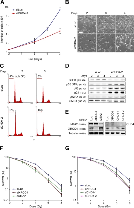
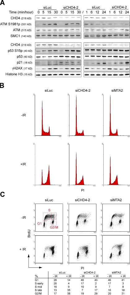
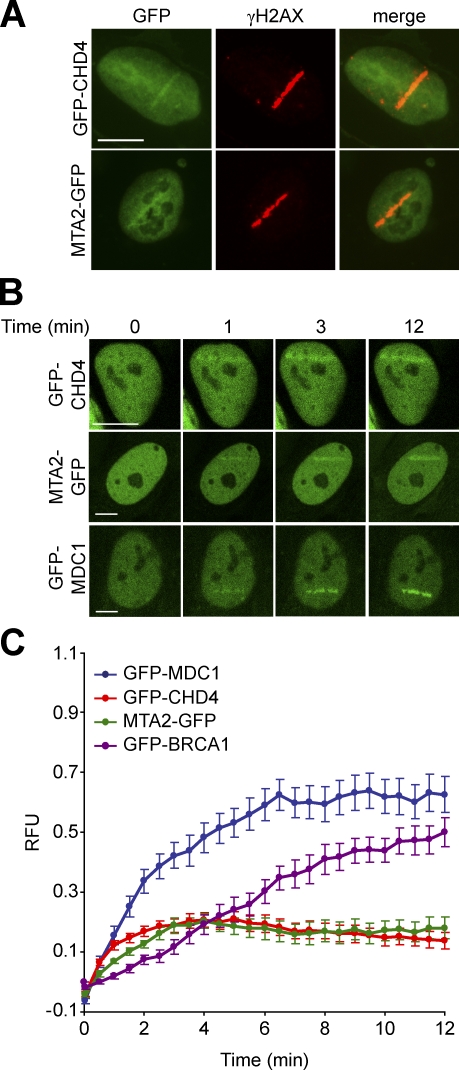
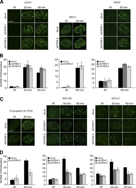
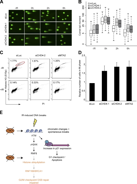
Cells respond to ionizing radiation (IR)-induced DNA double-strand breaks (DSBs) by orchestrating events that coordinate cell cycle progression and DNA repair. How cells signal and repair DSBs is not yet fully understood. A genome-wide RNA interference screen in Caenorhabditis elegans identified egr-1 as a factor that protects worm cells against IR. The human homologue of egr-1, MTA2 (metastasis-associated protein 2), is a subunit of the nucleosome-remodeling and histone deacetylation (NuRD) chromatin-remodeling complex. We show that knockdown of MTA2 and CHD4 (chromodomain helicase DNA-binding protein 4), the catalytic subunit (adenosine triphosphatase [ATPase]) of NuRD, leads to accumulation of spontaneous DNA damage and increased IR sensitivity. MTA2 and CHD4 accumulate in DSB-containing chromatin tracks generated by laser microirradiation. Directly at DSBs, CHD4 stimulates RNF8/RNF168-dependent formation of ubiquitin conjugates to facilitate the accrual of RNF168 and BRCA1. Finally, we show that CHD4 promotes DSB repair and checkpoint activation in response to IR. Thus, the NuRD chromatin-remodeling complex is a novel regulator of DNA damage responses that orchestrates proper signaling and repair of DSBs.


Figures

Figure 1.
Figure 1.
MTA2 or CHD4 depletion renders cells sensitive to IR. (A) Depletion of CHD4 reduces cell proliferation. U2OS cells were transfected with the indicated siRNAs. Cells were counted 0, 2, 3, and 4 d after siRNA transfection. (B) Pictures from representative areas of the cell dishes from A are shown. Bar, 200 µm. (C) FACS analysis of cells from A. PI, propidium iodide. (D) CHD4 depletion leads to enhanced levels of phosphorylated p53 (S15p), p53, p21, and γH2AX. Protein levels were monitored by Western blot analysis using WCEs from cells in A. SMC1 is a loading control. (E) CHD4, MTA2, and XRCC4 levels were monitored by Western blot analysis using WCEs of cells in F and G. Tubulin is a loading control. (F) MTA2 depletion renders cells hypersensitive to IR. VH10-SV40 cells were transfected with the indicated siRNAs. (G) CHD4 depletion renders cells hypersensitive to IR. As in F, except that siRNAs against CHD4 were used. Graphs represent the mean ± SEM of three independent experiments.
Figure 2.
Figure 2.
CHD4 controls IR-induced p53/p21 responses. (A) CHD4 depletion increases IR-induced p53 phosphorylation (S15p), p53, and p21 levels. U2OS cells were transfected with the indicated siRNAs and exposed to 6 Gy of IR. WCEs were prepared at the indicated time points and γH2AX, ATM S1981p, ATM, CHD4, p53 S15p, p53, and p21 levels were monitored by Western blot analysis. Histone H3 and SMC1 were loading controls. (B) CHD4- and MTA2-depleted cells accumulate in G1 phase and remain in G1 arrest after exposure to IR. U2OS cells were transfected with the indicated siRNAs for 72 h, exposed to 6 Gy of IR, and 12 h later, subjected to FACS. (C) As in B, except that cells were stained with BrdU. PI, propidium iodide.
Figure 3.
Figure 3.
CHD4 and MTA2 rapidly accumulate at sites of DNA damage. (A) CHD4 and MTA2 accumulate in DSB-containing regions marked by γH2AX. U2OS cells transiently expressing GFP-CHD4 or MTA2-GFP were subjected to laser microirradiation. After 15 min, cells were immunostained for γH2AX. (B) GFP-CHD4 and MTA2-GFP, like GFP-MDC1, rapidly accumulate in DSB-containing regions. U2OS cells transiently expressing GFP-CHD4, MTA2-GFP, or GFP-MDC1 were microirradiated as in A and subjected to real-time recording of protein assembly at the damaged area. (C) Quantitative representation of the results in B. U2OS cells transiently expressing GFP-BRCA1 were included. Relative fluorescence units (RFU) are plotted on a time scale. Graphs represent the mean ± SEM of at least 10 individual cells from at least two independent experiments. Bars, 10 µm.
Figure 4.
Figure 4.
CHD4 promotes histone ubiquitylation at DSBs to orchestrate the accumulation of RNF168 and BRCA1. (A) CHD4 depletion does not alter γH2AX, MDC1, and RNF8 IRIF formation. Cells were transfected with the indicated siRNAs, exposed to 1 Gy of IR, and 30 and/or 60 min later, immunostained for γH2AX, MDC1, or RNF8. (B) Quantitative analysis of γH2AX, MDC1, and RNF8 IRIF formation. More than 150 nuclei from cells in A were scored per time point in at least two independent experiments. (C) CHD4 depletion impairs ubiquitin, RNF168, and BRCA1 IRIF formation. As in A, except that cells were immunostained for conjugated ubiquitin (FK2), RNF168, and BRCA1. (D) Quantitative analysis of ubiquitin, RNF168, and BRCA1 IRIF formation (as in B). Error bars indicate mean ± SEM. Bars, 10 µm.
Figure 5.
Figure 5.
CHD4 promotes DSB repair and IR-induced activation of the G2/M checkpoint. (A) CHD4 depletion impairs DSB repair. VH10-TERT cells were transfected with the indicated siRNAs for 72 h, exposed to 20 Gy of IR, and subjected to neutral comet analysis at the indicated time points. Representative images are shown. Bar, 30 µm. (B) Quantification of tail moments using cells from A. Tail moments for each condition were calculated on a minimum of 300 cells for each data point. Results of four independent experiments are shown as a box and whisker plot. The ordinate is a cube root scale. Data were statistically analyzed using two-way analysis of variance, which revealed a significant delay in DSB repair for CHD4-depleted cells at 2 h after IR (P < 0.0001). (C) CHD4 or MTA2 depletion impairs IR-induced G2/M checkpoint activation. U2OS cells were transfected with the indicated siRNAs, exposed to 3 Gy of IR, and 1 h later, immunostained for phosphorylated histone H3 (S10p). Mitotic indexes were determined by FACS. A representative experiment is shown. PI, propidium iodide. Red circle indicates the fraction of M phase cells. (D) Graphical representation of relative mitotic index values. The ratio of index values from irradiated and unirradiated cells was calculated and normalized to that for control cells, which was set to 1. The mean ± SEM of four experiments is shown. (E) Model for the role of CHD4 in the maintenance of genome stability. See the last paragraph of Results and discussion for details.

References

    1. Ahel D., Horejsí Z., Wiechens N., Polo S.E., Garcia-Wilson E., Ahel I., Flynn H., Skehel M., West S.C., Jackson S.P., et al. 2009. Poly(ADP-ribose)-dependent regulation of DNA repair by the chromatin remodeling enzyme ALC1. Science. 325:1240–1243 10.1126/science.1177321 - DOI - PMC - PubMed
    1. Bekker-Jensen S., Lukas C., Kitagawa R., Melander F., Kastan M.B., Bartek J., Lukas J. 2006. Spatial organization of the mammalian genome surveillance machinery in response to DNA strand breaks. J. Cell Biol. 173:195–206 10.1083/jcb.200510130 - DOI - PMC - PubMed
    1. Chai B., Huang J., Cairns B.R., Laurent B.C. 2005. Distinct roles for the RSC and Swi/Snf ATP-dependent chromatin remodelers in DNA double-strand break repair. Genes Dev. 19:1656–1661 10.1101/gad.1273105 - DOI - PMC - PubMed
    1. Clapier C.R., Cairns B.R. 2009. The biology of chromatin remodeling complexes. Annu. Rev. Biochem. 78:273–304 10.1146/annurev.biochem.77.062706.153223 - DOI - PubMed
    1. Denslow S.A., Wade P.A. 2007. The human Mi-2/NuRD complex and gene regulation. Oncogene. 26:5433–5438 10.1038/sj.onc.1210611 - DOI - PubMed

Publication types

MeSH terms