. 2010 Sep 6;190(5):731-40.
doi: 10.1083/jcb.200912135. Epub 2010 Aug 30.

The chromatin-remodeling factor CHD4 coordinates signaling and repair after DNA damage

Affiliations

The chromatin-remodeling factor CHD4 coordinates signaling and repair after DNA damage

Dorthe Helena Larsen et al. J Cell Biol. .

Abstract

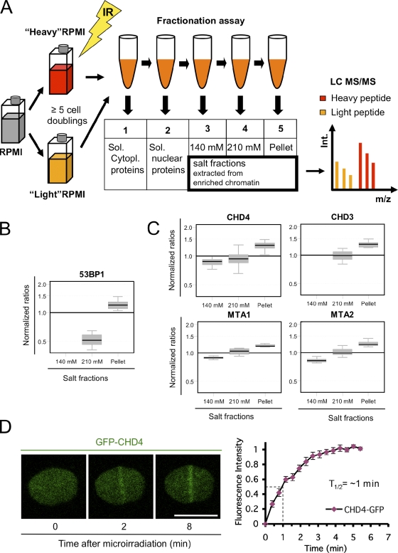
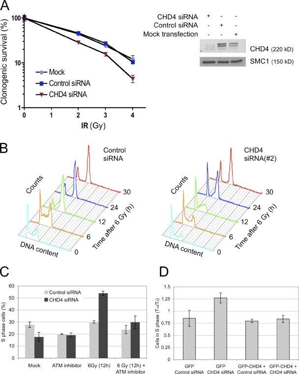
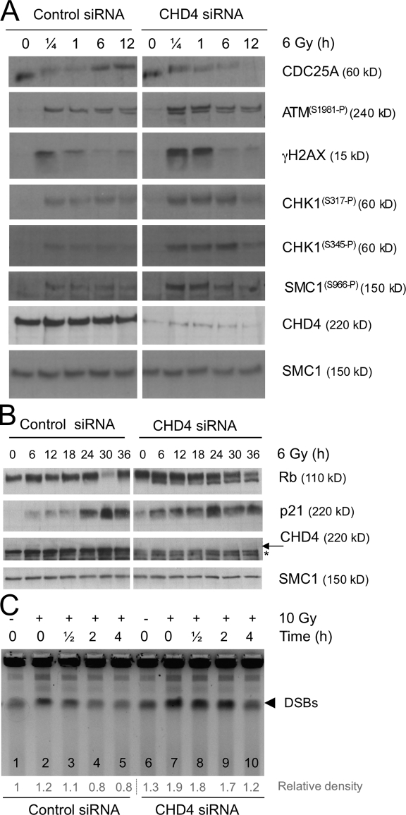
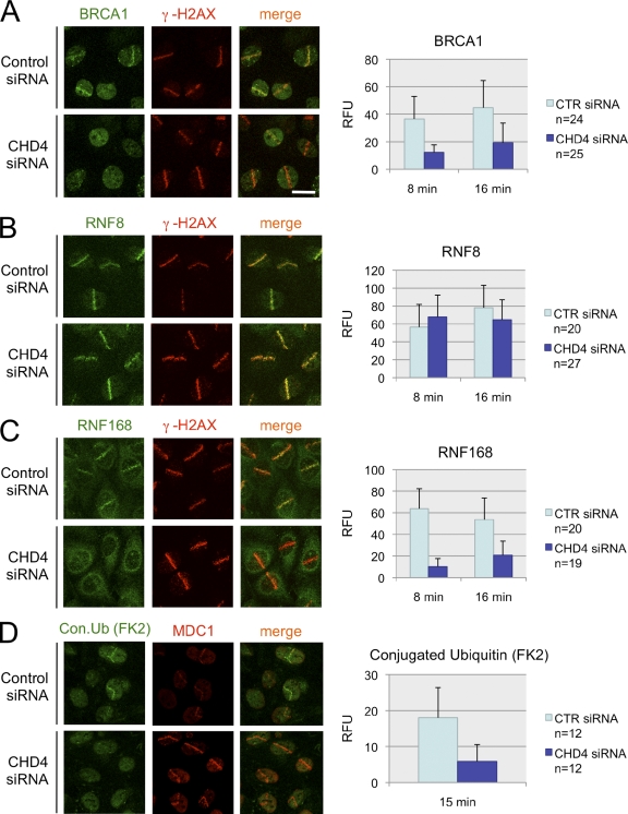
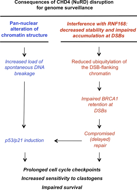
In response to ionizing radiation (IR), cells delay cell cycle progression and activate DNA repair. Both processes are vital for genome integrity, but the mechanisms involved in their coordination are not fully understood. In a mass spectrometry screen, we identified the adenosine triphosphate-dependent chromatin-remodeling protein CHD4 (chromodomain helicase DNA-binding protein 4) as a factor that becomes transiently immobilized on chromatin after IR. Knockdown of CHD4 triggers enhanced Cdc25A degradation and p21(Cip1) accumulation, which lead to more pronounced cyclin-dependent kinase inhibition and extended cell cycle delay. At DNA double-strand breaks, depletion of CHD4 disrupts the chromatin response at the level of the RNF168 ubiquitin ligase, which in turn impairs local ubiquitylation and BRCA1 assembly. These cell cycle and chromatin defects are accompanied by elevated spontaneous and IR-induced DNA breakage, reduced efficiency of DNA repair, and decreased clonogenic survival. Thus, CHD4 emerges as a novel genome caretaker and a factor that facilitates both checkpoint signaling and repair events after DNA damage.


Figures

Figure 1.
Figure 1.
Identification of CHD4 as a factor involved in the DDR. (A) Proteomic screening procedure. GM00130 lymphocytes were grown in heavy or light SILAC media, exposed to 10 Gy of IR, fractionated, and analyzed by tandem mass spectrometry (MS/MS). LC, liquid chromatography. (B) Box plot showing quantitative tandem mass spectrometry data for 53BP1 (positive control). Y axis, normalized ratios (IR peptide/control peptide) showing protein elution by progressive salt fractionation of irradiated lymphocytes relative to control lymphocytes. The box represents the central 50% of the distributions, and the whiskers approximate the 95% interval. (C) Tandem mass spectrometry data for NuRD subunits are shown. Box plots are as in B. (D) Accumulation of GFP-CHD4 at laser-generated DSBs (left) and real-time recruitment of GFP-CHD4 derived from 10 independent cells (right). Error bars indicate SEM. Bar, 10 µm.
Figure 2.
Figure 2.
Knockdown of CHD4 sensitizes cells to IR and deregulates cell cycle progression. (A) Clonogenic survival assay. U2OS cells were treated with control or CHD4 siRNAs (SMARTpool) for 72 h as indicated, irradiated, and colonies with >50 cells were counted. CHD4 down-regulation was monitored by immunoblotting. SMC1, loading control. (B) U2OS cells were treated with control or CHD4 siRNA (#2) for 72 h, irradiated (6 Gy), and analyzed at the indicated time points by flow cytometry. (C) U2OS cells were treated with control or CHD4 siRNAs (SMARTpool), treated with ATM inhibitor for 1.5 h, irradiated, and analyzed by flow cytometry. The efficiency of CHD4 siRNAs in B and C is shown in Fig. S3 B. (D) U2OS cell lines conditionally expressing GFP or GFP-CHD4 resistant to siRNA (#3) were treated with control or CHD4 siRNA (#3) as indicated. After 48 h, the transgenes were induced by addition of doxycycline after an additional 24 h, irradiated, and analyzed by flow cytometry. To compensate for minor differences in the starting S phase content in the two cell lines, the data were normalized and are presented as the ratios between the S phase content measured 10 h after IR (T10) and that in unirradiated cells (T0). The GFP-CHD4 cell line and the efficiency of siRNA (#3) are characterized in Fig. S1 E. Error bars indicate SEM.
Figure 3.
Figure 3.
Extended checkpoint signaling in the absence of CHD4. (A) U2OS cells were treated with control or CHD4 siRNAs (SMARTpool) for 72 h, irradiated, and analyzed by immunoblotting with antibodies and at the specified time points. (B) U2OS cells were treated with siRNAs as in A, irradiated, and analyzed by immunoblotting. Asterisk, nonspecific band; arrow, CHD4. (A and B) Total SMC1, loading control. (C) U2OS were treated with control or CHD4 siRNAs (SMARTpool) for 72 h and irradiated. The DNA breakage was analyzed by PFGE at the indicated time points (top). The relative densities of the DSB bands (bottom) were normalized to the value measured in nonirradiated cells treated with control siRNA (lane 1).
Figure 4.
Figure 4.
Impaired ubiquitylation and delayed accumulation of BRCA1 at the site of DSBs in the absence of CHD4. (A–D) U2OS cells were treated with control (CTR) or CHD4 siRNAs (SMARTpool) for 72 h, microirradiated by the laser, and immunostained with antibodies to BRCA1 (A), RNF8 (B), RNF168 (C), and conjugated ubiquitin (FK2 antibody; D). Cells were coimmunostained with antibodies to γ-H2AX (A–C) or MDC1 (D) to mark the DSB-containing tracks. (left) Representative fields for each DSB regulator (A–C, acquired 8 min after microirradiation; D, acquired 15 min after microirradiation) are shown. (right) Graphs show quantification of relative fluorescence intensities in the microirradiated areas subtracted by the background fluorescence in the undamaged parts of the nucleus. The efficiency of CHD4 siRNAs in A–D is shown in Fig. S3 B. RFU, relative fluorescence units. Error bars indicate SD. Bar, 10 µm.
Figure 5.
Figure 5.
A proposed model of CHD4 involvement in genome maintenance. See Results and discussion for details.

References

    1. Aebersold R., Mann M. 2003. Mass spectrometry-based proteomics. Nature. 422:198–207 10.1038/nature01511 - DOI - PubMed
    1. Bartek J., Lukas J. 2007. DNA damage checkpoints: from initiation to recovery or adaptation. Curr. Opin. Cell Biol. 19:238–245 10.1016/j.ceb.2007.02.009 - DOI - PubMed
    1. Bartek J., Lukas C., Lukas J. 2004. Checking on DNA damage in S phase. Nat. Rev. Mol. Cell Biol. 5:792–804 10.1038/nrm1493 - DOI - PubMed
    1. Bekker-Jensen S., Lukas C., Kitagawa R., Melander F., Kastan M.B., Bartek J., Lukas J. 2006. Spatial organization of the mammalian genome surveillance machinery in response to DNA strand breaks. J. Cell Biol. 173:195–206 10.1083/jcb.200510130 - DOI - PMC - PubMed
    1. Bowen N.J., Fujita N., Kajita M., Wade P.A. 2004. Mi-2/NuRD: multiple complexes for many purposes. Biochim. Biophys. Acta. 1677:52–57 - PubMed

Publication types

MeSH terms