. 2010 Nov;78(11):4560-9.
doi: 10.1128/IAI.00703-10. Epub 2010 Aug 30.

Role of Porphyromonas gingivalis phosphoserine phosphatase enzyme SerB in inflammation, immune response, and induction of alveolar bone resorption in rats

Affiliations

Role of Porphyromonas gingivalis phosphoserine phosphatase enzyme SerB in inflammation, immune response, and induction of alveolar bone resorption in rats

Brian Bainbridge et al. Infect Immun. 2010 Nov.

Abstract

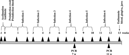
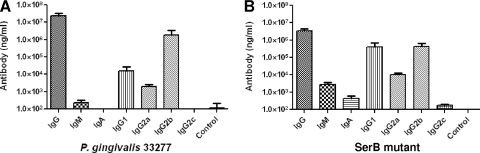
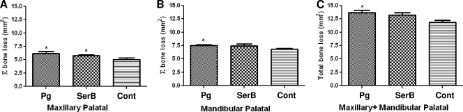
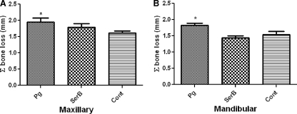
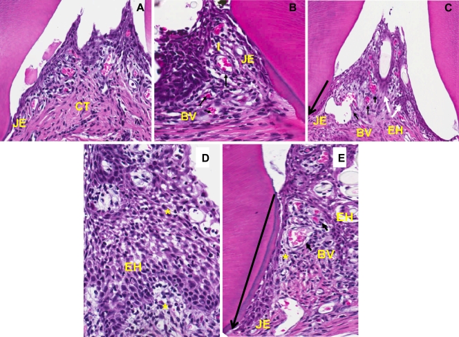
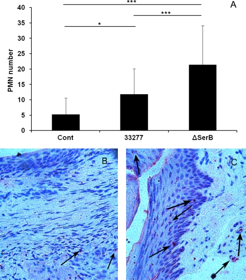
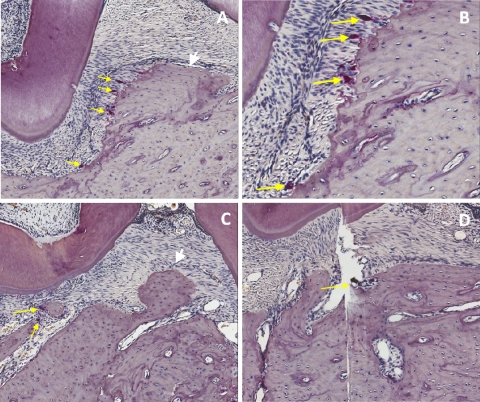
Porphyromonas gingivalis secretes a serine phosphatase enzyme, SerB, upon contact with gingival epithelial cells in vitro. The SerB protein plays a critical role in internalization and survival of the organism in epithelial cells. SerB is also responsible for the inhibition of interleukin-8 (IL-8) secretion from gingival epithelial cells infected with P. gingivalis. This study examined the ability of a P. gingivalis SerB mutant to colonize the oral cavity and induce gingival inflammation, immune responses, and alveolar bone resorption in a rat model of periodontal disease. Both P. gingivalis ATCC 33277 and an isogenic ΔSerB mutant colonized the oral cavities of rats during the 12-week experimental period. Both of the strains induced significant (P < 0.05) systemic levels of immunoglobulin G (IgG) and isotypes IgG1, IgG2a, and IgG2b, indicating the involvement of both T helper type 1 (Th1) and Th2 responses to infection. Both strains induced significantly (P < 0.05) higher levels of alveolar bone resorption in infected rats than in sham-infected control rats. However, horizontal and interproximal alveolar bone resorption induced by the SerB mutant was significantly (P < 0.05) lower than that induced by the parental strain. Rats infected with the ΔSerB mutant exhibited significantly higher levels of apical migration of the junctional epithelium (P < 0.01) and polymorphonuclear neutrophil (PMN) recruitment (P < 0.001) into the gingival tissues than rats infected with the wild type. In conclusion, in a rat model of periodontal disease, the SerB phosphatase of P. gingivalis is required for maximal alveolar bone resorption, and in the absence of SerB, more PMNs are recruited into the gingival tissues.


Figures

FIG. 1.
FIG. 1.
Schematic diagram illustrating the experimental design, including rat acclimation, antibiotics treatment, Peridex swabbing, preinfection, oral microbial sample collection, bacterial infection (6 infections), oral microbial sample collections (two times), PCR analysis, euthanasia, and gingival tissue and alveolar bone collection. For detailed information, see Materials and Methods.
FIG. 2.
FIG. 2.
IgG, IgG subclass (IgG1, IgG2a, IgG2b, and IgG2c), IgM, and IgA antibody levels in sera collected from rats at the end of 12 weeks of infection. (A) P. gingivalis wild-type-infected rats (n = 9); (B) P. gingivalis ΔSerB-infected rats (n = 9). The control used was sera obtained from sham-infected rats. Whole cells of the P. gingivalis wild type or SerB mutant (according to the infection condition) were used as the antigen in an ELISA. Error bars indicate standard deviations.
FIG. 3.
FIG. 3.
Maxillary (A), mandibular (B), and combined (C) morphometric horizontal area alveolar bone resorption in rats (n = 6) following infection with the P. gingivalis wild type or SerB mutant strain. Each bar indicates the mean level of horizontal area alveolar bone loss for the distance measured between the cementoenamel junction and alveolar bone crest of three molar teeth. Error bars indicate standard deviations. Asterisks denote significant differences from values obtained with sham-infected control rats (P < 0.05). Pg, P. gingivalis; SerB, ΔSerB; Cont, sham-infected control.
FIG. 4.
FIG. 4.
Maxillary (A) and mandibular (B) radiographic interproximal alveolar bone resorption in rats (n = 6) following infection with the P. gingivalis wild type or SerB mutant strain. Each bar indicates the mean level of interproximal alveolar bone loss for the distance measured between the cementoenamel junction and alveolar bone crest at mesial and distal sites of three molar teeth (6 sites). Error bars indicate standard deviations. Asterisks denote significant differences from values obtained with sham-infected control rats (P < 0.05). Pg, P. gingivalis; SerB, ΔSerB; Cont, sham-infected control.
FIG. 5.
FIG. 5.
Comparative histology (hematoxylin and eosin staining) of alveolar bone sections from the maxillae of rats after 12 weeks of infection with the P. gingivalis wild type or ΔSerB strain. (A) Section from a control rat displaying minimal inflammation, lack of migration of the junctional epithelium (JE), and minimal inflammation in the connective tissue (CT). (B) Section from a P. gingivalis wild-type-infected rat showing migration of the JE, an increase in the number of blood vessels (BV) (short black arrows), and dense inflammatory infiltrates (I). (C) Section from a SerB mutant-infected rat also exhibiting migration of the JE (long arrow shows direction of apical migration), prominent epithelial hyperplasia (EH) (white arrows), dilated blood vessels, and dense inflammation. Images are shown at 200× magnification. (D) P. gingivalis wild-type-infected tissue demonstrating extensive intra- and subepithelial edema (*), with prominent epithelial hyperplasia. (E) SerB mutant-infected tissue exhibiting extensive intra- and subepithelial edema (*), proliferation of blood vessels, epithelial hyperplasia, and migration of the JE. Images are shown at 600× magnification and are representative of over 5 rats from each group.
FIG. 6.
FIG. 6.
PMN recruitment in rat tissue following infection with the P. gingivalis wild type (33277) or ΔSerB. Cont, sham-infected control. (A) Mean PMN counts in a microscope field. Error bars indicate standard deviations (n = 30). *, P < 0.05; ***, P < 0.001. (B, C) P. gingivalis wild-type-infected (B) and SerB mutant-infected (C) rat tissue stained with anti-PMN antibody (200× magnification). Images are representative of 30 examined in each group.
FIG. 7.
FIG. 7.
Representative histological sections with TRAP staining showing osteoclastic activity from the right maxillae of rats. (A) P. gingivalis wild-type infection demonstrating an alveolar bone crest and tooth with numerous osteoclasts (yellow arrows) in the area of the alveolar bone crest (white arrow). Magnification of ×100. (B) Higher magnification (×200) of the same area as shown in panel A, demonstrating Howship's lacunae and osteoclasts. (C) Infection with the SerB mutant demonstrating the alveolar bone crest (white arrow), interdental area, and few osteoclasts (yellow arrow). Magnification of ×100. (D) Sham-infected control with minimal osteoclastic activity (yellow arrow). Magnification of ×100.

References

    1. Arbibe, L., D. W. Kim, E. Batsche, T. Pedron, B. Mateescu, C. Muchardt, C. Parsot, and P. J. Sansonetti. 2007. An injected bacterial effector targets chromatin access for transcription factor NF-kappaB to alter transcription of host genes involved in immune responses. Nat. Immunol. 8:47-56. - PubMed
    1. Baker, P. J., S. Carter, M. Dixon, R. T. Evans, and D. C. Roopenian. 1999. Serum antibody response to oral infection precedes but does not prevent Porphyromonas gingivalis-induced alveolar bone loss in mice. Oral Microbiol. Immunol. 14:194-196. - PubMed
    1. Baker, P. J., R. T. Evans, and D. C. Roopenian. 1994. Oral infection with Porphyromonas gingivalis and induced alveolar bone loss in immunocompetent and severe combined immunodeficient mice. Arch. Oral Biol. 39:1035-1040. - PubMed
    1. Byrne, S. J., S. G. Dashper, I. B. Darby, G. G. Adams, B. Hoffmann, and E. C. Reynolds. 2009. Progression of chronic periodontitis can be predicted by the levels of Porphyromonas gingivalis and Treponema denticola in subgingival plaque. Oral Microbiol. Immunol. 24:469-477. - PubMed
    1. Colombo, A. V., C. M. da Silva, A. Haffajee, and A. P. Colombo. 2007. Identification of intracellular oral species within human crevicular epithelial cells from subjects with chronic periodontitis by fluorescence in situ hybridization. J. Periodontal Res. 42:236-243. - PubMed

Publication types

MeSH terms

LinkOut - more resources