. 2010 Nov;30(21):5071-85.
doi: 10.1128/MCB.00396-10. Epub 2010 Aug 30.

The ubiquitin carboxyl hydrolase BAP1 forms a ternary complex with YY1 and HCF-1 and is a critical regulator of gene expression

Affiliations

The ubiquitin carboxyl hydrolase BAP1 forms a ternary complex with YY1 and HCF-1 and is a critical regulator of gene expression

Helen Yu et al. Mol Cell Biol. 2010 Nov.

Abstract

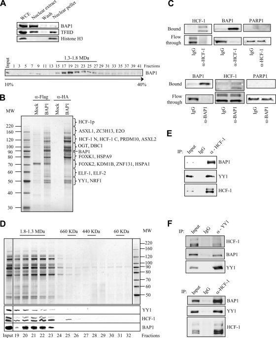
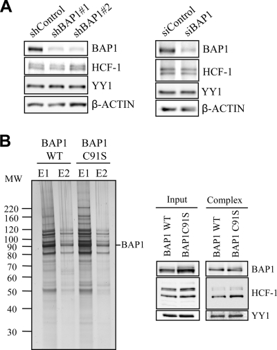
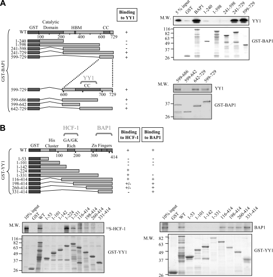
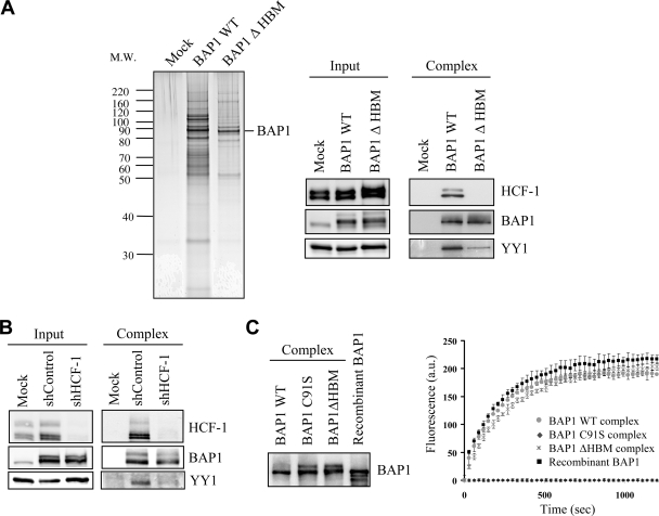
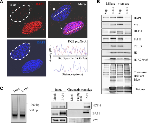
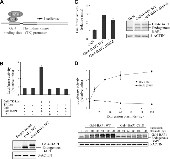
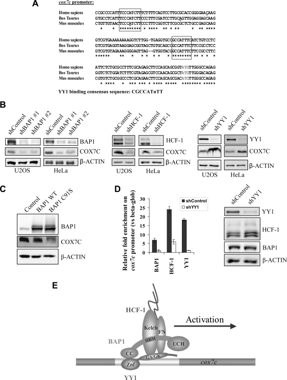
The candidate tumor suppressor BAP1 is a deubiquitinating enzyme (DUB) involved in the regulation of cell proliferation, although the molecular mechanisms governing its function remain poorly defined. BAP1 was recently shown to interact with and deubiquitinate the transcriptional regulator host cell factor 1 (HCF-1). Here we show that BAP1 assembles multiprotein complexes containing numerous transcription factors and cofactors, including HCF-1 and the transcription factor Yin Yang 1 (YY1). Through its coiled-coil motif, BAP1 directly interacts with the zinc fingers of YY1. Moreover, HCF-1 interacts with the middle region of YY1 encompassing the glycine-lysine-rich domain and is essential for the formation of a ternary complex with YY1 and BAP1 in vivo. BAP1 activates transcription in an enzymatic-activity-dependent manner and regulates the expression of a variety of genes involved in numerous cellular processes. We further show that BAP1 and HCF-1 are recruited by YY1 to the promoter of the cox7c gene, which encodes a mitochondrial protein used here as a model of BAP1-activated gene expression. Our findings (i) establish a direct link between BAP1 and the transcriptional control of genes regulating cell growth and proliferation and (ii) shed light on a novel mechanism of transcription regulation involving ubiquitin signaling.


Figures

FIG. 1.
FIG. 1.
BAP1 assembles high-molecular-weight multiprotein complexes containing the YY1 transcription factor. (A) (Top) Extraction of cellular BAP1 protein. HeLa nuclei isolated with hypotonic buffer were extracted with 300 mM KCl for 30 min in order to obtain the nuclear extract and the chromatin/nuclear matrix pellet fractions. The nuclear pellet was washed once. All fractions were resuspended in the same volume and were used for the immunodetection of BAP1. TFIID was detected as a marker for the transcriptional machinery and histone H3 as a marker for chromatin. WCE, whole-cell extract. (Bottom) Endogenous BAP1 migrates in high-molecular-weight fractions. A HeLa nuclear extract was fractionated using glycerol density gradient ultracentrifugation. Fractions collected from the top to the bottom were subsequently used for the immunodetection of BAP1. The gradient was calibrated with the previously purified CtBP complex, whose estimated molecular size is ∼1.3 to 1.5 MDa. (B) Purification of BAP1-associated proteins. A HeLa cell line stably expressing Flag-HA-BAP1 was used for sequential double immunopurification using anti-Flag antibody and anti-HA antibody columns. The Flag- or HA-eluted proteins were separated by SDS-PAGE and detected by silver staining. The mock purification was conducted using a stable cell line generated with the empty vector. Several regions were cut from the gel, and the polypeptides were identified by mass spectrometry. MW, molecular weight (in thousands). (C) Immunodepletion of HCF-1 (top) or BAP1 (bottom) from nuclear extracts using an excess of an anti-HCF-1 or anti-BAP1 polyclonal antibody. A nonrelevant anti-HA polyclonal antibody was used as a control IgG. BAP1 and HCF-1 were immunodetected in the beads and the flowthrough fractions. The nuclear protein PARP1 was detected as a negative control. (D) BAP1 forms high-molecular-weight multiprotein complexes. Fractionation of the BAP1-purified material was performed using a Superose 6 HR gel filtration column. The eluted complexes were detected by silver staining. BAP1, HCF-1, and YY1 were detected by immunoblotting. MW, molecular weight (in thousands). (E) Reciprocal immunoprecipitation. The Flag-purified BAP1 material was used as input for additional immunoprecipitations with a polyclonal antibody against HCF-1 or a nonrelevant anti-GFP antibody (IgG control). The immunocomplexes were extensively washed, and YY1, HCF-1, and BAP1 were detected by immunoblotting. (F) Interaction of endogenous HCF-1, BAP1, and YY1. A HeLa nuclear extract was used for immunoprecipitation with a polyclonal antibody against YY1 (top), a polyclonal antibody against HCF-1 (bottom), or a nonrelevant anti-GFP antibody (IgG control). The immunocomplexes were washed, and YY1, HCF-1, and BAP1 were detected by immunoblotting.
FIG. 2.
FIG. 2.
DUB activity is not required for the assembly of BAP1 complexes or for YY1 stability. (A) Depletion of BAP1 does not affect the steady-state levels of YY1 and HCF-1. (Left) HeLa cells were transfected with either a nontargeting control plasmid (shControl) or a BAP1 shRNA plasmid along with the pBABE puromycin resistance-encoding vector, and transfected cells were selected by addition of puromycin 2 days prior to harvesting for Western blotting using the indicated antibodies. (Right) The siRNA smart pools for human BAP1, or a nontarget control, were transfected into U2OS cells and expressed for 3 days prior to harvesting for Western blotting using the indicated antibodies. (B) BAP1 catalytic activity is not required for the formation of BAP1 complexes. A HeLa cell line stably expressing a Flag-HA-BAP1 catalytically inactive mutant (C91S) was used along with the wild-type control cells for double immunopurification of BAP1 complexes. (Left) Silver staining was conducted on fractions from two elutions (E1 and E2) with an HA peptide. (Right) Immunoblotting for YY1, HCF-1, and BAP1 was conducted.
FIG. 3.
FIG. 3.
YY1 interacts with HCF-1 and BAP1 in vitro. (A) Interaction between YY1 and BAP1 mutants in vitro. Various GST deletion fragments of BAP1 bound to glutathione beads were incubated with His-YY1 for 8 h, and following extensive washes, the bead-associated complexes were analyzed by Coomassie blue staining for GST-BAP1 fragments and by Western blotting for YY1. HBM, HCF-1 binding motif; CC, coiled-coil domain; M.W., molecular weight (in thousands). (B) Interaction between HCF-1 or BAP1 and various YY1 mutants in vitro. (Bottom left) Interaction between YY1 and HCF-1 in vitro. Various GST deletion fragments of YY1 bound to glutathione beads were incubated with in vitro-translated 35S-labeled HCF-1 for 8 h, and following purification, HCF-1 was analyzed by autoradiography. (Bottom right) Identification of the YY1 domain required for interaction with BAP1. Various GST deletion fragments of YY1 were incubated with His-BAP1 for 8 h, and the bead-associated complexes were analyzed by Coomassie blue staining and Western blotting for BAP1.
FIG. 4.
FIG. 4.
HCF-1 is required for the formation of a ternary complex with BAP1 and YY1 in vivo. (A) HCF-1 is required for the proper assembly of BAP1 complexes. A HeLa cell line stably expressing Flag-HA-BAP1 lacking the HBM was used for immunopurification with anti-Flag and anti-HA antibodies. (Left) The eluted material was used for SDS-PAGE and silver staining. (Right) Detection of BAP1, HCF-1, and YY1 by immunoblotting. WT BAP1 was used as a control. (B) Depletion of HCF-1 destabilizes the BAP1 interaction with YY1. A HeLa cell line stably expressing Flag-HA-BAP1 was transfected with either a nontargeting control plasmid (shControl) or an HCF-1 shRNA plasmid along with the pBABE puromycin resistance-encoding vector, and transfected cells were selected by addition of puromycin 2 days prior to harvesting for double immunopurification of BAP1. The eluted proteins were detected by Western blotting using the indicated antibodies. (C) Cleavage of Ub-AMC by various BAP1 complexes (WT, C91S, and ΔHBM) and recombinant BAP1. (Left) Equal quantities of BAP1 were used for deubiquitination reactions with 37.5 pmol of Ub-AMC. (Right) The release of AMC was monitored by fluorescence spectroscopy (excitation wavelength, 380 nm; emission wavelength, 460 nm). All experiments were repeated at least 3 times, and the data presented are means ± standard deviations. a.u., arbitrary units.
FIG. 5.
FIG. 5.
A BAP1/HCF-1/YY1 complex is associated with euchromatin regions. (A) Immunolocalization of BAP1 in U2OS cells, indicating that this DUB is mostly excluded from heterochromatic regions. To ensure the specificity of immunostaining, U2OS cells were transiently transfected with a siRNA against BAP1, and at 3 days posttransfection, cells were used for immunostaining with an anti-BAP1 monoclonal antibody. Following the acquisition of Z-stack images, RGB profiles were generated by the WCIF ImageJ program (NIH). Although most of the cells are depleted of BAP1, some were not transfected and show normal BAP1 expression. In the top left image, the cell delimited by the dashed line has been depleted of BAP1 by RNAi. The other cell shown presumably did not receive the siRNA and expresses normal levels of BAP1. The intensities of fluorescence signals for BAP1 (red) and DNA (blue) at the white bars in the top right image are shown in relative units (RU) at the bottom right. (B) BAP1 and other components of the BAP1 complexes are associated with euchromatin. The chromatin/nuclear matrix fraction was treated with micrococcal nuclease (MNase) to release nucleosomes. Proteins were detected in the soluble (Sup.) and pellet fractions by immunoblotting or Coomassie blue staining. (C) Purification of BAP1/HCF-1/YY1 from the chromatin fraction. The chromatin fraction of HeLa cells stably expressing Flag-HA-BAP1 was digested with MNase (3 U/ml) for 10 min. Following centrifugation at 13,000 × g for 10 min, an aliquot was used for phenol-chloroform extraction of DNA and agarose gel analysis (left). Immunopurification of BAP1 was conducted with the prepared chromatin fraction. The eluted proteins were detected using antibodies against BAP1, YY1, and HCF-1 (right).
FIG. 6.
FIG. 6.
BAP1 activates transcription in a DUB activity-dependent manner. (A) Schematic representation of the Gal4 transcription system. A transcription reporter assay was conducted by targeting BAP1 to the Gal4-TK-luciferase construct by using the Gal4-BAP1 fusion protein. In this process, a protein of interest, fused in frame to the GAL4 DNA binding domain, is targeted to the luciferase reporter driven by a promoter containing GAL4 binding sites and the thymidine kinase proximal promoter. (B) Gal4-BAP1 activates transcription. HeLa cells were transfected with 100 ng of a Gal4-BAP1, BAP1, or Gal4 expression plasmid along with 500 ng of the Gal4-TK-luciferase or TK-luciferase reporter plasmid. Equal expression of various BAP1 constructs was confirmed by Western blotting using anti-BAP1 (bottom), and luciferase activity was measured (top) at 2 days posttransfection. (C) HCF-1 is essentially dispensable for Gal4-BAP1 transcriptional activity. The Gal4 reporter assay was conducted using 500 ng Gal4-TK-luciferase and an equal amount of WT Gal4-BAP1 or Gal4-BAP1 ΔHBM. The expression of BAP1 constructs was monitored by Western blotting (right), and luciferase activity was measured (left) at 2 days posttransfection. (D) The catalytic activity of BAP1 is required for transcription activation. The Gal4 reporter assay was conducted using 500 ng Gal4-TK-luciferase and various amounts of WT Gal4-BAP1 or the catalytically inactive mutant (C91S). The expression of BAP1 constructs was monitored by Western blotting (bottom), and luciferase activity was measured (top) at 2 days posttransfection. All experiments were repeated at least 3 times, and results from a representative experiment are shown. Data are presented as means ± standard deviations.
FIG. 7.
FIG. 7.
BAP1 regulates the expression of genes involved in numerous cellular processes. (A) Functional analysis of genes deregulated following BAP1 depletion. The bar chart was generated by Ingenuity pathways analysis, version 8.5, using 1,244 genes deregulated by both shBAP1s (fold change, less than 0.7 or more than 1.5). P values were calculated using the Fisher exact test. The smaller the P value, the less likely that the association is random. The vertical line marks the cutoff for significance (P, 0.05). (B) RT-PCR analysis of selected genes. U2OS cells were transfected with either a nontargeting control plasmid or a BAP1 shRNA plasmid along with the pBABE puromycin resistance-encoding vector, and transfected cells were selected by addition of puromycin 24 h prior to synchronization at the G1/S border by the double-thymidine-block method. mRNA quantification was conducted by real-time RT-PCR analysis. All experiments were repeated at least 3 times, and the data are presented as means ± standard deviations.
FIG. 8.
FIG. 8.
YY1 recruits BAP1 to coactivate cox7c expression. (A) Alignment of cox7c promoter sequences from various mammalian species: Homo sapiens (NC000005.9 [reference assembly GRCH37]), Bos taurus (NC007305.4 [reference assembly Btau_4.2]), and Mus musculus (NC000079.5 [reference assembly C57BL/6J]). The YY1 binding sites are boxed. The transcription start site is underlined. (B) Expression of COX7C following depletion of BAP1, HCF-1, or YY1. COX7C protein levels following transfection with BAP1 (left), HCF-1 (center), YY1 (right), or nontarget control (shControl) shRNAs in U2OS or HeLa cells are shown. Following transfection and selection with puromycin for 2 days, cells were harvested for immunoblotting. (C) Downregulation of COX7C following the expression of catalytically inactive BAP1. U2OS cells were transduced with retroviral particles to overexpress either BAP1 or its catalytically inactive form (C91S). After 3 days, cells were harvested for Western blotting using the indicated antibodies. (D) cox7c promoter occupancy by YY1, BAP1, and HCF-1. A YY1 shRNA was expressed in U2OS cells by transfection and selection with puromycin for 2 days before harvesting for ChIP (left) or Western blotting (right). ChIP was conducted by using polyclonal antibodies against BAP1, HCF-1, or YY1. An IgG was used as a control. The enrichment of factors was calculated versus the β-globin promoter, used as a control. All experiments were repeated at least 3 times, and the results of a representative experiment are shown. Data are presented as means ± standard deviations. (E) Model representing the recruitment of BAP1 and HCF-1 to the cox7c promoter by the transcription factor YY1.

References

    1. Affar, E. B., F. Gay, Y. Shi, H. Liu, M. Huarte, S. Wu, C. Tucker, E. Li, and Y. Shi. 2006. Essential dosage-dependent functions of the transcription factor Yin Yang 1 in late embryonic development and cell cycle progression. Mol. Cell. Biol. 26:3565-3581. - PMC - PubMed
    1. Bottardi, S., J. Ross, V. Bourgoin, N. Fotouhi-Ardakani, E. B. Affar, M. Trudel, and E. Milot. 2009. Ikaros and GATA-1 combinatorial effect is required for silencing of human gamma-globin genes. Mol. Cell. Biol. 29:1526-1537. - PMC - PubMed
    1. Calvano, S. E., W. Xiao, D. R. Richards, R. M. Felciano, H. V. Baker, R. J. Cho, R. O. Chen, B. H. Brownstein, J. P. Cobb, S. K. Tschoeke, C. Miller-Graziano, L. L. Moldawer, M. N. Mindrinos, R. W. Davis, R. G. Tompkins, and S. F. Lowry. 2005. A network-based analysis of systemic inflammation in humans. Nature 437:1032-1037. - PubMed
    1. Chen, Z. J., and L. J. Sun. 2009. Nonproteolytic functions of ubiquitin in cell signaling. Mol. Cell 33:275-286. - PubMed
    1. Cho, Y. S., E. J. Kim, U. H. Park, H. S. Sin, and S. J. Um. 2006. Additional sex comb-like 1 (ASXL1), in cooperation with SRC-1, acts as a ligand-dependent coactivator for retinoic acid receptor. J. Biol. Chem. 281:17588-17598. - PubMed

Publication types

MeSH terms